DNA Genes Correspond to Physically Distinct Regions in
Plasmodium
falciparum
Julien Guizetti,a,bRafael Miyazawa Martins,a,bStéphanie Guadagnini,cAurélie Claes,a,bArtur Scherfa,b
Département de Parasitologie et Mycologie, Unité de Biologie des Interactions Hôte-Parasite, Institut Pasteur, Paris, Francea
; CNRS URA2581, Paris, Franceb ; Institut Pasteur, Plate-Forme de Microscopie Ultrastructurale, Imagopole, Paris, Francec
The human malaria parasitePlasmodium falciparummodifies the erythrocyte it infects by exporting variant proteins to the host cell surface. Thevargene family that codes for a large, variant adhesive surface protein calledP.falciparumerythrocyte mem-brane protein 1 (PfEMP1) plays a particular role in this process, which is linked to pathogenesis and immune evasion. A single member of this gene family is highly transcribed while the other 59 members remain silenced. Importantly,vargene transcrip-tion occurs at a spatially restricted, but yet undefined, perinuclear site that is distinct from repressedvargene clusters. To ad-vance our understanding of monoallelic expression, we investigated whether nuclear pores associate with thevargene expres-sion site. To this end, we studied the nuclear pore organization during the asexual blood stage using a specific antibody directed against a subunit of the nuclear pore,P.falciparumNup116 (PfNup116). Ring and schizont stage parasites showed highly polar-ized nuclear pore foci, whereas in trophozoite stage nuclear pores redistributed over the entire nuclear surface. Colocalization studies ofvartranscripts and anti-PfNup116 antibodies showed clear dissociation between nuclear pores and thevargene ex-pression site in ring stage. Similar results were obtained for another differentially transcribed perinuclear gene family, the ribo-somal DNA units. Furthermore, we show that in the poised state, thevargene locus is not physically linked to nuclear pores. Our results indicate thatP. falciparumdoes form compartments of high transcriptional activity at the nuclear periphery which are, unlike the case in yeast, devoid of nuclear pores.
A
s shown in yeast and mammals, nuclear pores can define compartments of high transcriptional activity at the nuclear periphery (1,2). Furthermore, relocation and tethering of specific genes to the nuclear pore complex provide transcriptional mem-ory that enables quicker reactivation of those genes (3). In the human malaria parasitePlasmodium falciparum, the major viru-lence factor, encoded by thevargene family, is transcribed at a yet undefined site at the nuclear periphery in a monoallelic fashion. Hence, the possibility that nuclear pores play a role in monoallelic vargene activation is an appealing hypothesis.P. falciparumcauses the most severe form of malaria, a para-sitic disease killing hundreds of thousands people a year (4,5). Clinical symptoms of malaria arise during its 48-h replication cycle within human red blood cells. After invasion of a blood cell by a merozoite, the parasite remains in ring stage for 24 h postin-vasion (hpi). In the trophozoite stage (25 to 38 hpi), it grows and replicates its genome. Segregation of the chromosomes into the newly forming daughter nuclei and cytokinesis occur in the schi-zont stage (38 to 48 hpi). Subsequently, 16 to 32 newly formed merozoites will be released upon rupture of the infected red blood cell and start a new infectious cycle.
This intracellular parasite expresses several proteins that are transported to the red blood cell surface and that have multiple functions, such as sequestrating infected red blood cells to the vascular endothelium (6). These proteins are exposed to recogni-tion by the host immune system. Hence, to prolong infecrecogni-tion and ensure effective transmission, the parasite has evolved sophisti-cated strategies for immune evasion. Antigenic variation allows the parasite to change its gene expression profile by switching between different members of gene families coding for surface proteins (7).
varis the best-studied variant gene family coding for the major virulence surface protein P. falciparumerythrocyte membrane protein 1 (PfEMP1). Monoallelic expression governs the tran-scription of only a single gene of the 60-member gene family, whereas all other genes remain silenced (8). Expression of thevar gene peaks around 10 to 14 hpi (9), but during later developmen-tal stages it remains in a state “poised” for reactivation in the next cycle (10). Recently, a histone methyltransferase, PfSet10, has been shown to colocalize specifically with the poisedvargene (11). Epigenetic regulation, such as histone posttranslational lysine modification, is an essential component in monoallelic expression (12). A second essential factor is the spatial regrouping ofvar genes at the nuclear periphery (13,14). Apparently, this organiza-tion is important to establish a default silencing pathway via fac-ultative heterochromatin.
Silent and activevargenes localize to the nuclear periphery, andvargene activation requires the relocation to a perinuclear site that remains undefined to date (13). This has raised the hypothesis that a predefinedvargene expression site, similar to the expression site body for variant surface glycoprotein (VSG) genes in
Trypano-Received1 February 2013Accepted27 February 2013
Published ahead of print8 March 2013
Address correspondence to Artur Scherf, [email protected].
Supplemental material for this article may be found athttp://dx.doi.org/10.1128
/EC.00023-13.
Copyright © 2013, American Society for Microbiology. All Rights Reserved.
doi:10.1128/EC.00023-13
The authors have paid a fee to allow immediate free access to this article.
on September 8, 2020 by guest
http://ec.asm.org/
soma brucei(15), might exist inP. falciparum. Besidesvargenes, rRNA is among the most highly expressed RNA species in plasmo-dia. rRNA genes, such as the 18S RNA, cluster in the nucleolus, a specific perinuclear subcompartment (16). Data from model or-ganisms suggested that proximity to the nuclear pore could enable more efficient transcription of highly expressed genes (17).
Ultrastructural analysis using focused-ion-beam scanning electron microscopy (EM) (“slice and view”) has enabled the first visualization of nuclear pore organization in three dimensions (3D) during theP. falciparumasexual blood stages (18). This tech-nique, however, does not allow labeling of molecular markers to link morphological features to functional studies.
In this study, we investigate nuclear pore distribution through-out the parasite’s blood stage development by immunofluores-cence using a specific antibody raised against a nuclear pore sub-unit, PfNup116. By combining immunofluorescence and RNA fluorescencein situhybridization (FISH), we tested the hypothesis whether nuclear pores are associated with thevargene expression site and ifvargene poising requires its sequestration to a nuclear pore. We show that perinuclear gene transcription sites andvar gene poising do not depend on nuclear pore association in P. falciparum.
MATERIALS AND METHODS
Parasite culture and synchronization.P. falciparumblood stage parasites were cultivated as described previously (19). Parasite culture was synchro-nized by sorbitol lysis during ring stage, subsequent plasmagel enrichment in schizont stage, and then by another sorbitol treatment at 6 h postinva-sion. Synchronized parasites were harvested at 4% hematocrit and⬃5% parasitemia. Parasite development was monitored by Giemsa staining.
Antibody.In order to produce specific antibodies against a plasmodial nuclear pore protein (PfNup116; PF3D7_1473700) rabbits were immu-nized with the synthetic peptide PQNTSSGTYQPQNTS conjugated to keyhole limpet hemocyanin (KLH) to generate a polyclonal anti-PfNup116 antibody according to standard protocols of GenScript (cata-log number SC1031). Serum from immunized rabbits was affinity puri-fied on a column with resin coupled to the antigenic peptide. After multiple washes, the eluate was collected, neutralized, and dialyzed with phosphate-buffered saline (PBS).
Western blotting.After lysis of infected red blood cells with 0.15% saponin, nuclear and cytoplasmic fractions of the isolated parasites were separated by mechanical douncing as described previously (20). After being washed in PBS, nuclei were directly extracted in Laemmli buffer. Protein extracts were separated on a 4 to 12% SDS-PAGE gel (Criterion, Bio-Rad) and analyzed by Western blotting using the rabbit anti-PfNup116 at 1:500. Horseradish peroxidase (HRP)-conjugated anti-P. falciparumaldolase (ab38905; Abcam) at 1:1,000 and rabbit anti-histone 3 (ab1791; Abcam) at 1:2,000 were used to assess purity of cytoplasmic and nuclear fractions as described previously (21). Secondary antibody detec-tion was done with anti-rabbit-HRP (GE health care) at 1:5,000. A Super-Signal West Pico ECL Western blotting kit (Thermo Scientific) was used to develop blots.
Freeze-fracture and cryo-scanning electron microscopy.Infected red blood cells were fixed in 2.5% glutaraldehyde in PBS overnight at 4°C and washed three times with PBS. One drop of blood cell pellet was pipetted into a cryo-freezer hat type B (G3467; Agar Scientific), a 100-mesh EM grid (G2410PD; Agar Scientific) was placed on top, and the sample was closed up by a second drop and cryo-freezer hat. Sandwiched samples were high-pressure frozen in an HPM 010 (Bal-Tec) freezing machine. After samples were frozen, they were transferred into an Alto 2500 cryo-transfer system (Gatan) and fractured manually. Sublimation was carried out at⫺105°C for 2 min. Then samples were coated with 9.5 nm of chromium in a 681 High Resolution Ion Beam Coater (Gatan).
Fractured cells were imaged with a JSM-6700F electron microscope (Jeol) using a yttrium aluminum garnet (YAG) detector at 5 kV and⫺120°C.
Immunofluorescence and RNA FISH.Infected red blood cells were lysed with 0.015% saponin in RPMI medium, and the released parasites were fixed in suspension with 4% paraformaldehyde in PBS overnight at 4°C. Parasites were then deposited on number 1.5 cover glasses, perme-abilized for 15 min with 0.1% Triton X-100, and subjected to immuno-fluorescence staining and RNA FISH as described previously (22). FISH probes were PCR amplified from genomic DNA using the primers 5=-CA GTAGTCATATGCTTGTC-3= and 5=-TAATGATCCTTCCGCAGG-3=
for 18S rRNA and 5=-CACACCACTGGACGTCTGCAA-3=and 5=-CTA GTGGATGAGGATTGGGTG-3=for PF3D7_0412700 mRNA and labeled by nick translation using a Fluorescein-High Prime kit (Roche). For se-quential immunofluorescence assay and RNA FISH, parasites were first hybridized with FISH probes at 37°C overnight. After they were washed, they were postfixed in 4% paraformaldehyde in PBS for 15 min and then subjected to immunofluorescence staining. Antibodies were diluted 1:200 in 0.5% pork skin gelatin in PBS for anti-PfNup116 and at 1:300 for mouse anti-hemagglutinin ([HA] ab18181; Abcam) and detected with Alexa Fluor 568-conjugated anti-rabbit IgG and Alexa Fluor 488-conju-gated anti-mouse IgG (Invitrogen) diluted 1:2,000. Images were captured using a Nikon Eclipse 80i microscope with a CoolSnap HQ2 camera (Pho-tometrics). NIS Elements, version 3.0, software (Nikon) was used for ac-quisition, and Fiji software (http://fiji.sc) was used for analysis.
Quantitative analysis of images.Nuclear pore polarization was quan-tified by scoring whether all detectable PfNup116 fluorescent dots local-ized within one quadrant of the nucleus (seeFig. 2C). Colocalization was scored in cell nuclei positive for both fluorescent signals when they over-lapped while using similar contrast adjustments throughout all images. Scoring was performed by direct optical observation using Fiji software (http://fiji.sc). For each analysis, images from three independent replicas were combined. For the movie in the supplemental material, az-stack was acquired and deconvolved using Huygens software (SVI).
RESULTS
Nuclear pore distribution changes throughout the developmen-tal blood stages.Scanning electron microscopy images of freeze-fractured infected red blood cells show clusters of pores on the nuclear surface (Fig. 1A). Their symmetric structure and diameter are consistent with the expected organization of the nuclear pore complex. Surprisingly, only one subunit protein of nuclear pores, PfNup100 (23,24), has been annotated in the genome ofP. falcip-arum (www.plasmoDB.org). To investigate nuclear pore posi-tioning, we evaluated a novel antibody against a putative nuclear pore protein (PF3D7_1473700). It shows homology with the Sac-charomyces cerevisiaenucleoporin Nup116 (34% identity and 50% similarity). Rat serum raised against this protein has been previ-ously used for nuclear pore labeling inP. falciparum(19,25). In this work, a polyclonal affinity-purified rabbit antibody was raised against a 15-amino-acid synthetic peptide from the central region of PfNup116 (Fig. 1B). We validated antibody specificity by West-ern blotting. In protein extracts from asynchronous parasite cul-ture, we detected a major band of 110 kDa, which migrates slightly lower than the predicted size of 142 kDA. This protein is highly enriched in the nuclear fraction (Fig. 1C). The efficient separation of the nuclear and cytoplasmic fractions was controlled for with anti-histone 3 and anti-aldolase antibodies, respectively (Fig. 1C, lower panels). To further characterize this nuclear pore protein, we carried out immunofluorescence localization studies on highly synchronized blood stage parasites throughout their intraerythro-cytic development. We compared immunofluorescence staining of nuclear pores in early ring stages (around 12 hpi) with tropho-zoite stages (around 32 hpi) and late, segmented schizont stages
on September 8, 2020 by guest
http://ec.asm.org/
(around 42 hpi) (Fig. 2A). The anti-PfNup116 signal concentrates in foci at the nuclear periphery. Interestingly, nuclear pores formed a polarized cluster in very early rings. Most ring stages showed a single dot, whereas about 30% showed two dots, and very few cells showed three dots (Fig. 2B). As parasites progress through their cycle, nuclear pores redistribute around the nuclear envelope, and signal intensity increases. During schizont stages the number of nuclear pore foci per nucleus diminishes again, most likely to a single dot, but due to spatial proximity the signals can only sometimes be attributed to individual nuclei (see Movie S1 in the supplemental material). While early-stage cells (n⫽74) have only one or two nuclear pore foci, the number of foci in-creases markedly in late-stage cells (n⫽170), whereas frequently individual clusters are not distinguishable any longer (Fig. 2A). Also, the polarization of nuclear pores in the ring stage is lost in the trophozoite stage (Fig. 2C).
Our immunofluorescence data using a nuclear pore marker are in agreement with previous ultrastructural data on nuclear pore distribution throughout the intraerythrocytic development cycle (18). This localization pattern indicates that our molecular pore marker is a valuable tool for functional studies inP. falciparum.
Activevargene and 18S RNA expression sites do not associ-ate with nuclear pores.Nuclear pores could form an “anchor” site required for monoallelicvargene expression. Further, proximity to nuclear pores could facilitate transcription and mRNA export of highly expressed genes. To test whether thevargene expression site colocalizes with nuclear pores, we combined RNA FISH of an activevargene with anti-PfNup116 immunostaining. rRNA, ex-pressed in the nucleolus, is the most highly transcribed RNA spe-cies inPlasmodium. Hence, we also tested whether nuclear pores would position closely to the 18S RNA transcription sites.
Asvargene expression peaks around 10 to 14 hpi, we ana-lyzed tightly synchronized ring stage parasites. We used the 3D7-G7 clone, which predominantly expresses the var gene PF3D7_0412700 against which our FISH probe was targeted (19). No significant colocalization of nuclear PF3D7_0412700 mRNA signal with the nuclear pore signal could be detected (Fig. 3A). Similarly, 18S RNA expression sites rarely colocalized with the PfNup116 signal (Fig. 3B). Since at this early stage the pores are still polarized, scoring colocalization with 18S RNA (n⫽32) and PF3D7_0412700 mRNA (n⫽33) was greatly facilitated (Fig. 3C). The frequency of random colocalization can be estimated by geo-metric calculation. The colocalization images we analyzed are two-dimensional. Ring stage nuclei have a diameter of 1m and therefore a circumference of 3.14m. The diameters of the fluo-rescence signals we see for the nuclear pore antibody and the FISH probe range from 0.2 to 0.3m. If both signals localize randomly on a circle of 3.14-m circumference (nuclear periphery), their
FIG 1Antibody targeting nuclear pore protein PfNup116. (A) Scanning elec-tron microscopy image of freeze-fractured infected red blood cells with a par-asite at the trophozoite stage. N, nucleus; C, parpar-asite cytoplasm; B, red blood cell. Scale bar, 500 nm. The nuclear surface with a nuclear pore cluster (boxed) is magnified in the lower panel. Scale bar, 100 nm. (B) Scheme showing posi-tion of the peptide within the PF3D7_1473700 (PfNup116) protein sequence used to immunize rabbits. (C) Western blot of nuclear and cytoplasmic para-site extracts detected with PfNup116 antibody. Anti-histone 3 and anti-aldolase antibody detection show efficient separation of cytoplasmic (C) and nuclear (N) fractions. aa, amino acids.
FIG 2Nuclear pore redistribution throughout the intraerythrocytic stages. (A) Immunofluorescence staining of nuclear pores using anti-PfNup116 at different intraerythrocytic stages. The schizont stage image is a maximum-intensity projection. The nucleus is stained with 4=,6= -diamidino-2-phenylin-dole (DAPI). Scale bar, 1m. (B) Fraction of nuclear pore-stained nuclei with different numbers of PfNup116 foci in the ring and trophozoite stages. (C) Fraction of cells with all PfNup116 foci within one quadrant of the nucleus in the ring and trophozoite stages. Schemata, which represent nuclei (blue) with two nuclear pore signals (red), illustrate polarized localization within one quadrant (upper circle) versus nonpolarized localization (lower circle).
on September 8, 2020 by guest
http://ec.asm.org/
predicted colocalization frequency ranges from 12 to 19%. These values are in the range of observed colocalization frequencies re-ported inFig. 3C. Our results strongly suggest that nuclear pores do not recruit or host the vargene expression site. Also, the other known perinuclear expression site, containing the ribosomal DNA (rDNA) genes, does not associate to nuclear pores.
var gene poising is not associated with nuclear pores.In yeast, it has been shown that certain regulated genes are tethered to the nuclear pore complex for extended periods of time, which enables rapid reactivation of transcription upon external stimula-tion (3). Similarly,vargenes remain in a poised state during the trophozoite and schizont stages before being reactivated at the start of the next cycle. We tested whethervargene poising occurs at the nuclear pore. A putative methyltransferase, PfSet10, which is expressed only in later stages, has recently been shown to colo-calize specifically with the poisedvargene (11).
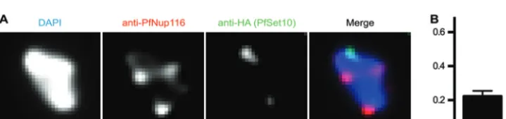
We used a parasite line expressing endogenous PfSet10 tagged with hemagglutinin (HA) to carry out double immunostaining with anti-HA and anti-PfNup116 antibodies on late-stage para-sites (11). For quantification, we selected cells positive for both signals but with a limited number of nuclear pore foci to clearly distinguish between colocalizing and noncolocalizing signals (Fig. 4A). In the parasites we scored (n⫽45), PfSet10-HA signal rarely colocalized with the PfNup116 signal (Fig. 4B). We conclude that vargene poising is not associated with nuclear pores.
DISCUSSION
This is, to our knowledge, the first study that investigates the role of nuclear pores in monoallelic gene expression.P. falciparumis a unique organism that uses perinuclear expression sites for gene families that are mutually exclusively expressed. So far, no specific molecular marker has been identified that characterizes these sites. Given the recent evidence for roles of nuclear pores in regu-lating gene expression, we explored the idea of whether pores as-sociate with thevargene expression site.
Our study supports and expands the ultrastructural data on the unusual distribution pattern of nuclear pores inP. falciparumby immunofluorescence staining (18). Strong polarization of pores, found in early-stage parasites, has been very rarely described in other cell types and organisms. In late development of mouse spermatids, nuclear pores are clustered at the caudal pole of the nucleus, opposite to the acrosomal cap (26). In budding yeast, deletion of Nup133, a nucleoporin, causes extensive clustering of nuclear pores (27). A recent paper shows that motor proteins and cytoskeletal elements are important for uniform distribution of nuclear pores in fungal nuclei, suggesting that an active process prevents their clustering (28). Weiner et al. argue that ring and schizont stage nuclei are in close contact with the plasma mem-brane, and therefore nuclear pores polarize to the side of the nu-clear envelope with free access to the cytoplasm (18). Even though
FIG 3Colocalization of gene expression sites with nuclear pores. (A) Combination of anti-PfNup116 immunofluorescence staining with RNA FISH using a probe against the activevargene expressed in the 3D7-G7 clone (PF3D7_0412700). The nucleus is stained by DAPI. Scale bar, 1m. (B) The experiment as described for panel A but with a FISH probe against 18S rRNA. (C) Fraction of cells with an overlap of FISH and nuclear pore foci.
FIG 4Colocalization of PfSet10-HA with nuclear pores. (A) Combination of anti-PfNup116 and anti-HA immunofluorescence staining on an early-tropho-zoite-stage parasite expressing recombinant PfSet10-HA. The nucleus is stained by DAPI. Scale bar, 1m. (B) Fraction of cells with an overlap of PfSet10-HA and nuclear pore foci.
on September 8, 2020 by guest
http://ec.asm.org/
data on nuclear pore biology ofP. falciparumis still very scarce, a recent study of theP. falciparumnuclear proteome yielded inter-esting candidate proteins, whose further characterization can im-prove understanding of nuclear pore organization (23).
Ribosomal DNA genes and var genes are among the most highly transcribed RNA species inP. falciparum(16). As neither of them is expressed in the proximity of the nuclear pore, this shows that the spatial proximity is not limiting for efficient RNA export into the cytoplasm, as has been suggested by models andin vivo diffusion measurements in other organisms (29). As 18S RNA is expressed in the nucleolus, we can conclude that nucleolar posi-tioning is independent of nuclear pore localization.
What defines thevargene expression site at the nuclear periph-ery is a long-standing question in the field. Since nuclear pores have been previously associated with transcriptional activation, as proposed first in the gene-gating hypothesis (30), they are attrac-tive candidates for recruitment sites of the activatedvargene. Our observations, however, show that tethering to the nuclear pore does not occur for activevarand rDNA genes. The only factor known to spatially organizevargenes remains actin, which con-tributes to the tethering of centralvargenes to the nuclear periph-ery (25). A number of other chromatin components have been identified that are enriched at the nuclear periphery; however, no functional data are yet available that demonstrate a role of these proteins in the spatial organization of silent or activevargenes through, e.g., chromosome end clustering (31,32). Recently, it has been shown that spatial segregation is also important for activa-tion of other clonally variant genes. As relocating the silent PfRh4 gene to a transcriptionally active site increases its probability of being switched on, the authors hypothesized that translocation precedes activation (33).
Gene poising is another important aspect of antigenic varia-tion. Keeping a single gene marked for reactivation reduces switching to another member of the family and therefore prevents premature depletion of the “immunogenic repertoire.” Poten-tially, the poised gene could be maintained at a defined site, marked by PfSet10 (11), throughout the late parasite stages. This would provide positional memory for reactivation of the samevar gene. If, however, positional memory exists in the context ofvar gene poising, it is not associated with the nuclear pore.
In conclusion, our work points to a distinctive perinuclear compartment involved in monoallelic exclusion. The identifica-tion of molecular markers associated to this expression site is of key importance to gain new insight into the virulence process of malaria parasites.
ACKNOWLEDGMENTS
We thank Jose Juan Lopez-Rubio for providing the 3D7-G7 clone as well as Jennifer Volz and Alan Cowman for providing the Set10-HA clone.
This work was supported by an ERC Advanced Grant (PlasmoEscape 250320), ANR grant (number 09-BLAN-0274-01), the French parasitol-ogy network of excellence ParaFrap, and a Human Frontier Science Pro-gram fellowship to J.G.
REFERENCES
1.Arib G, Akhtar A.2011. Multiple facets of nuclear periphery in gene expression control. Curr. Opin. Cell Biol.23:346 –353.
2.Dieppois G, Stutz F.2010. Connecting the transcription site to the nu-clear pore: a multi-tether process that regulates gene expression. J. Cell Sci.
123:1989 –1999.
3.Brickner DG, Brickner JH.2012. Interchromosomal clustering of active genes at the nuclear pore complex. Nucleus3:487– 492.
4.Snow RW, Guerra CA, Noor AM, Myint HY, Hay SI.2005. The global distribution of clinical episodes ofPlasmodium falciparummalaria. Na-ture434:214 –217.
5.Murray CJ, Rosenfeld LC, Lim SS, Andrews KG, Foreman KJ, Haring D, Fullman N, Naghavi M, Lozano R, Lopez AD.2012. Global malaria mortality between 1980 and 2010: a systematic analysis. Lancet379:413– 431.
6.Maier AG, Cooke BM, Cowman AF, Tilley L. 2009. Malaria parasite proteins that remodel the host erythrocyte. Nat. Rev. Microbiol.7:341– 354.
7.Scherf A, Lopez-Rubio JJ, Riviere L.2008. Antigenic variation in Plas-modium falciparum. Annu. Rev. Microbiol.62:445– 470.
8.Scherf A, Hernandez-Rivas R, Buffet P, Bottius E, Benatar C, Pouvelle B, Gysin J, Lanzer M.1998. Antigenic variation in malaria: in situ switch-ing, relaxed and mutually exclusive transcription ofvargenes during in-tra-erythrocytic development inPlasmodium falciparum. EMBO J.17: 5418 –5426.
9.Schieck E, Pfahler JM, Sanchez CP, Lanzer M.2007. Nuclear run-on analysis of var gene expression inPlasmodium falciparum. Mol. Biochem. Parasitol.153:207–212.
10. Lopez-Rubio JJ, Gontijo AM, Nunes MC, Issar N, Hernandez Rivas R, Scherf A.2007. 5=Flanking region ofvargenes nucleate histone modifi-cation patterns linked to phenotypic inheritance of virulence traits in malaria parasites. Mol. Microbiol.66:1296 –1305.
11. Volz JC, Bartfai R, Petter M, Langer C, Josling GA, Tsuboi T, Schwach F, Baum J, Rayner JC, Stunnenberg HG, Duffy MF, Cowman AF.2012. PfSET10, aPlasmodium falciparummethyltransferase, maintains the ac-tivevargene in a poised state during parasite division. Cell Host Microbe
11:7–18.
12. Scherf A, Riviere L, Lopez-Rubio JJ.2008. SnapShot:vargene expression in the malaria parasite. Cell134:190.
13. Ralph SA, Scheidig-Benatar C, Scherf A.2005. Antigenic variation in
Plasmodium falciparumis associated with movement ofvarloci between subnuclear locations. Proc. Natl. Acad. Sci. U. S. A.102:5414 –5419. 14. Freitas-Junior LH, Hernandez-Rivas R, Ralph SA, Montiel-Condado D,
Ruvalcaba-Salazar OK, Rojas-Meza AP, Mancio-Silva L, Leal-Silvestre RJ, Gontijo AM, Shorte S, Scherf A.2005. Telomeric heterochromatin propagation and histone acetylation control mutually exclusive expres-sion of antigenic variation genes in malaria parasites. Cell121:25–36. 15. Navarro M, Gull K.2001. A pol I transcriptional body associated with
VSG mono-allelic expression inTrypanosoma brucei. Nature414:759 – 763.
16. Mancio-Silva L, Zhang Q, Scheidig-Benatar C, Scherf A.2010. Cluster-ing of dispersed ribosomal DNA and its role in gene regulation and chro-mosome-end associations in malaria parasites. Proc. Natl. Acad. Sci. U. S. A.107:15117–15122.
17. Akhtar A, Gasser SM.2007. The nuclear envelope and transcriptional control. Nat. Rev. Genet.8:507–517.
18. Weiner A, Dahan-Pasternak N, Shimoni E, Shinder V, P von Huth Elbaum M, Dzikowski R.2011. 3D nuclear architecture reveals coupled cell cycle dynamics of chromatin and nuclear pores in the malaria parasite
Plasmodium falciparum. Cell Microbiol.13:967–977.
19. Lopez-Rubio JJ, Mancio-Silva L, Scherf A.2009. Genome-wide analysis of heterochromatin associates clonally variant gene regulation with peri-nuclear repressive centers in malaria parasites. Cell Host Microbe5:179 – 190.
20. Chene A, Vembar SS, Riviere L, Lopez-Rubio JJ, Claes A, Siegel TN, Sakamoto H, Scheidig-Benatar C, Hernandez-Rivas R, Scherf A.2012. PfAlbas constitute a new eukaryotic DNA/RNA-binding protein family in malaria parasites. Nucleic Acids Res.40:3066 –3077.
21. Dastidar EG, Dzeyk K, Krijgsveld J, Malmquist NA, Doerig C, Scherf A, Lopez-Rubio JJ.2013. Comprehensive histone phosphorylation analysis and identification of pf14-3-3 protein as a histone h3 phosphorylation reader in malaria parasites. PLoS One8:e53179. doi:10.1371/journal.pone .0053179.
22. Mancio-Silva L, Scherf A. 2013.In situ fluorescence visualization of transcription sites and genomic loci in blood stages ofPlasmodium falcip-arum. Methods Mol. Biol.923:335–351.
23. Oehring SC, Woodcroft BJ, Moes S, Wetzel J, Dietz O, Pulfer A, Dekiwadia C, Maeser P, Flueck C, Witmer K, Brancucci NM, Nieder-wieser I, Jenoe P, Ralph SA, Voss TS. 2012. Organellar proteomics reveals hundreds of novel nuclear proteins in the malaria parasite
on September 8, 2020 by guest
http://ec.asm.org/
dium falciparum. Genome Biol. 13:R108. doi:10.1186/gb-2012-13-11 -r108.
24. Volz J, Carvalho TG, Ralph SA, Gilson P, Thompson J, Tonkin CJ, Langer C, Crabb BS, Cowman AF.2010. Potential epigenetic regulatory proteins localise to distinct nuclear sub-compartments inPlasmodium falciparum. Int. J. Parasitol.40:109 –121.
25. Zhang Q, Huang Y, Zhang Y, Fang X, Claes A, Duchateau M, Namane A, Lopez-Rubio JJ, Pan W, Scherf A.2011. A critical role of perinuclear filamentous actin in spatial repositioning and mutually exclusive expres-sion of virulence genes in malaria parasites. Cell Host Microbe10:451– 463.
26. Ho HC.2010. Redistribution of nuclear pores during formation of the redundant nuclear envelope in mouse spermatids. J. Anat.216:525–532. 27. Doye V, Wepf R, Hurt EC.1994. A novel nuclear pore protein Nup133p
with distinct roles in poly(A)⫹RNA transport and nuclear pore distribu-tion. EMBO J.13:6062– 6075.
28. Steinberg G, Schuster M, Theisen U, Kilaru S, Forge A, Martin-Urdiroz M.2012. Motor-driven motility of fungal nuclear pores organizes
chro-mosomes and fosters nucleocytoplasmic transport. J. Cell Biol.198:343– 355.
29. Gorski SA, Dundr M, Misteli T.2006. The road much traveled: traffick-ing in the cell nucleus. Curr. Opin. Cell Biol.18:284 –290.
30. Blobel G.1985. Gene gating: a hypothesis. Proc. Natl. Acad. Sci. U. S. A.
82:8527– 8529.
31. Freitas-Junior LH, Bottius E, Pirrit LA, Deitsch KW, Scheidig C, Guinet F, Nehrbass U, Wellems TE, Scherf A.2000. Frequent ectopic recombi-nation of virulence factor genes in telomeric chromosome clusters ofP.
falciparum. Nature407:1018 –1022.
32. Figueiredo LM, Freitas-Junior LH, Bottius E, Olivo-Marin JC, Scherf A.2002. A central role forPlasmodium falciparumsubtelomeric re-gions in spatial positioning and telomere length regulation. EMBO J.
21:815– 824.
33. Coleman BI, Ribacke U, Manary M, Bei AK, Winzeler EA, Wirth DF, Duraisingh MT.2012. Nuclear repositioning precedes promoter accessi-bility and is linked to the switching frequency of aPlasmodium falciparum
invasion gene. Cell Host Microbe.12:739 –750.