Involved in Regulating the Position, Movement, and Function of
Woronin Bodies in
Aspergillus oryzae
Pei Han, Feng Jie Jin, Jun-ichi Maruyama, Katsuhiko Kitamoto
Department of Biotechnology, The University of Tokyo, Bunkyo-ku, Tokyo, Japan
The Woronin body is a Pezizomycotina-specific organelle that is typically tethered to the septum, but upon hyphal wounding, it plugs the septal pore to prevent excessive cytoplasmic loss. Leashin (LAH) is a large Woronin body tethering protein that con-tains highly conserved N- and C-terminal regions and a long (⬃2,500-amino-acid) nonconserved middle region. As the involve-ment of the nonconserved region in Woronin body function has not been investigated, here, we functionally characterized indi-vidual regions of the LAH protein ofAspergillus oryzae(AoLAH). In anAolahdisruptant, no Woronin bodies were tethered to the septum, and hyphae had a reduced ability to prevent excessive cytoplasmic loss upon hyphal wounding. Localization analysis revealed that the N-terminal region of AoLAH associated with Woronin bodies dependently on AoWSC, which is homologous to Neurospora crassaWSC (Woronin body sorting complex), and that the C-terminal region was localized to the septum. Elastic movement of Woronin bodies was observed when visualized with an AoLAH N-terminal-region– enhanced green fluorescent protein (EGFP) fusion protein. An N- and C-terminal fusion construct lacking the nonconserved middle region of AoLAH was sufficient for the tethering of Woronin bodies to the septum. However, Woronin bodies were located closer to the septum and exhibited impaired elastic movement. Moreover, expression of middle-region-deleted AoLAH in theAolahdisruptant did not restore the ability to prevent excessive cytoplasmic loss. These findings indicate that the nonconserved middle region of AoLAH has functional importance for regulating the position, movement, and function of Woronin bodies.
F
ilamentous fungi grow via a polarized tip extension, which forms tubular filaments called hyphae that are further divided into distinct cells by the formation of a septum. Although filamen-tous fungi are classified as multicellular organisms, septa do not completely separate adjacent hyphal cells due to the presence of a septal pore, which allows the passage of cytoplasm and organelles between adjacent cells (1–3). Cytoplasmic continuity through the septal pore is associated with the catastrophic risk of cytoplasmic loss in the event of hyphal wounding. Therefore, Pezizomycotina species have evolved a septal pore-associated organelle known as the Woronin body (4), which plugs the septal pore upon hyphal damage to limit the loss of cytoplasm.Jedd and Chua (5) first identified Hex1 as a major protein of the Woronin body inNeurospora crassa, and the encoding gene,
hex1, is conserved in Pezizomycotina species (5–10). Hex1 self-assembles to form the core of the Woronin body and provides mechanical resistance against cytoplasmic streaming pressure (5,
11). Deletion of thehex1gene leads to a defect of Woronin body formation and severe cytoplasmic bleeding upon hyphal wound-ing (5,9,12). Although in a limited number of species, including
N. crassa, the hexagonal crystal structure of the Woronin body is
clearly visible by light microscopy, the fusion of Hex1 with a flu-orescent protein is necessary to visualize and track Woronin bod-ies in most Pezizomycotina specbod-ies (9,10). Such localization stud-ies have revealed that Woronin bodstud-ies reversibly close the septal pore during normal growth and impede the cytoplasmic continu-ity between adjacent cells, thereby creating hyphal heterogenecontinu-ity with regard to gene expression activity (13).
In most Pezizomycotina species, Woronin bodies are tethered to the septum at a distance of 100 to 200 nm (9,14); however, in a small group of species, which includes members of the genera
NeurosporaandSordaria, Woronin bodies exhibit a delocalized
pattern of cortex association (15). InN. crassa, aleashinlocus, consisting of the adjacent genesleashin-1(lah-1) andleashin-2
(lah-2), is required for the cortex association of Woronin bodies
(3). LAH-1 binds to Woronin bodies through its N-terminal re-gion via the membrane protein Woronin sorting complex (WSC), while the C-terminal region of LAH-1 mediates the cell cortex association of Woronin bodies (3). In contrast, LAH-2 is localized to the hyphal tip and septum and is not functionally related to Woronin bodies.
Optical trapping experiments withNectria haematococcahave revealed that Woronin bodies are tethered to the septum by an elastic filament (16). Woronin bodies reversibly plug the septal pore during normal growth but quickly plug the septal pore upon hyphal wounding. These processes require the proper positioning of the Woronin body and sufficient flexibility of the tethering linker. For species with Woronin bodies tethered to the septum, it is speculated that thelahgene locus expresses a single polypeptide with the capacity to bind to both Woronin bodies and the septum (3,15). Although Hex1 localization studies withAspergillus
fu-migatusdemonstrated that deletion of the C-terminal region of
LAH impairs the tethering of Woronin bodies to the septum (17),
Received1 March 2014Accepted1 May 2014 Published ahead of print9 May 2014
Address correspondence to Jun-ichi Maruyama, [email protected].
Supplemental material for this article may be found athttp://dx.doi.org/10.1128 /EC.00060-14.
Copyright © 2014, American Society for Microbiology. All Rights Reserved. doi:10.1128/EC.00060-14
on September 8, 2020 by guest
http://ec.asm.org/
it is not known whether septal tethering by the LAH protein alone is sufficient for Woronin body function.
For the filamentous fungusAspergillus oryzae, a quantitative assay for evaluating Woronin body function upon hyphal wound-ing has been established (9). In this assay, hyphal tip bursting is induced by floodingA. oryzaecolonies with water, and the ability of hyphal cells to prevent the excessive loss of cytoplasm is as-sessed. This ability is lost in hyphae defective for Woronin body formation (9), and excessive cytoplasmic loss also partially occurs as a result of a deficiency in Woronin body differentiation from peroxisomes (18). Using this quantitative assay, here, the roles of
theA. oryzaeLAH (AoLAH) protein in the tethering and septal
plugging functions of Woronin bodies were investigated.
MATERIALS AND METHODS
Strains, DNA materials, and media.A. oryzaewild-type strain RIB40 (19) was used as a DNA donor.Escherichia coliDH5␣was used for DNA ma-nipulation.A. oryzaeNSRKu70-1-1 (18) was used as a host strain for gene disruption. Strains and primers used in this study are listed inTable 1and Table S1 in the supplemental material, respectively. DPY medium (2% dextrin, 1% polypeptone, 0.5% yeast extract, 0.5% KH2PO4, and 0.05% MgSO4· 7H2O [pH 5.5]) was used for liquid cultivation and growth analyses of theA. oryzaestrains. Czapek Dox (CD) medium (2% glucose, 0.3% NaNO3, 0.2% KCl, 0.1% KH2PO4, 0.05% MgSO4· 7H2O, 0.002% FeSO4· 7H2O [pH 5.5]), M medium [2% glucose, 0.2% NH4Cl, 0.1% (NH4)2SO4, 0.05% KCl, 0.05% NaCl, 0.1% KH2PO4, 0.05% MgSO4· 7H2O, 0.002% FeSO4· 7H2O (pH 5.5)], and their methionine-supple-mented media were used for transformation and growth analyses ofA. oryzae. Transformation ofA. oryzaewas carried out as described previ-ously (20). Potato dextrose (PD) agar medium (Nissui, Tokyo, Japan) was used for the harvesting of conidia.
Construction of anAolahdisruptant.For disruption of theAolah gene, plasmid pgdAoLAH was constructed by using the MultiSite
Gate-way cloning system (Invitrogen, Carlsbad, CA). The 1.5-kb upstream re-gion of theAolahgene was amplified by PCR using primers (attB4)Aolah-up-F and (attB1)Aolah-up-R, and the 1.5-kb downstream region was amplified by using primers (attB2)Aolah-down-F_2 and (attB3)Aolah-down-R_2. The upstream and downstream DNA fragments of theAolah gene were cloned into the entry vectors P4-P1R and pDONR-P2R-P3, respectively, using the BP clonase reaction of the MultiSite Gate-way system. The obtained 5=and 3=entry clones and the center entry clone pgEaA, containing theadeAmarker gene (21), were combined for the LR clonase reaction of the MultiSite Gateway system with the destination vector pDEST R4-R3 to obtain plasmid pgdAoLAH. A deletion DNA frag-ment for theAolahgene was amplified by PCR using plasmid pgdAoLAH as the template and primers (attB4)Aolah-up-F and (attB3)Aolah-down-R_2. The obtained PCR product was introduced into strain NSRKu70-1-1 (18), and M agar medium supplemented with 0.15% methionine was used for the selection ofadeA⫹transformants. All sixAolahdisruptants ob-tained in this study showed nearly identical phenotypes, and strain NSK-⌬lah2 was used as the representativeAolahdisruptant. Strain NSRKu70-1-1A, which was constructed by introducing plasmid pAdeA, containing theadeAmarker gene, into strain NSRKu70-1-1 (18), was used as a wild-type strain. AnAohex1disruptant strain, NSK-⌬hex1-1, was obtained by using strain NSRKu70-1-1, as previously described (13), and was used for comparative phenotype analysis in the same genetic background as that for theAolahdisruptant.
Construction of strains expressing AoLAH[1–2039]-EGFP.A fusion protein consisting of the first 2,039 amino acids of AoLAH and enhanced green fluorescent protein (EGFP) (AoLAH[1–2039]-EGFP) was con-structed as follows.Aolah[1–2039]was amplified by PCR using primers (attB1)Aolah_1-F and (attB2)Aolah_6243-R and cloned into the entry vector pDONR221 using the BP clonase reaction, generating the center entry clone pgAoLAH[1–2039]. The 5=entry clone pg5=PaB (PamyB) (22), center entry clone pgAoLAH[1–2039], 3=entry clone pg3=E (egfp) (22), and destination vector pgDSO, containing theA. oryzae sCgene as a selectable marker (18), were subjected to the LR clonase reaction. The
NSK-⌬lah-LAH-3HA NSK-⌬lah2 niaD sC adeA ⌬argB::adeA ⌬ku70::argB⌬Aolah::adeApgAoLAHN (P-amyB::Aolah-3⫻HA:: T-amyB::niaD)
NSK-⌬lah-LAH[1–2039]G-LAH-3HA NSK-⌬lah-LAH[1–2039]G niaD⫺sC⫺adeA⫺⌬argB::adeA⫺⌬ku70::argB⌬Aolah::adeApgAoLAH[1–2039]G
(P-amyB::Aolah[1–2039]-egfp::T-amyB::AosC) pgAoLAHN (P-amyB::Aolah-3⫻HA::T-amyB::
niaD)
NSK-LAH[(1–2039)⫹(4710–5727)]-3HA NSK-⌬lah2 niaD⫺sC⫺adeA⫺⌬argB::adeA⫺⌬ku70::argB⌬Aolah::adeApgAoLAH[(1–2039)⫹(4710–5727)]N (P-amyB::Aolah[(1–2039)⫹(4710–5727)]-3⫻HA::T-amyB::niaD)
NSK-LAH[1–2039]G-LAH[(1–2039)
⫹(4710–5727)]-3HA
NSK-⌬lah-LAH[1–2039]G niaD⫺sC⫺adeA⫺⌬argB::adeA⫺⌬ku70::argB⌬Aolah::adeApgAoLAH[1–2039]G (P-amyB::Aolah[1–2039]-egfp::T-amyB::AosC) pgAoLAH[(1–2039)⫹(4710–5727)]N (P-amyB::Aolah[(1–2039)⫹(4710–5727)]-3⫻HA::T-amyB::niaD)
NSRKu70-1-1AN NSRKu70-1-1A niaD⫺sC⫺adeA⫺⌬argB::adeA⫺⌬ku70::argBpAdeA (adeA) pNR10 (niaD)
NSK-⌬lahN NSK-⌬lah2 niaD⫺sC⫺adeA⫺⌬argB::adeA⫺⌬ku70::argB⌬Aolah::adeApNR10 (niaD)
NSK-LAH[1–2039]G-DPTS1 NSK-⌬lah-LAH[1–2039]G niaD⫺sC⫺adeA⫺⌬argB::adeA⫺⌬ku70::argB⌬Aolah::adeApgAoLAH[1–2039]G (P-amyB::Aolah[1–2039]-egfp::T-amyB::AosC) pgDPTS1N
(P-amyB::mdsred-pts1::T-amyB::niaD)
aSee reference19. b
See reference18.
on September 8, 2020 by guest
generated plasmid, designated pgAoLAH[1–2039]G, was introduced into the wild-type strain (NSRKu70-1-1A) and theAolahdisruptant (NSK-⌬lah2), which were then plated onto M agar medium for the selection of transformants. The resulting transformants were named NSK-LAH[1– 2039]G and NSK-⌬lah-LAH[1–2039]G, respectively.
Construction of strains for visualization of Woronin bodies and peroxisomes.A DNA fragment for mDsRed (monomeric DsRed) fused with PTS1 (peroxisomal targeting signal 1) was amplified by PCR using primers (attB1)-DsRed-M-F and (attB2)-PTS1-R and cloned into pDONR221 by the BP clonase reaction, generating the center entry clone pgmDsRed-PTS1. The 5=entry clone pg5=PaB, containing the amyBpromoter; center entry clone pgmDsRed-PTS1; 3=entry clone pg3=TaN, containing theamyBterminator andniaDmarker; and des-tination vector pDEST R4-R3 were mixed for the LR clonase reaction. The generated plasmid was named pgDPTS1N and introduced into strain NSK-LAH[1–2039]G. One of the resulting transformants was named NSK-LAH[1–2039]G-DPTS1 and was used in experiments to allow the visualization of Woronin bodies and peroxisomes.
Construction of strains expressing AoLAH[4710 –5727]-EGFP.A fusion protein consisting of the C-terminal region of AoLAH (amino acids 4710 to 5727) and EGFP was constructed as follows.Aolah[4710 – 5727]was PCR amplified by using primers Aolah-Cter-F(14254) and Aolah-Cter-R(17400) and cloned into pDONR221 by using the BP clon-ase reaction, generating the center entry clone pgAoLAH[4710 –5727]. The 5=entry clone pg5=PaB (PamyB), center entry clone pgAoLAH[4710 – 5727], 3=entry clone pg3=E (egfp), and destination vector pgDN (22), containing theA. oryzae niaDgene as a selectable marker, were subjected to the LR clonase reaction. The generated plasmid, designated pgAoLAH[4710 –5727]G, was introduced into the wild-type strain (NSRKu70-1-1A) and theAolahdisruptant (NSK-⌬lah2), which were then plated onto CD agar medium supplemented with 0.0015% methio-nine for the selection of transformants. The resulting transformants were named NSK-LAH[4710 –5727]G and NSK-⌬lah-LAH[4710 –5727]G, re-spectively.
Complementation of theAolahdisruptant.For complementation of the Aolah disruptant (NSK-⌬lah2), a plasmid expressing full-length AoLAH was constructed. Briefly, theAolahcoding region (not including the stop codon) was PCR amplified by using primers (attB1)Aolah_1-F and Aolah-Cter-R(17400) and then cloned into the entry vector pDONR-P221 by the BP clonase reaction. The obtained center entry clone; 5=entry clone pg5=PaB, containing the amyB promoter; and 3= entry clone pg3=HA3, containing the 3⫻hemagglutinin (3⫻HA) gene, were subjected to the LR clonase reaction with the destination vector pgDN to obtain plasmid pAoLAHN, which containedniaDas a selectable marker. Plasmid pgAoLAHN was introduced into strains NSK-⌬lah2 and NK-⌬ lah-LAH[1–2039]G, and positive transformants were selected on CD agar medium supplemented with 0.0015% methionine and unsupplemented CD agar medium, respectively. Plasmid pNR10, containing the niaD marker, was transformed into wild-type strain NSRKu70-1-1A andAolah disruptant strain NSK-⌬lah2 as controls.
Construction of a strain expressingAolahwith a truncated middle region.DNA fragments for AoLAH[1–2039] and AoLAH[4710 –5727] were amplified by using primers (attB1)Aolah_1-F and Aolah_N-R-fu-sion and primers Aolah_C-F-fuAolah_N-R-fu-sion and Aolah-Cter-R(17400), respec-tively. The two DNA fragments were fused by PCR using primers (attB1)Aolah_1-F and Aolah-Cter-R(17400), and the resulting DNA frag-ment for AoLAH[(1–2039)⫹(4710 –5727)] was cloned by the BP clonase reaction into the entry vector pDONR-P221. The obtained center entry clone was mixed with the 5=entry clone pg5=PaB, containing theamyB promoter, and 3=entry clone pg3=HA3, containing the 3⫻HA gene, for the LR clonase reaction in the presence of the destination vector pgDN to obtain plasmid pAoLAH[(1–2039)⫹(4710 –5727)]N, containingniaDas a selectable marker. Plasmid pAoLAH[(1–2039)⫹(4710 –5727)]N was in-troduced into theAolahdisruptant strains NSK-⌬lah2 and NSK-⌬ lah-LAH[1–2039]EGFP, and positive transformants were selected on CD agar
medium supplemented with 0.0015% methionine and unsupplemented CD agar medium, respectively. The expression of truncated AoLAH was confirmed by Western blotting (see the supplemental material).
Construction of anAowscdisruptant.For disruption of theAowsc gene, plasmid pgdAoWSC was constructed by using the MultiSite Gate-way cloning system. Briefly, the 1.5-kb upstream and downstream regions of theAowscgene were amplified by PCR using primers (attB4)Aowsc-up-F and (attB1)Aowsc-up-R and primers (attB2)Aowsc-down-F and (attB3)Aowsc-R, respectively. The amplified upstream and down-stream DNA fragments were cloned into the entry vectors pDONR-P4-P1R and pDONR-P2R-P3, respectively, by the BP clonase reaction. The obtained 5=and 3=entry clones were mixed with the center entry clone pgEaA, containing theadeAmarker gene, for the LR clonase reaction in the presence of the destination vector pDEST R4-R3 to obtain plasmid pgdAoWSC. The deletion fragment for theAowscgene was amplified by PCR using plasmid pgdAoWSC as the template and primers (attB4)Aowsc-up-F and (attB3)Aowsc-down-R. The PCR product was introduced into strain NSRKu70-1-1 (18), and anadeA⫹transformant, designated NSK-⌬wsc1, was selected on M agar medium supplemented with 0.15% methionine.
Transmission electron microscopy (TEM).Mycelia grown on DPY agar medium at 30°C for 4 days were fixed in 0.1 M phosphate buffer (pH 7.0) containing 4% glutaraldehyde at 4°C for 4 h and were then washed three times in 0.1 M phosphate buffer (pH 7.0) for 10 min. The washed samples were treated with a solution containing 1% osmium tetroxide, 1% potassium bichromate, and 0.85% sodium chloride (adjusted to pH 7.2 with potassium hydroxide) for 1 h. The fixed mycelia were washed with water at 4°C for 2 h and then incubated in 30%, 50%, 70%, 80%, 90%, and 100% ethanol for 10 min. After incubation overnight in a mix-ture of dehydrated acetone and epoxy resin (4:6), the samples were em-bedded in epoxy resin and heated at 40°C, 50°C, 60°C, and 70°C for 1 day. Ultrathin sections were prepared by using an ultramicrotome fitted with a glass knife and then stained with uranyl acetate and lead citrate. Samples were observed with a JEM-1010 transmission electron microscope (JEOL, Tokyo, Japan), and images were analyzed with ImageJ v. 1.44 software (National Institutes of Health, Bethesda, MD [http://rsb.info.nih.gov/ij]). The nearest distance of the Woronin body margin from the septum was measured, and 44 and 48 independent Woronin bodies were analyzed for the strains expressing full-length AoLAH and truncated AoLAH, respec-tively.
Hypotonic shock experiment.To induce hyphal tip bursting, 1 ml water was added toA. oryzaecolonies cultured on a thin layer of DPY agar medium at 30°C for 24 h. Fifty randomly selected hyphae showing hyphal tip bursting in each culture were observed by differential interference contrast (DIC) microscopy, as described previously (9). The ability to prevent the excessive loss of cytoplasm was analyzed by judging whether the cytoplasm in the cell adjacent to the burst tip cell was retained.
Fluorescence microscopy.Conidia (1⫻103) of the strain expressing an EGFP fusion protein were preinoculated into 100l CD liquid me-dium supplemented with 0.0015% methionine in a glass-bottom dish. After incubation at 30°C for 18 h, the cultures were observed by confocal laser scanning microscopy using an IX71 inverted microscope (Olympus, Tokyo, Japan) equipped with a 100⫻Neofluor objective lens (1.40 nu-merical aperture), a 488-nm semiconductor laser (Furukawa Electric, To-kyo, Japan), GFP filters (Nippon Roper, Chiba, Japan), a CSU22 confocal scanning system (Yokogawa Electronics, Tokyo, Japan), and an Andor iXon cooled digital charge-coupled-device (CCD) camera (Andor Tech-nology PLC, Belfast, United Kingdom). Images were analyzed with Andor iQ software (Andor Technology PLC) and Meta-morph software (Molec-ular Devices, Sunnyvale, CA).
RESULTS
Identification of theAolahgene.A gene (AO090011000895) en-coding a protein with similarity to theN. crassaLAH-1 and LAH-2 proteins was identified in the A. oryzae genome database
on September 8, 2020 by guest
http://ec.asm.org/
(DOGAN [Database of the Genomes Analyzed at NITE {National Institute of Technology and Evaluation, Japan}] [http://www.bio .nite.go.jp/dogan/Top]). The encoding region of this gene, desig-natedAolah, was again predicted to contain an additional 1,198 amino acids extending from the N terminus based on comparison with the gene sequences for otherAspergillusLAH proteins (see Fig. S1 in the supplemental material). In contrast to theN. crassa
leashin(lah) locus, which consists of two unidirectionally aligned
genes for LAH-1 and LAH-2 (3), theAolahgene was predicted to encode a single polypeptide of 5,727 amino acids (Fig. 1A). The N-terminal region of AoLAH shared 12% identity with that ofN.
crassaLAH-1, whereas the C-terminal region displayed 26%
iden-tity with that of LAH-2, and few conserved sequences were found in the middle region of AoLAH. Dot plot analysis revealed that the N- and C-terminal regions of AoLAH shared homologies with those of A. fumigatus and Aspergillus nidulans (Fig. 1B). The AoLAH N-terminal region had 42% and 30% identities with those of the LAH proteins ofA. fumigatusandA. nidulans, respectively, whereas the C-terminal region had 60% and 55% identities with the respective LAH proteins. However, the middle region of
AoLAH had low identities with the LAH proteins ofA. fumigatus
andA. nidulans. Rectangular patterns between the middle regions
in dot plots (Fig. 1B) indicate that several polylysine stretches are commonly present in these proteins (see Fig. S2 in the supplemen-tal material).
Phenotypic analysis of anAolahdisruptant.To investigate the function of AoLAH, theAolahgene was disrupted by replacing the 17.4-kb open reading frame (ORF) with theadeAmarker gene, and the successful disruption was confirmed by Southern blotting (see Fig. S3 in the supplemental material).
The localization of Woronin bodies in theAolahdisruptant (NSK-⌬lah2) was analyzed by transmission electron microscopy (TEM) (Fig. 2). In the wild-type strain, Woronin bodies were ob-served as electron-dense spherical structures located near the sep-tum. In contrast, in theAolahdisruptant, only untethered Wo-ronin bodies located away from the septum were found (Fig. 2). Based on these imaging findings, it was concluded that AoLAH is required for the tethering of Woronin bodies to the septum.
We next investigated whether the absence of AoLAH led to the excessive loss of cytoplasm upon hyphal wounding. Hyphal tip FIG 1Comparison between AoLAH and LAH proteins of other fungal species (Neurospora crassa,Aspergillus fumigatus, andAspergillus nidulans). (A) Identities between AoLAH andN. crassaLAH proteins. The ClustalW program (http://www.genome.jp/tools/clustalw/) was used for sequence identity analysis. (B) Dot plot analysis and identities between AoLAH (vertical axis) and LAH proteins ofA. fumigatusandA. nidulans(horizontal axis). Amino acid sequence data for LAH proteins ofA. fumigatusandA. nidulanswere taken from theAspergillusGenome Database (AspGD) (http://www.aspgd.org/). Sequence similarity comparisons were performed with Dotter software (33). When the residues of compared sequences match, a dot is plotted at the corresponding position. Note that diagonal lines are seen in N- and C-terminal regions, while in middle regions, rectangular patterns were found. The scale is in amino acid (aa) residues. Red lines indicate borders between the N-terminal, middle, and C-terminal regions.
on September 8, 2020 by guest
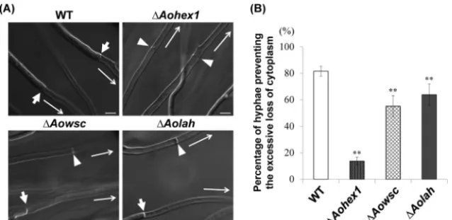
bursting was induced by the hypotonic shock of colonies grown on agar medium (9,18), and cytoplasmic loss in the cells adjacent to the burst tip cell was examined by DIC microscopic observa-tion. In this assay, it was considered that the septal pore was effec-tively plugged by the Woronin bodies if the cytoplasm was re-tained by the cells, and conversely, the excessive loss of cytoplasm was attributed to Woronin body dysfunction (Fig. 3A). Although 81% of wild-type cells retained their cytoplasmic constituents upon hypotonic shock, only 63% of cells of theAolahdisruptant retained their cytoplasmic constituents (Fig. 3B). As expected, the
Aohex1disruptant, which lacks Woronin bodies, showed a
signif-icantly impaired ability (14%) to prevent the excessive loss of cy-toplasm (Fig. 3B).
TheAowscgene (AO090102000111), which encodes a protein
with identity to WSC proteins fromN. crassa(50%),A. fumigatus
(80%), andA. nidulans(71%), was also disrupted inA. oryzae(see Fig. S4 in the supplemental material). Disruption of theAowsc
gene was confirmed by Southern blotting (see Fig. S5 in the sup-plemental material). TheAowscdisruptant showed a reduced abil-ity (55%) to prevent the excessive loss of cytoplasm compared to wild-type cells (Fig. 3B), suggesting that AoWSC is involved in Woronin body function. Taken together, these results indicate that AoLAH-mediated tethering of Woronin bodies to the septum is involved in their ability to prevent the excessive loss of cyto-plasm upon hyphal wounding.
Localization analysis of AoLAH N- and C-terminal regions. AoLAH was predicted to be a large protein consisting of 5,727 amino acid residues. To analyze the contribution of individual regions of AoLAH to its localization, the AoLAH protein was di-vided into N-terminal (amino acids 1 to 2039), middle (amino acids 2040 to 4709), and C-terminal (amino acids 4710 to 5727) regions based on the results of dot plot analysis (Fig. 1B).
InN. crassa, LAH-1 binds and tethers Woronin bodies to the
cell cortex (3). As the N-terminal region of AoLAH (AoLAH[1– 2039]) showed homology to LAH-1, the localization of an AoLAH[1–2039]-EGFP fusion protein was examined in the wild-type strain. The fluorescence of AoLAH[1–2039]-EGFP was in-tensely localized at both sides of the septum and was also observed as punctate structures in the cytoplasm (Fig. 4A, left), a pattern that is reminiscent of the localization of Woronin bodies shown by TEM (Fig. 2). To determine whether the N-terminal region of AoLAH was associated with Woronin bodies, we induced hyphal tip bursting by hypotonic shock to observe whether AoLAH[1– 2039]-EGFP moves together with Woronin bodies to the septal pore upon hyphal wounding. Upon hyphal tip bursting, the fluo-rescence of AoLAH[1–2039]-EGFP was detected at the septal pore (Fig. 4A, right). This localization behavior is similar to that ob-served during septal plugging by Woronin bodies, as previously demonstrated by the expression of AoHex1 fused with a fluores-cent protein (9).
FIG 2Transmission electron microscopic images of the wild-typeA. oryzaeandAolahdisruptant strains. Images around the septal pore are magnified. Arrows indicate Woronin bodies. Bars, 500 nm.
on September 8, 2020 by guest
http://ec.asm.org/
Woronin bodies differentiate from the peroxisome via a bud-ding process (2,18,23). In previous reports onA. oryzaeandA.
fumigatus, the Hex1 protein fused with a fluorescent protein was
found to localize near the septum, but the fusion protein often colo-calized or associated with peroxisomes (10,18), demonstrating that its localization does not always correspond to Woronin bodies. To examine whether the localization of AoLAH[1–2039]-EGFP was in-dependent of peroxisomes, mDsRed fused with peroxisomal target-ing signal 1 (PTS1) was coexpressed with AoLAH[1–2039]-EGFP
in A. oryzae. Fluorescence microscopic analysis revealed that
AoLAH[1–2039]-EGFP was localized near the septum and in the cy-toplasm independently of peroxisomes (Fig. 4B, left). It was previ-ously reported that inN. crassa, hyphal apical cells contain undif-ferentiated Woronin bodies that associate with peroxisomes (2). Here, the peripheral association of AoLAH[1–2039]-EGFP with peroxisomes was also observed, in addition to numerous fluores-cent punctate structures in the cytoplasm (Fig. 4B, right). These findings demonstrated that AoLAH[1–2039]-EGFP was localized independently of peroxisomes, with the exception of the observed association in apical cells, which likely represented actively bud-ding Woronin bodies.
AsN. crassaLAH-1 binds to Woronin bodies via WSC (3), we
also investigated whether the localization patterns of the AoLAH N-terminal region were dependent on AoWSC. In theAowsc dis-ruptant, no intense localization of AoLAH[1–2039]-EGFP was detected in the vicinity of the septum; rather, the fusion protein was completely dispersed in the cytoplasm, and no punctate struc-tures were observed (Fig. 4C, left). This result suggested that the recruitment of the AoLAH N-terminal region to Woronin bodies is dependent on AoWSC, a finding that is in agreement with data
forN. crassareported previously by Ng et al. (3). If AoLAH[1–
2039]-EGFP localization represents the septal tethering of Wo-ronin bodies, such localization would not occur in theAolah dis-ruptant background (Fig. 2). As expected, no fluorescence was observed near the septum when AoLAH[1–2039]-EGFP was
ex-pressed in the Aolah disruptant, although punctate structures were found in the cytoplasm (Fig. 4C, right). Time-lapse tracking revealed that the punctate structures found in the cytoplasm ran-domly moved in the wild-type andAolahdisruptant strains (see Videos S1 and S2, respectively, in the supplemental material), in-dicating that they were not attached to the septum. This finding demonstrated that the presence of endogenous AoLAH protein is needed for the localization of AoLAH[1–2039]-EGFP near the septum. As the AoLAH N-terminal region exhibited localization patterns typical of those of Woronin bodies, the AoLAH[1–2039]-EGFP fusion protein was used as a marker of Woronin bodies in subsequent experiments.
The C-terminal conserved region of AoLAH shares similarity
withN. crassaLAH-2, which is closely associated with the septal
pore (3). To examine its localization, the C-terminal region of AoLAH tagged with EGFP (AoLAH[4710 –5727]-EGFP) was ex-pressed in the wild-type andAolahdisruptant strains. As shown in
Fig. 4D, AoLAH[4710 –5727]-EGFP was localized to the septal pore in both strains, indicating that the C-terminal region of AoLAH associates with the septal pore.
Woronin body tethering mediated by AoLAH lacking the middle region.According to the results of the localization analysis described above, it can be assumed that the AoLAH N- and C-ter-minal fusion construct without the nonconserved middle region may be functionally sufficient to tether Woronin bodies to the septum. To test this speculation, a middle-region-deleted AoLAH construct {AoLAH[(1–2039)⫹(4710 –5727)]} and full-length AoLAH were expressed in theAolahdisruptant. The two con-structs were expressed with a 3⫻HA tag, which was verified by Western blotting (see Fig. S6 in the supplemental material). Full-length AoLAH-3⫻HA was predicted to be a polypeptide of⬃620 kDa, and a band was detected at a mass much higher than the maximum molecular mass (460 kDa) of the protein marker. Mid-dle-region-deleted AoLAH-3⫻HA was predicted to be⬃330 kDa but was detected at around 460 kDa.
FIG 3Ability ofAolahdisruptant cells to prevent excessive loss of cytoplasm upon hyphal wounding. (A) Differential interference contrast images of wounded hyphae capable or incapable of preventing the excessive loss of cytoplasm from the second cell upon hyphal tip bursting. A colony grown on agar medium was subjected to hypotonic shock by flooding with water to induce hyphal tip bursting, and the first septum adjacent to the burst tip cell was observed by differential interference contrast microscopy. Arrows indicate the first septa in the hyphae preventing the excessive loss of cytoplasm, and arrowheads show the septa in the hyphae not preventing excessive cytoplasmic loss. Long arrows indicate the directions toward the burst hyphal tips. Bars, 10m. (B) Assay of the ability of cells to prevent excessive loss of cytoplasm upon hyphal wounding. The wild-type (WT) strain andAohex1,Aowsc, andAolahgene disruptant strains were subjected to hypotonic shock, and hyphae with cells adjacent to the burst tip cell that retained the cytoplasm were counted by differential interference contrast microscopy. Average percentages of hyphae preventing the excessive loss of cytoplasm are shown in the graph. Fifty randomly selected hyphae with burst hyphal tips were observed for each culture. The error bars indicate standard deviations. **,P⬍0.01 (by one-way analysis of variance with Dunnett’spost hoctest for comparison with the wild-type strain;n⫽9).
on September 8, 2020 by guest
TEM revealed that Woronin bodies were located near the sep-tal pore of the Aolah disruptant strain expressing full-length AoLAH (Fig. 5A). Similarly, in the strain expressing AoLAH[(1– 2039)⫹(4710 –5727)], the Woronin bodies were also observed near the septal pore (Fig. 5A), indicating that the N- and C-termi-nal fusion construct without the middle region is sufficient to tether Woronin bodies to the septum.
To more precisely investigate the effect of the deletion of the middle region of AoLAH on the tethering of Woronin bodies, the average distance of Woronin bodies from the septum was mea-sured by TEM (Fig. 5B). In theAolahdisruptant strain expressing full-length AoLAH, Woronin bodies were located approximately 99 nm from the septum, whereas in the strain expressing
middle-region-deleted AoLAH, the position of Woronin bodies had shifted approximately 50 nm closer to the septum (Fig. 5B). These results suggest that the middle region of AoLAH has a role in regulating the distance of Woronin bodies from the septum.
Elastic movement of Woronin bodies visualized with an AoLAH N-terminal-region–EGFP fusion protein.The tethering of Woronin bodies to the septum inN. haematococcawas previ-ously reported to exhibit elasticity, as demonstrated by the return of the Woronin bodies to the septum after their physical separa-tion by laser trapping (16). As shown inFig. 5B, the distance of Woronin bodies from the septum was more variable in theAolah
disruptant strain expressing full-length AoLAH than in the strain expressing the middle-region-truncated form of AoLAH. Based FIG 4Localization of AoLAH N- and C-terminal regions fused with EGFP. (A) Localization of AoLAH[1–2039]-EGFP in the wild-type background around the septum (left) and upon hyphal wounding (right). Note that the fluorescent signal was located at a distance from the septum during normal growth (see left). The right image shows the localization of AoLAH[1–2039]-EGFP to the septal pore upon hyphal wounding. Hyphal tip bursting was induced by hypotonic shock, and the septal pore adjacent to the burst tip cell was observed by fluorescence microscopy. Arrows indicate the septum, and arrowheads show the localization of AoLAH[1–2039]-EGFP to the septal pore. Bars, 5m. (B) Subcellular localization analysis of AoLAH[1–2039]-EGFP and peroxisomes. mDsRed fused with PTS1 was expressed in anA. oryzaestrain expressing AoLAH[1–2039]-EGFP. Fluorescence microscopic analysis revealed an independent distribution of the two fluorescent fusion proteins around the septum (left) and in the apical cell (right). The septum is shown by arrows. A peripheral association of AoLAH[1–2039]-EGFP with the peroxisome was also observed in the apical cell, as shown by the arrowhead. Bars, 1m. (C) Localization of AoLAH[1–2039]-EGFP in theAowsc (left) andAolah(right) disruptants. Arrows indicate the septum. Bars, 5m. (D) Localization of AoLAH[4710 –5727]-EGFP in the wild-type strain (left) and Aolahdisruptant (right). Arrows indicate the septum, and arrowheads show the close association of AoLAH[4710 –5727]-EGFP with the septal pore. Bars, 5m.
on September 8, 2020 by guest
http://ec.asm.org/
on this finding, it was hypothesized that the middle region of AoLAH might confer the observed elastic characteristics of the Woronin body tether.
To answer this question, the movement of Woronin bodies in living cells expressing AoLAH[1–2039]-EGFP was examined by fluorescence microscopy (Fig. 6). In the strain expressing middle-region-deleted AoLAH, Woronin bodies were located near the septum, and fluorescent dots were closer together across the sep-tum than in the strain expressing full-length AoLAH (Fig. 6A). Time-lapse analysis revealed that Woronin bodies located near the septum exhibited positional changes in cells expressing full-length AoLAH (Fig. 6B). Microscopic observation of the first septum of hyphae expressing full-length AoLAH showed that at least one of the tethered Woronin bodies moved rapidly back and forth be-tween two positions in approximately 5 s (see Video S3 in the supplemental material). Two distinct movement patterns of Wo-ronin bodies were observed: movement toward the septal pore and then back to the starting position (Fig. 6B, top) and move-ment away from the septal pore and then back to the starting position (Fig. 6B, middle). These observational analyses demon-strated that tethered Woronin bodies are capable of elastic move-ment. In the strain expressing middle-region-deleted AoLAH, however, such movement was not observed for Woronin bodies tethered to the septum (Fig. 6B, bottom; see also Video S4 in the supplemental material). This finding indicates that the middle
region of AoLAH is required for the elastic movement of Woronin bodies.
Involvement of the AoLAH middle region in Woronin body function. As it was demonstrated that the middle region of AoLAH influences the positional variation and elastic motility of tethered Woronin bodies (Fig. 5Band6B), we next investigated if the function of Woronin bodies was affected by deleting the mid-dle region of AoLAH. Complementation analysis of theAolah
disruptant was performed with various AoLAH constructs, and marker-introduced strains in the wild-type andAolahdisruptant backgrounds were used as positive and negative controls, respec-tively. The ability of cells to prevent the excessive loss of cytoplasm was analyzed by inducing hyphal tip bursting in each strain through hypotonic shock. Although 82% of wild-type cells pre-vented the excessive loss of cytoplasm in cells adjacent to the hy-phal tip upon lysis, cells of theAolahdisruptant showed a reduced ability (63%). Cells of theAolahdisruptant expressing full-length AoLAH displayed a restored ability (81%) (Fig. 7A) that was com-parable to that of the wild-type strain, indicating the functional complementation of the disruption. However, the expression of middle-region-deleted AoLAH in theAolah disruptant did not restore the resistance of cells to cytoplasmic loss through the sep-tum (62%) (Fig. 7A). These results indicate that the middle region of AoLAH is needed for proper Woronin body position to prevent the excessive loss of cytoplasm upon hyphal wounding.
FIG 5Transmission electron microscopy of Woronin bodies in theAolahdisruptant strain expressing full-length or middle-region-deleted AoLAHs. (A) Transmission electron microscopy of Woronin bodies located around the septum in the strain expressing full-length or middle-region-deleted AoLAH. Arrows indicate Woronin bodies. Bars, 200 nm. (B) Distance of Woronin bodies from the septum in the strain expressing full-length or middle-region-deleted AoLAH. The data are presented in the box plot chart. The top, bottom, and middle lines correspond to the 75th percentile, 25th percentile, and median, respectively; bars represent the limits of the upper (top) and lower (bottom) quartiles. The whiskers show the highest and lowest readings within a 1.5⫻interquartile range. The outliers are indicated byŒ(n⫽44 and 48 for strains expressing full-length AoLAH and middle-region-deleted AoLAH, respectively; ***,P⬍0.001).
on September 8, 2020 by guest
As the Woronin bodies visualized with the EGFP fusion of the AoLAH N-terminal region plugged the septal pore upon hyphal wounding (Fig. 4A, right), we also examined whether Woronin bodies plug the septal pore in strains expressing full-length or middle-region-deleted AoLAH. In theAolahdisruptant, although Woronin bodies did not localize near the septum (Fig. 4C, right), a fluorescent dot was observed in the septal pore upon hyphal wounding (Fig. 7B), suggesting that untethered Woronin bodies were pushed into the septal pore by cytoplasmic flow. In the strain expressing full-length AoLAH, a fluorescent dot was also observed in the septal pore (Fig. 7B), a finding that is consistent with the results presented inFig. 4A. In theAolahdisruptant and the strain complemented with full-length AoLAH, almost no fluorescence was detected near the septum, with the exception of the dot plug-ging the septal pore. In contrast, in the strain expressing middle-region-deleted AoLAH, Woronin bodies tethered to the septum were frequently observed after hyphal wounding, in addition to a fluorescent dot within the septal pore (Fig. 7B). Taken together, these results indicated that the nonconserved middle region of AoLAH is involved in the proper spacing of tethered Woronin bodies around the septal pore, and this spacing is important for efficient plugging of the septal pore upon hyphal wounding.
DISCUSSION
In this study, we investigated the role of the AoLAH protein in the tethering of and septal plugging by Woronin bodies. TEM analysis clearly demonstrated that AoLAH is required for the tethering of
Woronin bodies to the septum (Fig. 2). Moreover, the excessive loss of cytoplasm was observed in anAolahdisruptant upon hy-phal wounding induced by the hypotonic shock ofA. oryzae col-onies grown on agar media (9).Aohex1gene disruption causes the loss of Woronin bodies and leads to a significant defect in the ability of cells to prevent the loss of cytoplasm (Fig. 3B) (9). Here, disruption of theAolahgene also reduced this protective ability but to a lesser extent than that observed for theAohex1gene dis-ruptant (Fig. 3B). This finding indicates that the tethering of Wo-ronin bodies to the septum by AoLAH is important for their effi-ciency in plugging the septal pore upon hyphal wounding and thereby preventing the excessive loss of cytoplasm.
To functionally characterize the extremely large AoLAH pro-tein, which is⬎5,000 amino acids, it was divided into N-terminal, middle, and C-terminal regions according to amino acid sequence homology with the LAH proteins of otherAspergillusspecies (Fig. 1B). The N-terminal conserved region of AoLAH (amino acids 1 to 2039) fused with EGFP was detected at a distance from both sides of the septal pore, and the C-terminal conserved region of AoLAH (amino acids 4710 to 5727) fused with EGFP was closely associated with the septal pore (Fig. 4), localizations that are in agreement with a recent report onA. fumigatus(17). The fluores-cence of AoLAH[1–2039]-EGFP was observed at the septal pore upon hyphal wounding (Fig. 4A, right), which also induces the DsRed2-AoHex1 fusion protein to plug the septal pore (9). These data suggest that the N-terminal region of AoLAH exhibits a be-havior typical of Woronin bodies. AoHex1 has been used as a FIG 6Fluorescence microscopy of Woronin bodies visualized with the AoLAH N-terminal-region–EGFP fusion protein in theAolahdisruptant strain expressing full-length or middle-region-deleted AoLAH. (A) Woronin bodies visualized by expression of AoLAH[1–2039]-EGFP near the septum in the strain expressing full-length or middle-region-deleted AoLAH. Arrows indicate the septum. Bars, 1m. (B) Back-and-forth movement of the Woronin bodies visualized by using AoLAH[1–2039]-EGFP. Woronin body movement at the first hyphal septum was analyzed by time-lapse analysis with a time interval of 550 ms. Tracked Woronin bodies are indicated by arrowheads in the first frame of time-lapse analysis. Blue, red, green, and yellow dotted lines indicate the positions of Woronin bodies in individual frames. Note that the back-and-forth movement was not clearly detected in the strain expressing middle-region-deleted AoLAH.
Bars, 1m.
on September 8, 2020 by guest
http://ec.asm.org/
Woronin body marker (9); however, fluorescent protein-tagged AoHex1 only partially complements anAohex1 disruptant and often colocalizes to peroxisomes (18). Here, nearly all AoLAH[1– 2039]-EGFP fluorescent dots observed inA. oryzaecells were in-dependent of peroxisomes (Fig. 4B), and expression of the AoLAH[1–2039]-EGFP fusion protein did not affect the ability of cells to prevent the excessive loss of cytoplasm (data not shown).
N. crassaLAH-1 is associated with Woronin bodies via binding to
the WSC protein (3). Similarly, AoLAH[1–2039]-EGFP was dis-persed in the cytoplasm of theAowscdisruptant and did not ex-hibit dot-like localization patterns (Fig. 4C, left), confirming the AoWSC-dependent recruitment of the AoLAH N-terminal region to Woronin bodies. A model was recently proposed where WSC might assemble into a pore-like structure and cytosolically expose the N-terminal end of Hex1 for the recruitment of LAH to Wo-ronin bodies inA. fumigatus(17), which could be in the same line of our genetic data. However, it remains to be determined whether Hex1 or WSC directly binds to LAH. Taken together, the findings from this study demonstrate that AoLAH[1–2039]-EGFP is a more reliable marker for the observation of Woronin bodies inA.
oryzae.
In addition to the characterization of the conserved N- and C-terminal regions of AoLAH, the functional roles of the noncon-served middle region of AoLAH were also examined. When the AoLAH-EGFP fusion construct lacking the middle region (⬃2,700 amino acids) of AoLAH was expressed in theAolah dis-ruptant, Woronin bodies were retethered to the septum (Fig. 5A), revealing that middle-region-deleted AoLAH is sufficient for the tethering of Woronin bodies to the septum. However, the Wo-ronin bodies were located approximately 50 nm closer to the septum than those in the strain expressing full-length AoLAH
(Fig. 5B). Based on the fact that the giant protein titin, which is 4 MDa, is 1m in length (24), the length of the deleted 2,670 amino acids of AoLAH is theoretically estimated to be 70 nm, which is consistent with the reduced distance of Woronin bodies from the septum that was observed for theAolah disruptant expressing middle-region-deleted AoLAH.
In theAolahdisruptant, although no Woronin bodies were tethered to the septum, untethered Woronin bodies were found to plug the septal pore after hyphal wounding (Fig. 7B) and were likely pushed into position by cytoplasmic flow. However, unteth-ered Woronin bodies were not able to plug the septal pore as quickly as the tethered Woronin bodies, which may explain why the loss of cytoplasm was greater in theAolahdisruptant than in the wild-type strain but was less than that in theAohex1disruptant (Fig. 3Band7A). This is similar to the fact that anA. fumigatus lah
mutant defective in the septal tethering of Woronin bodies exhib-its no increased sensitivity to calcofluor white, whereas thehex1
disruptant is sensitive (17). Unexpectedly, the ability to prevent the excessive loss of cytoplasm was not restored by expressing the middle-region-deleted AoLAH construct in theAolahdisruptant (Fig. 7A), even though Woronin bodies were tethered to the sep-tum in this strain (Fig. 5Aand6A). We specifically examined the septal plugging state by visualizing Woronin bodies bound to the AoLAH N-terminal-region–EGFP fusion protein. Upon hyphal wounding, nearly all Woronin bodies tethered to the septum con-tributed to septal plugging in the wild-type and full-length AoLAH-expressing strains (Fig. 4A, right, and7B). However, the Woronin bodies remained tethered at a distance from the septal pore upon hyphal wounding in the strain expressing middle-re-gion-deleted AoLAH (Fig. 7B), indicating that they failed to plug the septal pore. It was likely that the untethered Woronin bodies FIG 7Functional analysis of Woronin bodies in theAolahdisruptant strain expressing middle-region-deleted AoLAH. (A) Colonies formed on agar medium were subjected to hypotonic shock, and the ability of hyphae to prevent the excessive loss of cytoplasm was analyzed as described in the legend ofFig. 3B. The error bars indicate standard deviations. **,P⬍0.01 (by one-way analysis of variance with Dunnett’spost hoctest for comparison with the wild-type strain;n⫽9). (B) Woronin body localization around the septum upon hyphal wounding in theAolahdisruptant strain andAolahdisruptant strains expressing full-length AoLAH or middle-region-deleted AoLAH. Woronin bodies were visualized by using AoLAH[1–2039]-EGFP. Arrows indicate the septum, and arrowheads show the septal pore. Bars, 5m.
on September 8, 2020 by guest
plugged the septal pore, as was observed for theAolahdisruptant. This result may explain why the cells expressing middle-region-deleted AoLAH had a reduced ability to prevent the excessive loss of cytoplasm (Fig. 7A), although it should be noted that the mid-dle region is not essential for the overall ability of Woronin bodies to plug the septal pore.
According to the TEM observations, the distance between Wo-ronin bodies and the septum displayed greater variability in the presence of full-length AoLAH (Fig. 5B), suggesting that Woronin bodies have positional flexibility. This speculation is consistent with the reported elasticity of the Woronin body tether inN.
haematococca, as demonstrated by laser capturing experiments
(16). It was recently reported that limited lateral mobility of Wo-ronin bodies tethered at the septum was found forA. fumigatus
(17). In this study, by visualizing Woronin bodies with an AoLAH N-terminal-region–EGFP fusion protein, we observed that some tethered Woronin bodies displayed dynamic movement that may reflect the presence of an elastic tether, a characteristic that is not seen in the strain expressing middle-region-deleted AoLAH (Fig. 6B). This finding suggests that the middle region of AoLAH con-fers elastic movement activity to Woronin bodies. Moreover, the fixed tethering of Woronin bodies to the septum (Fig. 7B) is at-tributable to the excessive loss of cytoplasm upon hyphal wound-ing and is due to decreased movement activity by deletion of the middle region of AoLAH. Collectively, the results presented here suggest that efficient septal plugging requires not only that Wo-ronin bodies be tethered to the septum but also that they must have the movement activity, which is conferred by the middle region of AoLAH.
A number of intrinsically disordered proteins are localized to the septum, where they are often involved in intercellular connec-tivity (25). AoLAH is also predicted to be highly disordered, par-ticularly in the N-terminal and middle regions (see Fig. S7 in the supplemental material). Detection of the full-length and middle-region-deleted AoLAHs with a higher molecular mass than that predicted by Western blotting (see Fig. S6 in the supplemental material) may be caused by the presence of intrinsically disor-dered characteristics in the N-terminal and middle regions, which is in agreement with previous reports (26,27). The middle region of AoLAH does not share high similarity with other fungal LAH proteins (Fig. 1B); however, several polylysine stretches are com-monly present in these proteins, although their interval length is not conserved (see Fig. S2 in the supplemental material). Gluta-mate was the most frequently found amino acid in the middle region of AoLAH (see Fig. S8 in the supplemental material), a feature that is also seen in otherAspergillusspecies. The giant protein titin exhibits molecular spring-like elasticity via its intrin-sically disordered region and contributes to the contraction-relax-ation cycle of skeletal muscle (28). Calcium reduces the length of titin, and this calcium-dependent process requires the presence of a glutamate-rich motif in the disordered region (29). The high content of glutamate in the middle region of AoLAH raises the possibility that this protein may also be calcium sensitive, given the fact that the calcium concentration would be increased upon hyphal wounding, as has been reported for other organisms (30). We speculate that glutamates frequently found in the middle re-gion of AoLAH may be involved in the extension and contraction of the Woronin body tether. An increasing number of intrinsically disordered proteins have recently been reported to have many biological roles (31). However, with the exception of a few studies
(25,32), little knowledge about the function of disordered pro-teins in filamentous fungi has been accumulated. Here, we unex-pectedly discovered the physiological importance of a noncon-served region in AoLAH that is predicted to be disordered and potentially functions as a molecular spring, although further anal-yses of this protein are needed to confirm these findings.
ACKNOWLEDGMENTS
This work was supported by a grant-in-aid for young scientists from the Japan Society for the Promotion of Science (JSPS) and by funding from the Noda Institute for Scientific Research.
We are grateful to Fumiko Ishizuna for her help with transmission electron microscopy.
REFERENCES
1.Lew RR.2005. Mass flow and pressure-driven hyphal extension in Neu-rospora crassa. Microbiology 151:2685–2692.http://dx.doi.org/10.1099 /mic.0.27947-0.
2.Tey WK, North AJ, Reyes JL, Lu YF, Jedd G. 2005. Polarized gene expression determines Woronin body formation at the leading edge of the fungal colony. Mol. Biol. Cell16:2651–2659.http://dx.doi.org/10.1091 /mbc.E04-10-0937.
3.Ng SK, Liu F, Lai J, Low W, Jedd G.2009. A tether for Woronin body inheritance is associated with evolutionary variation in organelle position-ing. PLoS Genet. 5: e1000521. http://dx.doi.org/10.1371/journal.pgen .1000521.
4.Markham P, Collinge AJ.1987. Woronin bodies of filamentous fungi. FEMS Microbiol. Rev. 46:1–11. http://dx.doi.org/10.1111/j.1574-6968 .1987.tb02448.x.
5.Jedd G, Chua NH.2000. A new self-assembled peroxisomal vesicle re-quired for efficient resealing of the plasma membrane. Nat. Cell Biol. 2:226 –231.http://dx.doi.org/10.1038/35008652.
6.Asiegbu FO, Choi W, Jeong JS, Dean RA.2004. Cloning, sequencing and functional analysis ofMagnaporthe grisea MVP1gene, ahex-1homolog encoding a putative ‘woronin body’ protein. FEMS Microbiol. Lett.230: 85–90.http://dx.doi.org/10.1016/S0378-1097(03)00858-9.
7.Curach NC, Te’o VS, Gibbs MD, Bergquist PL, Nevalainen KM.2004. Isolation, characterization and expression of the hex1 gene from Trichoderma reesei. Gene331:133–140.http://dx.doi.org/10.1016/j.gene .2004.02.007.
8.Soundararajan S, Jedd G, Li X, Ramos-Pamplona M, Chua NH, Naqvi NI.2004. Woronin body function inMagnaporthe griseais essential for efficient pathogenesis and for survival during nitrogen starvation stress. Plant Cell16:1564 –1574.http://dx.doi.org/10.1105/tpc.020677. 9.Maruyama J, Juvvadi PR, Ishi K, Kitamoto K.2005. Three-dimensional
image analysis of plugging at the septal pore by Woronin body during hypotonic shock inducing hyphal tip bursting in the filamentous fungus Aspergillus oryzae. Biochem. Biophys. Res. Commun.331:1081–1088. http://dx.doi.org/10.1016/j.bbrc.2005.03.233.
10. Beck J, Ebel F.2013. Characterization of the major Woronin body protein HexA of the human pathogenic moldAspergillus fumigatus. Int. J. Med. Microbiol.303:90 –97.http://dx.doi.org/10.1016/j.ijmm.2012.11.005. 11. Yuan P, Jedd G, Kumaran D, Swaminathan S, Shio H, Hewitt D, Chua
NH, Swaminathan K.2003. A HEX-1 crystal lattice required for Woronin body function inNeurospora crassa. Nat. Struct. Biol.10:264 –270.http: //dx.doi.org/10.1038/nsb910.
12. Tenney K, Hunt I, Sweigard J, Pounder JI, McClain C, Bowman EJ, Bowman BJ.2000. Hex-1, a gene unique to filamentous fungi, encodes the major protein of the Woronin body and functions as a plug for septal pores. Fungal Genet. Biol. 31:205–217. http://dx.doi.org/10.1006/fgbi .2000.1230.
13. Bleichrodt RJ, van Veluw GJ, Recter B, Maruyama J, Kitamoto K, Wösten HA.2012. Hyphal heterogeneity inAspergillus oryzaeis the result of dynamic closure of septa by Woronin bodies. Mol. Microbiol.86:1334 – 1344.http://dx.doi.org/10.1111/mmi.12077.
14. Momany M, Richardson EA, Van Sickle C, Jedd G. 2002. Mapping Woronin body position inAspergillus nidulans. Mycologia94:260 –266. http://dx.doi.org/10.2307/3761802.
15. Plamann M.2009. Cytoplasmic streaming inNeurospora: disperse the plug to increase the flow? PLoS Genet.5:e1000526.http://dx.doi.org/10 .1371/journal.pgen.1000526.
on September 8, 2020 by guest
http://ec.asm.org/
TE, Fedorova ND, Gotoh O, Horikawa H, Hosoyama A, Ichinomiya M, Igarashi R, Iwashita K, Juvvadi PR, Kato M, Kato Y, Kin T, Kokubun A, Maeda H, Maeyama N, Maruyama J, Nagasaki H, Nakajima T, Oda K, Okada K, Paulsen I, Sakamoto K, Sawano T, Takahashi M, Takase K, Terabayashi Y, Wortman JR, Yamada O, Yamagata Y, Anazawa H, Hata Y, Koide Y, Komori T, Koyama Y, Minetoki T, Suharnan S, Tanaka A, Isono K, Kuhara S, Ogasawara N, Kikuchi H.2005. Genome sequencing and analysis ofAspergillus oryzae. Nature438:1157–1161.http://dx.doi .org/10.1038/nature04300.
20. Maruyama J, Kitamoto K.2011. Targeted gene disruption inkojimold Aspergillus oryzae. Methods Mol. Biol.765:447– 456.http://dx.doi.org/10 .1007/978-1-61779-197-0_27.
21. Jin FJ, Watanabe T, Juvvadi PR, Maruyama J, Arioka M, Kitamoto K. 2007. Double disruption of the proteinase genes,tppAandpepE, increases the production level of human lysozyme byAspergillus oryzae. Appl. Microbiol. Biotechnol.76:1059 –1068.http://dx.doi.org/10.1007/s00253 -007-1088-4.
22. Mabashi Y, Kikuma T, Maruyama J, Arioka M, Kitamoto K. 2006. Development of a versatile expression plasmid construction system for Aspergillus oryzaeand its application to visualization of mitochondria. Biosci. Biotechnol. Biochem. 70:1882–1889. http://dx.doi.org/10.1271 /bbb.60052.
23. Liu F, Ng SK, Lu Y, Low W, Lai J, Jedd G.2008. Making two organelles
Biochim. Biophys. Acta 1804:1231–1264. http://dx.doi.org/10.1016/j .bbapap.2010.01.017.
28. Li H, Oberhauser AF, Redick SD, Carrion-Vazquez M, Erickson HP, Fernandez JM.2001. Multiple conformations of PEVK proteins detected by single-molecule techniques. Proc. Natl. Acad. Sci. U. S. A.98:10682– 10686.http://dx.doi.org/10.1073/pnas.191189098.
29. Labeit D, Watanabe K, Witt C, Fujita H, Wu Y, Lahmers S, Funck T, Labeit S, Granzier H.2003. Calcium-dependent molecular spring ele-ments in the giant protein titin. Proc. Natl. Acad. Sci. U. S. A.100:13716 – 13721.http://dx.doi.org/10.1073/pnas.2235652100.
30. McNeil PL, Kirchhausen T.2005. An emergency response team for mem-brane repair. Nat. Rev. Mol. Cell Biol.6:499 –505.http://dx.doi.org/10 .1038/nrm1665.
31. Rauscher S, Pomès R.2012. Structural disorder and protein elasticity. Adv. Exp. Med. Biol.725:159 –183.http://dx.doi.org/10.1007/978-1-4614 -0659-4_10.
32. Hurley JM, Larrondo LF, Loros JJ, Dunlap JC.2013. Conserved RNA helicase FRH acts nonenzymatically to support the intrinsically disordered Neurospora clock protein FRQ. Mol. Cell52:832– 843.http://dx.doi.org /10.1016/j.molcel.2013.11.005.
33. Sonnhammer EL, Durbin R.1995. A dot-matrix program with dynamic threshold control suited for genomic DNA and protein sequence analysis. Gene167:GC1–GC10.

![FIG 4 Localization of AoLAH N- and C-terminal regions fused with EGFP. (A) Localization of AoLAH[1–2039]-EGFP in the wild-type background around the(left) andfluorescent fusion proteins around the septum (left) and in the apical cell (right)](https://thumb-us.123doks.com/thumbv2/123dok_us/8698187.1737686/7.594.123.462.68.462/localization-terminal-regions-localization-background-anduorescent-proteins-septum.webp)
