INTERNATIONAL RESEARCH JOURNAL OF PHARMACY
www.irjponline.com
ISSN 2230
–
8407
Research Article
EVALUATION OF SILVER NANOPARTICLES AS A COMPARATIVE STUDY AND
DETERMINATION OF ITS EFFECT ON VIGNA RADIATA
Kalaiselvi Mani *, Subbaiya Ramasamy, Ayyappadasan Ganesan and Rubavathi Subbaiyan
Department of Biotechnology, K.S. Rangasamy College of Technology, Tiruchengode, Namakkal, Tamil Nadu, India
*Corresponding Author Email: [email protected]
Article Received on: 30/11/16 Revised on: 27/12/16 Approved for publication: 23/01/17
DOI: 10.7897/2230-8407.08017
ABSTRACT
The silver nanoparticles (AgNP’s) were synthesized using green synthesis and chemical synthesis method. The nanoparticles synthesized using green synthesis (boiled extract and normal extract) were compared for the difference in various properties after characterization u sing X-ray diffraction (XRD), Fourier Transform Infra-Red spectroscopy (FT-IR) and Atomic Force Microscopy (AFM). There is not much difference between the AgNP’s synthesized using both the methods. The chemically synthesized AgNP’s were also characterized. FTIR analysis revealed that there is a strong peak of absorbance at 1379 cm-1 on comparison with the control. XRD analysis is used to find the nature and size of the silver nanoparticle. The silver
nanoparticles are found to be more stable in distilled water than the deionized water and tap water. The antimicrobial activity is high for the boiled extract sample when compared with other samples. The effect of AgNP’s on Vigna radiata is studied by exposing the seeds in time intervals and allowing them to germinate. The comparison was done by estimation of chlorophyll and protein. Based on the studies it could b e said that if the exposure to silver nanoparticles increases the content of the plant will be affected.
Keywords: Green synthesis, Chemical Synthesis, Fourier Transform Infra-Red spectroscopy, X-Ray Diffraction, Atomic Force Microscope.
INTRODUCTION
Nanotechnology is a field which is gaining wide application in all the areas nowadays. The application of nanotechnology is eternal with a multidisciplinary feature including drug delivery and molecular diagnostics1. In such field, the development of metal nanoparticles of well-defined shape, size and composition is being a big challenge2. Because of the distinct property, larger surface area to volume ratio the metal nanoparticles are considered as the building blocks of nanotechnology3.
The compatibility of this method lies on the fact that they do not use any toxic chemicals for the synthesis of metal nanoparticles4. Nanotechnology is also used in agricultural field to obtain nano formulations. Nanotoxicology is also emerging to study the effect of various metal nanoparticles on the human cell lines5.Marine organisms are used for the synthesis of AgNP’s in which AgNO3 is reduced to Ag ions by a nitrate dependent reductase6. Magnetotactic bacteria are also used for the synthesis of silver nanoparticles. Attempts were also made to synthesize the nanoparticles using human cells. The process has been tried in both cancerous and non-cancerous cells7.
The synthesis of silver nanoparticles using the plant parts is known as green synthesis. The various parts of plant like fruit, leaf and flower are used for the synthesis. Almost all plants can be used for the synthesis process. The formation of silver nanoparticles will be crystalline nature. The water soluble organic materials present in the plants are responsible for the reduction of silver nitrate to silver nanoparticles8. Although chemical synthesis is considered as a toxic method for silver nanoparticles synthesis, the process of chemical reduction is a
simple method when compared to the other three processes of synthesis. It is less time consuming when compared to the other methods. This method involves the use of biomolecules especially amino acids as reducing agents9.
Parthenium hysterophorus L. (P. hysterophorus) also known as congress grass, carrot weed, bitter weed etc., is an invasive, noxious weed. It is an annual herb of necrotropicals origin. It is the crop which is more tolerant to herbicides. Therefore, it is ranked fourth most serious crop weed10. Nowadays this crop is found widely in the tropical and subtropical regions. This weed is used as an effective remedy for amoebiasis, neuralgia and certain types of rheumatism4.
The silver nanoparticles (AgNP’s) are widely used because of their antimicrobial activity. AgNP’s are used in antimicrobial dressing because of its reaction with the moisture in the skin and the fluid of the wounds11. They are also used against the plant pathogens. The effectiveness of AgNP’s can be improved by applying them well before the penetration and colonization of fungal spores within the plant tissues12.
Silver nanoparticles are used in non-linear optics, spectrally selective coating for solar energy absorption, biolabelling, intercalation materials for electrical batteries and so on 13. Though the silver nanoparticles and all other metal nanoparticles offer several advantages, the deposition and toxicity of those nanoparticles prevents their use. Therefore, various studies are being performed to check the toxicity of the nanoparticles on the cells. The nanoparticles would enter the human system through inhalation, dermal contact, ingestion and penetration14.
MATERIALS AND METHODS Green Synthesis
Collection and preparation of leaf extract
The leaves of Parthenium hysterophorus were collected from the rural areas of Tiruchengode, Namakkal, Tamil nadu, India. The latitude and longitude of the location is 11.3805° N, 77.8951° E. Leaf extracts were prepared in two different forms. One of the extracts was obtained by boiling the leaves of
Parthenium hysterophorus L. at 50̊ C for 15 minutes. The other extract was obtained by grinding the leaves4.
Synthesis of silver nanoparticles
The methodology of Vyom et al. (2009) was followed with slight modification. 1mM of 90 mL silver nitrate (AgNO3) solution was prepared. To the prepared solution 10 mL of
Parthenium hysterophorus L. leaf extract was added. The solution is then incubated for 24 hours in dark. The solution was observed for color change.
Chemical Synthesis
Equal volumes of 1 mM silver nitrate (AgNO3) and 1 mM L-tyrosine solution were mixed. The solution was then diluted to fivefold using deionized water. The diluted form is then heated to 100̊ C. After that 1 mL of 0.1 M potassium hydroxide solution was added and boiled till the yellow color was obtained. Then the sample is dialyzed for 24 hours to obtain pure silver nanoparticles. The solution is then dried at 40-50̊̊ C for 24 hours15.
Characterization of synthesized silver nanoparticles
The synthesized silver nanoparticles were characterized by UV- Visible spectrophotometer (Hitachi U-2900), Fourier Transform Infrared Spectroscopy (FTIR) (Perkin Elmer Spectra-100), X-Ray Diffraction (XRD) (Bruker AXS D4 Endeavor X-ray diffractometer) and Atomic force microscopy (AFM) (Nanosurf Easyscan 2).
Evaluation of silver nanoparticles
The synthesized silver nanoparticles were evaluated for various properties like stability, antibacterial activity and toxicity.
Stability testing
The stability of the synthesized nanoparticles was tested in distilled water for different time intervals and the change in the stability was noted using UV-Visible spectrophotometer at 420 nm. The nanoparticles solution of 1 mg/mL concentration was prepared and exposed in room temperature. The initial absorbance was noted as 0th hour reading. Subsequent readings were taken in different time intervals namely 2nd, 4th and 24th hour respectively16.
Antibacterial activity
The antibacterial activity for the nanoparticles was checked using the Mueller Hinton agar medium by good diffusion
method. The antimicrobial activity was checked for four different species namely Escherichia coli (MTCC 1692), Salmonella typhi (MTCC 733), Staphylococcus aureus (MTCC 7443) and Klebsiella pneumoniae (MTCC 7407).
Effect of silver nanoparticles on Vigna radiata
The toxicity of the silver nanoparticles on the growth and contents of V. radiata was studied by treating the seeds with silver nanoparticles for two different time intervals, 2 hours and 24 hours respectively.
Treatment of Vigna radiata seeds
The seeds of Vigna radiata L. were treated with different concentrations like 200 µg/ mL,400 µg/ mL, 600 µg/ mL, 800 µg/ mL and 1000 µg/ mL concentration of silver nanoparticles solution synthesized by chemical method and green method for two different timings, 2 hours and 24 hours. The seeds soaked in the solution for different time intervals were filter paper dried and weighed to compare the difference in the weight of the seeds before and after soaking. After weighing the seeds were seeded. The growth of the seeds was noted from the day immediately after seeding. The percentage germination of the seeds was noted to determine the effect of silver nanoparticles18.
Analysis of the plants to evaluate the effect of silver nanoparticles
The leaves of the plants treated with two different samples for two different timings were extracted and the chlorophyll estimation19 and protein estimation20.
Chlorophyll estimation
The chlorophyll contents of the plants grown after treatment with five different concentrations of the silver nanoparticles synthesized by two different methods exposed for two different timings19.
Chlorophyll a = [(12.7x A663)-(2.63x A645)]/ (Wt in g * 1000) --- (Eq. 1)
Chlorophyll b = [(22.9x A645)-(4.48x A663)]/ (Wt in g * 1000) --- (Eq. 2)
Total chlorophyll = [(20.2x A645) + (8.02 x A663)]/ (Wt in g * 1000)--- (Eq. 3)
Protein estimation
The leaf extracts of the two-different timing treated plants were prepared using phosphate buffer. The protein was estimated following Lowry et al., method using Bovine Serum Albumin (BSA) as standard20.
Statistical analysis
Boiled extract sample, Grounded extract sample, Chemical sample
Figure 1. UV- Visible spectra of the synthesized silver nanoparticles indicating maximum peak of absorbance
Control, Boiled extract sample, Grounded extract sample,
Chemical sample
Figure 2. FTIR spectra of the synthesized silver nanoparticles indicating the wave numbers of dominant peaks obtained
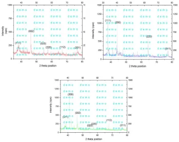
Figure 3: Representative XRD pattern of silver nanoparticles synthesized using boiled extract sample (a), grounded extract sample (b) and chemical synthesis sample (c).
Boiled extract sample, Chemical sample, Grounded extract sample
Figure 5: Absorption spectra of the synthesized silver nanoparticles at 420 nm indicating their stability at different time intervals
Figure 6: Effect of AgNP’s synthesized by green method (a) and chemical method (b) on the chlorophyll contents of the plants (p < 0.001 ) which indicates the values obtained are significant for both time intervals (i.e.) 2 hours and 24 hours treated
Table 1: The zone of inhibition obtained for the silver nanoparticles against four different bacteria
Name of the organisms
Zone of inhibition in [mm]
Tetracyclin Boiled extract sample Grounded extract sample Chemical sample
Escherichia coli 15 13 11 12
Salmonella typhi 25 21 20 23
Staphylococcus aureus 23 19 14 17
Klebsiella pneumoniae 27 20 13 17
RESULTS AND DISCUSSION Green synthesis
The silver nanoparticles (AgNP’s) formation from the metal precursor silver nitrate by the reducing power Parthenium hysterophorus leaf extract was confirmed by the color change observed after an incubation period of 24 hours. There was a color change in the solution of both the boiled extract and grounded extract of P. hysterophorus leaf extract (Figure 1). The color change in the solution prepared using the boiled leaf extract of the plant was not like that of the color obtained by Vyom et al.4. The color obtained by them after incubation was light brown whereas in this case the color was dark brown. But the color change obtained for the grounded leaf extract is in line with that of a previous work4result.
Chemical synthesis
The silver nitrate and L-tyrosine could react under boiling temperature. The change in the color observed by mixing the equal volume mixture of silver nitrate (1 mM) and L-tyrosine (1 mM) when boiled at 1000 C for one hour showed the interaction. The reduction silver nitrate by tyrosine happened under alkaline condition was observed by the formation of yellow color when potassium hydroxide was added and the solution was boiled for
30 minutes. The color indicating the formation of silver nanoparticles is in accordance with the result of Srivastava et al.15. This result is also in line with that of previous study 9 in which they have said that the reducing ability of tyrosine will occur only in alkaline pH.
Characterization of synthesized silver nanoparticles UV- Visible spectrophotometer analysis
The peak of absorbance was measured for the green synthesized and chemically synthesized samples. The wavelength was set from 300-600 nm. The maximum peak of absorbance was noted for each sample and the values are given in the Figure 1. The wavelength with the maximum peak of absorbance for the boiled extract of Parthenium hysterophorus was 422.5 nm and grounded extract was 428.5 nm which was contrasting with the result of Vyom et al.4 as the maximum peak of absorbance obtained by them was at 474 nm. But the maximum peak of absorbance obtained by the similar work21 and that of this sample was similar.
Fourier Transform Infrared spectroscopy (FTIR)
The silver nanoparticles synthesized by two different approaches were analyzed for their chemical nature using FTIR spectroscopy. To identify the functional group variations in the synthesized particles they were compared with the FTIR spectra of the control (AgNO3). On comparison with the control all the three samples showed a very strong peak of absorbance within a range of 1350-1480 cm-1. A strong peak of absorbance was also found at the wave numbers in the range of 3200-3600 cm-1, 1620-1680 cm-1 and 1080-1360 cm-1. Some other slight peak of absorbance was found within the ranges of 2850-3000 cm-1 and 675-1000 cm-1 (Figure 2). The result was found to correlate with the previous work22 for their work on electrolytic synthesis and characterization of silver nano powder. They have concluded that the very strong peak of absorbance and strong peak absorbance within the ranges 1350-1480 cm-1, 3200-3600 cm-1 and 1620-1680 cm-1 indicates the presence of NO2 which may be from AgNO3 and in addition to that it is said that the peak at the 1620-1680 cm-1 has a very strong visible intensity.
The peak of absorbance at this range 1350-1480 cm-1 would have strong visible intensity due to stretching of –C=H bond which was also determined by 22. A strong peak of absorbance at the wave number in the range of 2850-3000 cm-1 is because of the strong interaction of water with the surface of silver leading to O-H stretching mode. This result is also in accordance with that of 23 which indicated the presence of various functional groups in the silver nanoparticles.
The result obtained was also in line with the previous work24 which showed peak of absorbance at 3420-3371 cm-1 (due to N–H stretching, amides), 2931–2925 cm-1(due to C-H stretching, alkanes), 1383-1371 cm-1 (characteristic of hydroxyl groups, phenolic hydroxyl), 1051-1044 cm−1 (due to C-stretching, ether groups). The synthesized particles were also compared with the control for functional group difference. A wide range of difference was observed in all the three samples when compared with the control (AgNO3).
X-Ray Diffraction studies (XRD)
The XRD study was performed to determine the nature of the synthesized silver nanoparticles. The XRD pattern of all the samples was obtained to determine the nature and size of the synthesized silver nanoparticles. The obtained pattern was compared with Joint Committee on Powder Diffraction Standards (JCPDS) standard to determine the lattice position and peak index of the synthesized nanoparticles. The XRD pattern of the silver nanoparticles synthesized using the boiled extract of Parthenium hysterophorus was found to have six intense peaks corresponding 2θ positions 38.020, 46.240, 54.620, 57.600, 66.790 and 76.770 respectively. The obtained pattern was compared with JCPDS file No. 870598 standard to identify the peak indices. On comparison with the standard it was found that the indices to the corresponding positions were 111, 002, 020, 220, 112 and 201. The same standard JCPDS file was used to study the peak indices of silver nanoparticles synthesized from the grounded extract of Parthenium hysterophorus. The 2θ positions with intense peaks were 38.670, 41.340, 46.150, 54.910, 57.320, 67.600 and 76.610 with the corresponding peak indices 311,200, 002, 020, 220,112 and 201.
For the silver nanoparticles synthesized using chemical method the XRD pattern was compared with JCPDS file No. 893722 since both patterns were quite similar. The four intense peak positions and the corresponding indices found were 38.060,
44.070, 64.360 and 77.420 with indices 111, 200, 020, 220 and 311 respectively.
Based on the XRD patterns the synthesized silver nanoparticles were found to be cubic in shape with face centered lattice. Therefore, the synthesized nanoparticles are face centered cubic (FCC). The size of the nanoparticles was calculated by Debye-Scherrer formula as follows:
Dp = 0.94 ƛ/ β1/2 Cosθ --- (Eq. 4)
Where,
‘ƛ’ is wave length of X-Ray (0.1541 nm), ‘β’ is FWHM (full width at half maximum),
‘θ’ is the diffraction angle and ‘D’ is particle diameter size.
The average size of the nanoparticles obtained using Debye-Scherrer formula was 10.4 nm for boiled extract synthesized sample, 10.73 nm for grounded extract sample and 11.7 nm for chemically synthesized sample. The size of the nanoparticles obtained from the boiled extract sample is in accordance with the result of Ananda25. The size of the nanoparticles obtained by them is 9.3 nm whereas the same for the boiled extract sample is 10.4 nm with a standard deviation of 1.01 nm. The size of AgNP’s obtained from the grounded extract sample 10.73 nm with a standard deviation of 0.78 nm which is in contrast with Ananda25 whereas it is in line with the previous result reported.26. The average size of AgNP’s obtained through chemical synthesis method with a standard deviation of 1.07 nm is not on par with the result of Selvakannan9 in which the size obtained is 22 nm by following the similar procedure.
The AgNP’s synthesized from boiled and grounded extracts of
P. hysterophorus had showed more peak of indices at different 2θ positions when compared to the same prepared from
Parthenium hysterophorus25 and from Cissus quadrangularis27. The peak indices were obtained at 2θ positions of 7.90, 11.90, 17.880, 30, 380 and 440 whereas the same for my samples were at 38.670, 41.340, 46.150, 54.910, 57.320, 67.600 and 76.610. Though there were differences in the peak indices compared to their work the same results correlate nearly with the Ashok Kumar21 work consisted of XRD pattern for the AgNP’s synthesized from Parthenium hysterophorus.
Atomic force microscopy (AFM)
The AFM analysis had revealed that the synthesized nanoparticles were spherical in shape (Figure 4). Therefore, the spheres are arranged in face centered cubic (FCC) system when it is correlated with XRD data.
The result obtained is in accordance with the already reported work28 in which the spherical nanoparticles were formed with a size of 20–50 nm. This result is also coinciding with29 in which the obtained particles were spherical in shape with a tendency to form aggregates.
Evaluation of silver nanoparticles Stability testing
The stability of the synthesized silver nanoparticles was tested in distilled water. Initially the chemically synthesized particles were tested for their stability in tap water, distilled water and deionized water for different time intervals as 0 hour, 2 hours, 4 hours and 24 hours (Figure 5). The sample was found to be more stable in distilled water. Therefore, all the samples were further analyzed for their stability in distilled water alone as it is going to be used widely for all applications.
The stability of the nanoparticles decreased with increase in exposure to distilled water (1 mg/mL). As the time proceeds the nanoparticles will start to agglomerate in the water with the increase in size. Due to the formation of large size agglomerates the stability of the nanoparticles decreases. The result obtained is in accordance with the result reported by the earlier reseacher16. In that work they have attempted to study the stability of the silver nanoparticles in sea water and distilled water. Because of his work it was concluded that the silver nanoparticles were more stable in distilled water than in sea water. It was found that the stability of the boiled extract synthesized sample and the chemically synthesized sample were found to be better when compared to the AgNP’s synthesized from grounded extract. Further the boiled extract possessed higher stability than chemical synthesis sample.
Antibacterial activity
The antibacterial activity of AgNP’s (1 mg/mL) was tested by good diffusion method against four bacterial species namely
Escherichia coli, Salmonella typhi, Staphylococcus aureus and
Klebsiella pneumoniae. The Tetracyclin was used as control antibiotic disc (Table 1).
The zone of inhibition by AgNP’s synthesized by boiled, grounded extract as well chemical synthesis against all the chosen microbes is comparable with that of the standard antibiotic disc. The inhibition exhibited by green synthesized samples against the Staphylococcus aureus for the samples is contrasting with27 as the zone of inhibition obtained by them was 12 mm. whereas the antibacterial effect shown by chemical synthesis sample nearly coincides with.30. For Escherichia coli the zone of inhibition obtained for all the three samples contrast completely with25 as the result obtained for the nanoparticles against the same species was 27 mm but the results of Klebsiella pneumonia was on par with the same publication. Against
Salmonella typhi, the zone of inhibition for boiled extract synthesized AgNP’s was 21 mm and the same for the boiled extract synthesized sample was 20 mm. But the zone of inhibition obtained for Salmonella typhi by the chemically synthesized sample was 17 mm. The result obtained for all the samples against Salmonella typhi is in line with the result of31 in which the zone obtained was also 21 mm with the boiled extract of Parthenium hysterophorus.
Among the antibacterial activity analyzed for the three samples, it was found that the boiled extract synthesized sample showed better activity when compared to other samples. Therefore, the based on the stability and the antibacterial evaluation it was confirmed that the boiled could be used for the synthesis of silver than the grounded extract. So, this work is further focused on the application of boiled extract sample (green synthesized) and chemically synthesized sample.
Effect of silver nanoparticles on Vigna radiata Treatment of Vigna radiata seeds
The seeds of Vigna radiata were treated with the green synthesized AgNP’s and the chemically synthesized AgNP’s for two different timings namely two hours and 24 hours were allowed to germinate and the germination of the seeds were noted from the day immediately after initial germination. From the growth of the plants and the percentage of germination the toxicity of AgNP’s on the Vigna radiata L. seeds were determined. After the effect on germination was noted the plants were analyzed for various biochemical parameters to determine the effect under stress condition.
The concentrations of the AgNP’s were 200 to 1000 µg/mL with an increasing value of 200 µg/mL till it reaches 1000 µg/mL. The germination of the seeds treated for two hours with AgNP’s was not affected when compared to that of 24 hours treated seeds. The seeds treated with low concentrations like 200 µg/mL and 400 µg/mL showed 100% germination on the third day of seedling whereas the seeds with other higher concentrations showed 60-80% germination for both the green synthesized and chemically synthesized AgNP’s treated seeds.
The percentage germination of the seeds treated for 24 hours was only 60-80% even for the lower concentrations like 200 µg/mL and 400 µg/mL in third day of germination. And at the higher concentrations there was no germination during the third day indicating that treatment with higher concentrations affects the rate of germination period.
The result obtained is in accordance with32 for their work on AgNP’s mediated growth enhancement of the plant Brassica juncea. They had reported that at lower concentrations like 25 ppm and 50 ppm the growth of the plants was enhanced. But at the concentration of 400 ppm a decline of 22% was noted which is stating that the obtained result increased concentration of silver nanoparticles affects the germination rate of the plants is correct.
But the result of reported33 had stated that the biosynthesized silver nanoparticles did not show any effect on the plant Bacopa monneiri even at 100 ppm concentration which is contrary to the result obtained in this work and as well to the result32. There were no results stating the effect of silver nanoparticles when treated with seeds for 24 hours.
Analysis of the plants to evaluate the effect of silver nanoparticles
Chlorophyll estimation
The chlorophyll estimation was performed following19 procedure using the acetone extract of plant leaves. The chlorophyll content of all the treated plants in different time intervals was given in the Figure 6.
The chlorophyll contents of the grown plants were compared for the treated and not treated plants to determine the effect of silver nanoparticles. The chlorophyll content of all the plants treated with different concentrations of silver nanoparticles synthesized by two different methods at two different time intervals was found to increase with increase in the concentration till an optimum concentration but at concentrations greater than the optimum one the chlorophyll content decreased. In this work, it has been observed that the optimum concentration of AgNP’s synthesized by two different approaches in which the chlorophyll content reached a maximum was 600 µg/mL whereas at later concentrations decline in the chlorophyll content was observed.
It was also observed that even at the optimum concentration there was a difference in the chlorophyll contents of the plants treated with green synthesized AgNP’s and chemically synthesized AgNP’s. The plants treated with green synthesized AgNP’s showed an increased value of total chlorophyll when compared to the chemically synthesized AgNP’s treated plants at 600 µg/mL for both time intervals of treatment.
pigment concentration increased up to a concentration of 40 mg/L and started to decrease with the further increasing concentrations. But the obtained result is contrary to32 work in which it was stated that there was increase in chlorophyll concentration up to 40% even at a concentration of 100 ppm.
Protein estimation
The protein content of the plants treated with different samples at different time intervals was analyzed20. The amount of protein was found to increase with the increased concentration of silver nanoparticles synthesized by both the methods when the time exposure was 2 hours. But the same decreased with the increase in the concentration of silver nanoparticles synthesized by both the methods when the time exposure was 24 hours.
The result obtained for 24 hours treated sample was like 33 where it was stated that the content of the protein in the treated plants was lesser when compared to the not treated plants. It has been also stated the protein content of the plants increased for 5 days from the day of exposure which could be correlated to the result obtained for 2 hours treated plants. So, based on the above results and statements it could be confirmed that if the time of exposure to silver nanoparticles increase the effect would also increase. The amount of protein in all the samples which were obtained from 2 hours treated plants was in accordance with the result34. In that work it was stated that the protein amount also increased up to certain and started to decrease beyond that concentration.
CONCLUSION
The silver nanoparticles were synthesized using green synthesis and the chemical synthesis methods. The green synthesis method was performed using boiled extract as well grounded extract of the Parthenium hysterophorus L. leaf extract. The silver characterized nanoparticles revealed that the all the silver nanoparticles were spherical in shape arranged in a face centered cubic lattice (FCC) with little variations in size. The size of the silver nanoparticles obtained from the boiled extract was 10.4 nm and that of grounded extract was 10.7 nm. The chemically synthesized AgNP’s were of 18 nm in size. The boiled extract synthesized sample and the chemical sample were more stable than the grounded extract sample with the stability of boiled extract greater than the chemical sample. The antibacterial activity was also higher for the boiled extract synthesized sample when compared with the others. It was also found that the boiled extract synthesized AgNP’s induces less stress on the plants when compared to the chemical synthesis sample. The adverse effect of the silver nanoparticles on the plants was studied by chlorophyll and protein estimation studies which showed that the adverse effect increases with the increase in exposure time. Therefore, it was concluded that the boiled extract method could be used for the synthesis of AgNP’s from the plants and the effect of AgNP’s on the plant increases with the increase in exposure even if they are not incorporated in to the plant system. Thus, the green synthesis method is ecofriendly, cheap and effective method for nanoparticles synthesis.
ACKNOWLEDGEMENTS
The authors are sincerely thankful to the institute, K. S.Rangasamy College of Technology, Tamil Nadu for providing the facility and support for doing our research.
REFERENCES
1. Sankar R, Karthika A, Prabu A, Karthik S, Shivashangari S, Ravikumar V. Origanum vulgare mediated biosynthesis of silver nanoparticles for its antibacterial and anticancer activity. Colloid Surfaces B2013; 108:80-84.
2. Gericke M, Pinches A. Biological synthesis of metal nanoparticles. Hydrometallurgy 2006; 83:132-140.
3. Leela A, Vivekanandan M. Tapping the unexploited plant resources for the synthesis of silver nanoparticles. African Journal of Biotechnology 2008; 7:3162-3165.
4. Vyom P, Rashmi P, Bechan S, Avinash, P. Parthenium leaf extract mediated synthesis of silver nanoparticles: a novel approach towards weed utilization. Digest Journal of Nanomaterial and Biosciences 2009; 4:45-50.
5. Asharani PV, KahMun GL, Hande MP, Suresh V. Cytotoxicity and genotoxicity of silver nanoparticles in human Cells. American Chemical Society Nanoletter. 2009; 3:279-290.
6. Mubarak Ali D, Sasikala M, Gunasekaran M, Thajuddin N. Biosynthesis and characterization of silver nanoparticles using marine cyanobacterium Oscillatoria willei NTDM01. Digest Journal of Nanomaterial and Biosciences. 2011; 6:385-390.
7. Sadowski Z, Maliszewska IH, Grochowalska B, Polowczyk I, Koźlecki, T. Synthesis of silver nanoparticles using microorganisms.Material Sciences-Poland 2008; 26: 419-424.
8. Sridhara V, Pratima K, Krishnamurthy G, Sreekanth B. Vegetable assisted synthesis of silver nanoparticles and its antibacterial activity against two human pathogens. Asian Journal of Pharmacy and Clinical Research2013;6:53-57. 9. Selvakannan PR, Swami A, Srisathiyanarayanan D, Shirude
PS, Pasricha R, Mandale AB, et al., Synthesis of aqueous Au core-Ag shell nanoparticles using tyrosine as a pH-dependent reducing agent and assembling phase-transferred silver nanoparticles at the air-water interface. Langmuir 2004; 20:7825-7836.
10.Anuj K, Verma VC, Gond SK, Kumar V, Kharwar RN. Biocontrol potential of Cladosporium sp. (MCPL - 461) against a noxious weed Parthenium hysterophorus L. Journal of Environmental Biology.2009; 30:307-312. 11.Mahendra R, Alka Y, Aniket G. Silver nanoparticles as a
new generation of antimicrobials. Biotechnology Advances. 2009; 27:76-83.
12.Remya N, Saino HV, Baiju GN, Maekawa, T, Yoshida Y, Sakthi Kumar D. Nanoparticulate material delivery to plants. Plant Sciences. 2010; 179:154-163.
13.Mohanpuria P, Nisha K, Rana S, Yadav K. Biosynthesis of nanoparticles: Technological concepts and future application. Journal of Nanoparticle Research. 2008; 10:507-517.
14.Vivek R, Thangama R, Muthuchelian K, Gunasekaran P, Kaveri K, Kannan S. Green biosynthesis of silver nanoparticles from Annona squamosa leaf extract and its in vitro cytotoxic effect on MCF- cells. Process Biochemistry. 2012; 47: 2405-2410.
Srivastava M, Singh S, William TS. Exposure to silver nanoparticles inhibits selenoprotein synthesis and the activity of thioredoxin reductase. Environmental Health Perspectives. 2012; 120:56-61.
15.Hangyue Z. Physicochemical properties of protein-modified silver nanoparticles in seawater. Inernational Nano Letters. 2013; 3:1-5.
17.Rezvani N, Sorooshzadeh A, Farhadi N. Effect of nano silver on growth of Saffron in flooding stress. World Academy of Science, Engineering and Technology. 2012; 61:606-611.
18.Arnon DI. Copper enzymes in isolated chloroplasts, polyphenoxidase in Beta vulgaris. Plant Physiology, 1949; 24:1-15.
19.Lowry OH, Rosebrough NJ, Farr AL, Randall RJ. Protein measurement with the Folin phenol reagent. Journal of Biology and Chemistry. 1951; 193:265.
20.Ashok Kumar D. Rapid and green synthesis of silver nanoparticles using the leaf extracts of Parthenium hysterophorus: A novel biological approach. International Research Journal of Pharmcy. 2012; 3:169-173.
21.Theivasanthi T, Alagar M. Electrolytic synthesis and characterizations of silver nanopowder. Nano Biomedicine and Engineering. 2012; 4:58-65.
22.Sridhar S, Anarkali J, Vijaya Raj D, Rajathi K. Biological synthesis of silver nanoparticles by using Mollugo nudicaulis extract and their antibacterial activity. Archives of Applied Science Research. 2012; 4:1436-1441.
23.Sun Q, Caia X, Li J, Zheng M, Chen Z, Yu CP. Green synthesis of silver nanoparticles using tea leaf extract and evaluation of their stability and antibacterial activity. Colloids and Surfaces A., 2014; 444:226-231.
24.Ananda S. Exploitation of Parthenium Hysterophorus L. for the rapid biosynthesis of silver nanoparticles and evaluation of their anti-microbial activity. Indian Journal of Applied Research. 2013; 7:79-83.
25.Rebecca T, Parvathi L, Roopa H, Fenali P, Glory F, Sourabh M. et al., A facile method for green synthesis of stabilized silver nanoparticles and its in vitro antagonistic applications. Journal of Natural Products and Plant Resources 2013; 3:36-40.
26.Alagumuthu G, Kirubha R. Green synthesis of silver nanoparticles using Cissus quadrangularis plant extract and their antibacterial activity. International Journal of Nanomaterials and Biostructures. 2012; 2:30-33
27.Gopinath K, Gowri S, Arumugam A. Phytosynthesis of silver nanoparticles using Pterocarpus santalinus leaf extract and their antibacterial properties. Journal of nanostructure in chemistry 2013; 3:1-7.
28.Christensen L, Vivekanandhan S, Misra M, Mohanty AK. Biosynthesis of silver nanoparticles using Murraya koenigii
(curry leaf): An investigation on the effect of broth concentration in reduction mechanism and particle size.
Advanced Material Letters. 2011; 2:429-434.
29.Malabadi RB, Mulgund GS, Meti NT, Nataraja K, Vijaya Kumar S. Antibacterial activity of silver nanoparticles synthesized by using whole plant extracts of Clitoria ternatea. Research in Pharmacy 2012; 2:10-21.
30.Kalaiselvi M, Subbaiya R, Selvam M. Synthesis and characterization of silver nanoparticles from leaf extract of
Parthenium hysterophorus and its anti-bacterial and antioxidant activity. International Journal of Current Microbiology and Applied Sciences. 2013; 2:220-227. 31.Priyadarshini S, Deepesh P, Zaidi MGH, Pardha SP, Khanna
PK, Sandeep A. Silver nanoparticle mediated enhancement in growth and antioxidant status of Brassica juncea. Applied Biochemistry and Biotechnology.2012; 167:2225-2233. 32.Krishnaraj C, Jagan EG, Ramachandran R, Abirami SM,
Mohan N, Kalaichelvan PT. Effect of biologically synthesized silver nanoparticles on Bacopa monnieri (Linn.) wettst. plant growth metabolism. Process Biochemistry. 2012; 47:651-658.
33.Hatami M, Ghorbanpour M. Defense enzyme activities and biochemical variations of Pelargonium zonale in response to nanosilver application and dark storage. Turkish Journal of Biology. 2014; 38:130-139.
34.Salama MH. Effects of silver nanoparticles in some crop plants, common bean (Phaseolus vulgaris L.) and corn (Zea mays L.). International Research Journal of Biotechnology.
2012; 3: 190-197.
Cite this article as:
Kalaiselvi Mani, Subbaiya Ramasamy, Ayyappadasan Ganesan and Rubavathi Subbaiyan. Evaluation of silver nanoparticles as a comparative study and determination of its effect on Vigna radiata. Int. Res. J. Pharm. 2017;8(1):33-40 http://dx.doi.org/ 10.7897/2230-8407.08017
Source of support: Nil, Conflict of interest: None Declared