Open Access Emergency Medicine
Correlation of CT findings remote from prime
area of interest: a multitrauma study
Miguel Bardon1 Noel Young1 Poppy Sindhusake2 Theresa Lee1 Ken Le1
1Department of Radiology, Centre
for Biomedical Imaging Research and Development, Westmead Hospital, Sydney, NSW, Australia; 2Centre
Health Sciences, University of Sydney, Sydney, NSW, Australia
Correspondence: Miguel Bardon 3a/27 Sutherland Crescent, Darling Point, NSW 2027, Australia Tel +61 2 9328 4446
Email [email protected]
Background: Multitrauma patients represent a difficult cohort of patients from a diagnostic standpoint. Current trauma recommendations do not advise whole-body computed tomography (CT) in hemodynamically stable patients.
Objective: To measure the prevalence of abnormal CT findings in areas other than the prime area of clinical interest in multitrauma patients.
Method: The records of 462 consecutive adult patients who underwent whole-body CT scans between 2004 and 2005 at Westmead Hospital, a Level 1 trauma center, were assessed. Clinical parameters, including suspected clinical injury, regional tenderness, bruising, loss of consciousness, scalp laceration, and unequal chest air entry, were examined. Correlation was made with CTs performed from the brain to symphysis pubis, on a Toshiba 16 slice machine, with evaluation of clinically significant findings.
Results: The prevalence of abnormal CT findings distant to the prime area of concern varied between anatomical areas: brain (10.3%–88.7%), skull (6.7%–39.7%), facial bones (4.4%–54.3%), cervical spine (5.6%–13.7%), thoracolumbar spine (5.6%–26.7%), chest (30.8%–54.4%), and abdomen/pelvis (20%–27.2%).
Conclusion: There is a high prevalence of injuries remote from the prime area of clinical concern in multitrauma patients. Whole-body CT is a rapid, accurate, and systematic imaging modality that provides an early, complete, clinical picture for the treating physician.
Keywords: CT, trauma, radiology, multitrauma, whole-body imaging
Introduction
Multitrauma patients can be difficult to assess fully in the early stages of their presentation and have been shown to have a high prevalence of occult injuries remote from the prime area of interest.1 In the past decade trauma management has
been evolving toward an advanced trauma life support (ATLS) model, a system that uses a multidisciplinary approach as a way of standardizing management of trauma patients to benefit outcome.2 Current ATLS guidelines proposed by the American
College of Surgeons do not address the multitrauma scenario specifically; rather, they deal with individual areas. Computed tomography (CT) is recommended as a first-line imaging modality for some areas, such as the brain and abdomen/pelvis; however, it is recommended mainly as a secondary or adjunct method for spinal cord injury.3
Within this system, investigations are dependent upon the attending physician tailoring investigations according to their acute clinical findings. Current recommendations do not recommend whole-body CT as a matter of course in multitrauma patients.3
Dovepress
O R I g I N A L R E S E A R C H
open access to scientific and medical research
Open Access Full Text Article
Open Access Emergency Medicine downloaded from https://www.dovepress.com/ by 118.70.13.36 on 26-Aug-2020
For personal use only.
Number of times this article has been viewed
This article was published in the following Dove Press journal: Open Access Emergency Medicine
This reliance on clinical information can be problematic as clinical findings have been found to be equivocal or misleading in 20% to 50% of blunt multitrauma victims.1
Further to this, there is a well-described phenomenon known as “satisfaction of search” that describes the tendency of detection of one radiographic abnormality to interfere with the detection of injuries in remote anatomical areas.4 Injuries
to the pelvis or spine as well as intra-abdominal injuries have all been identified as regions particularly likely to be missed by standard radiographic and clinical examination.5
Since the inception of multidetector CT technology, a number of studies have been conducted to assess the potential for whole-body CT in a multitrauma setting to increase the sensitivity of the secondary survey. CT has been shown to be more sensitive and specific than other diagnostic investigations (sensitivity 94%, specificity 100%, and accuracy 97%) and is significantly faster (average total scanning time 20 minutes), involving considerably less maneuvering of the patient.6,7 Of particular interest was a
large retrospective study conducted by the German Trauma Group that showed that survival was significantly increased in patients who underwent whole-body CT in the resuscitation phase compared to those who did not.2 However, what was
not found in the literature was a clear delineation of the variety and correlation of injuries in this setting and how CT had affected the diagnosis.
The aim of this study is to measure the prevalence of abnormal CT findings on whole-body imaging and to assess how remote findings correlate to the injury of primary concern.
Materials and methods
This retrospective study was conducted at Westmead Hospital, an adult Level 1 trauma center servicing a population of 1.5 million people, with over 50,000 emergency presentations per year. The medical records of consecutive patients who underwent whole-body CT (brain, skull, facial bones, cervical/thoracic/lumbar spine, chest, and abdomen/ pelvis) over 2 years from 2004 to 2005 were reviewed.
Trauma patients are subject to a standard primary, secondary, and tertiary survey protocol in alignment with current ATLS guidelines. The secondary and tertiary survey are performed by the attending emergency physician and supervised by a consultant. Depending on the injury type, the tertiary survey is carried out on the ward by medical staff or in the recovery bay by surgical staff.
Trauma in the hospital is classified as either category 2 (motor vehicle 60 km, cyclist/pedestrian versus vehicle,
assault to the head or torso, fall .2 m), which is dealt with by the attending emergency physicians, or category 1 (penetrating injury, .20% burns, spinal cord injury, pregnant .20 weeks, major crush injury, amputation, major long bone/pelvic fracture) that necessitates the attention of the trauma team who are available 24 hours a day, 7 days a week. The trauma team consists of registrars from the respective departments: intensive care, anesthetics, obstetrics and gynecology, ortho-pedic surgery, and the acute surgical team.
Ethics approval was obtained through the Westmead Hospital ethics committee before the study commenced. Sixteen years was considered to be the minimum age for consent into the study.
Technical aspects
A Toshiba 16-slice CT (Aquilon 16 TSX-101A/6C; Toshiba Medical Systems, Tochiai, Japan) was used for all scans. All patients were positioned supine and scanned from the top of the skull to the symphysis pubis. No contrast was used in CT scans of the skull, brain, or facial bones. In the chest a postcontrast scan (arterial enhancement phase) was used from the supraclavicular region to aortic bifurcation. For the abdomen and pelvis, a postcontrast scan (portovenous enhancement, 70-second delay) was used to cover from above the diaphragm to synthesis pubis. In all cases, nonionic contrast was used by pump injection of 100 mL at 3–4 mL per second. The estimated effective radiation dose of this examination is about 12 mSv as a weighted average of doses to all organs. Standard pediatric protocol in place in the department ensured lower radiation doses for patients under 18 years, using a lower kVp, mA, and automatic tube current modulation.
Radiological classification
All studies were reported on workstations by the on-duty radiology registrar with immediate feedback to the clinical team. Reports were then later reviewed and verified by a consultant radiologist. CT findings were defined as being significant if they required immediate therapeutic intervention or close monitoring and observation. Scans were reviewed on workstations on multiplanar reformats with three-dimensional reconstruction being performed for those cases that needed surgical intervention.
Radiological classification was done retrospectively by review of the official reports adapting elements of a number of classification schema (Marshall brain CT criteria; Tile classification of pelvic fractures; Dingman and Natvig classification of facial fractures; and the American Association for the Surgery of Trauma’s injury scaling
Dovepress
Bardon et al
Open Access Emergency Medicine downloaded from https://www.dovepress.com/ by 118.70.13.36 on 26-Aug-2020
score classification of injuries of the thorax, abdomen, and pelvis).8–12
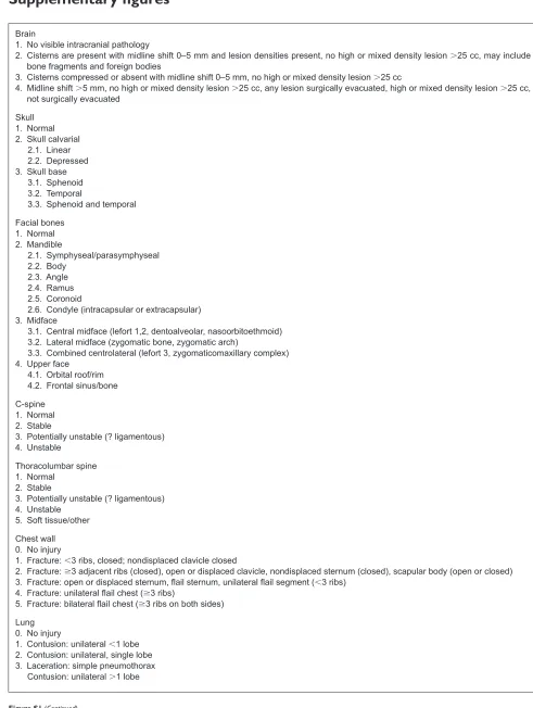
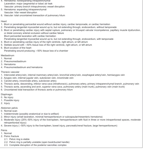
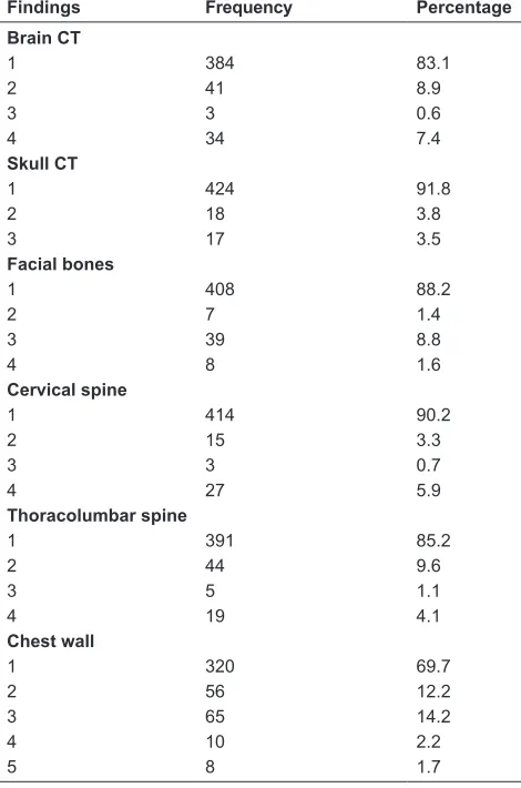
Classification was devised by the research team where appropriate precedent was not found in the literature. A detailed outline of the exact classification criteria can be found in Figure S1, with corresponding frequency data in Figure S2.
Clinical information
Clinical parameters in all anatomical regions were recorded. These included suspected clinical injury, regional tenderness, bruising, loss of consciousness, scalp laceration, and unequal air entry. Emergency specialist staff performed focused assessment sonography for trauma (FAST) with free intraperitoneal fluid being deemed a positive result and no free intraperitoneal fluid a negative result.
Injury severity was measured using an Abbreviated Injury Scale (AIS) and then totaling the sum of the squares of these scores to calculate an Injury Severity Score (ISS). The Glasgow Coma Scale was also recorded retrospectively and categorized into two groups, 15 and ,15.
For this study’s purposes, the prime area of interest was determined by factors of recorded clinical examination findings and associated investigations (eg, FAST). In cases where more than one region was of significant clinical interest, for evaluation purposes we evaluated each region as a separate item.
Results
There were 331 males and 131 females with a median age of 40.2 years, ranging from 16 to 100 years. Table 1 details the mechanism of injury.
Table 2 shows the other region prevalence of significant abnormal CT findings where brain injury was considered to be the prime area of interest. Over 30% of patients with significantly abnormal brain CT were also found to have abnormal CT findings in the skull, facial bones, and chest.
Table 3 shows the other region prevalence of significant abnormal CT findings where the skull was considered to
be the prime area of interest. Over 30% of patients with significantly abnormal skull CT were also found to have abnormal CT findings in the facial bones and chest, and over 80% of these patients were found to have abnormal brain CT findings.
Table 4 shows the other region prevalence of significant abnormal CT findings where the facial bones were considered to be the prime area of interest. Over 30% of patients with significantly abnormal facial bones CT were also found to have abnormal CT findings in the brain, skull, and chest.
Table 5 shows the other region prevalence of significant abnormal CT findings where the cervical spine was considered to be the prime area of interest. Over 20% of these patients had significant abnormalities in the brain thoracolumbar spine and abdomen/pelvis, and over 40% had abnormalities in the chest.
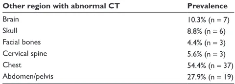
Table 6 shows the other region prevalence of significant abnormal CT findings where the thoracolumbar spine was considered to be the prime area of interest. Twenty-eight percent of these patients had significant abnormality on abdominal/pelvis CT, and over 50% had abnormalities seen in the chest.
Table 7 shows the other region prevalence of significant abnormal CT findings where the chest was considered to be the prime area of interest. Over 20% of these patients had significantly abnormal findings on thoracolumbar spine and abdominal/pelvis CT. However, what is perhaps
Table 1 Mechanism of injury
Mechanism of injury Number of patients Percentage
Motor vehicle accident 208 44.9
Motorcycle accident 55 11.8
Fall 56 12.1
Assault 38 8.9
Pedestrian 51 11.0
Other 49 10.3
Table 2 Prevalence of significant abnormal CT findings in the 78 patients with significantly abnormal brain CTs
Other region with abnormal CT Prevalence
Skull 39.7% (n = 31)
Facial bones 35.9% (n = 28)
Cervical spine 14.1% (n = 11)
Thoracolumbar spine 9.0% (n = 7)
Chest 30.8% (n = 24)
Abdomen/pelvis 24.4% (n = 19)
Abbreviation: CT, computed tomography.
Table 3 Prevalence of significant abnormal CT findings in the 35 patients with significantly abnormal skull CTs
Other region with abnormal CT Prevalence
Brain 88.6% (n = 31)
Facial bones 54.3% (n = 19)
Cervical spine 8.6% (n = 3)
Thoracolumbar Spine 17.1% (n = 6)
Chest 34.3% (n = 12)
Abdomen/pelvis 25.7% (n = 9)
Abbreviation: CT, computed tomography.
Dovepress Correlation of CT findings remote from prime area of interest
Open Access Emergency Medicine downloaded from https://www.dovepress.com/ by 118.70.13.36 on 26-Aug-2020
most clinically relevant is that 17.3% of these patients had significant abnormalities visualized in the brain.
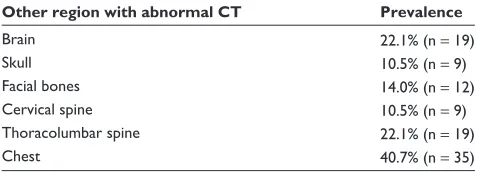
Table 8 shows the other region prevalence of significant abnormal CT findings where the abdomen/pelvis was considered to be the prime area of interest. Over 20% of these patients had significant abnormalities on brain and thoracolumbar spine CT, and over 40% had significant chest pathology demonstrated.
Discussion
The best outcome management of multitrauma patients is dependent on rapid and accurate assessment of multiple anatomical areas. Although urgent intervention in unstable patients should not be delayed by imaging, in hemodynamically stable patients there is a clear need for a systematic method of identifying significant injuries to prevent against the develop-ment of complications and to avoid or minimize fixation to one area at the expense of other injured regions.
This paper evaluates whole-body assessment for significant trauma in sites other than those of prime clinical concern. It is our understanding that no previous publication has explored this particular aspect of whole-body CT in trauma.
Brain
The current standard investigation for investigating intrac-ranial pathology is CT as it is extremely sensitive and is
a helpful baseline for intervention. A retrospective study conducted by Wu et al13 concluded that concomitant head
and intra-abdominal injuries are rare (1% of 1478 patients assessed). Our research also shows a significant correlation with abnormal brain CT and abnormal CT findings in the chest (30.8% abnormal chest CTs in the presence of abnor-mal brain CT), a finding supported by Karaaslan et al,14 who
showed similar prevalence rates in a comparison between CT and plain chest radiographs in trauma patients.
Skull and facial bones
Injury to the skull and facial bones that are not causing an acute complication (eg, airway compromise) can often be overlooked and, if left untreated, can lead to significant deformity, impacting negatively on the patient.15 This study
showed that not only are these types of injuries common in multitrauma patients (70 out of 462 patients with either a skull fracture, facial bone fractures, or both) but they are routinely associated with injuries in remote anatomical sites. There was an understandably high correlation with intracranial pathology (88.6% prevalence of abnormal brain CT in the presence of abnormal skull findings), similar to findings by Rehm and Ross in the early 1990s.15
Spine
It has been well documented that multidetector CT exami-nations of the cervical and thoracolumbar spine are more Table 4 Prevalence of significant abnormal CT findings in the
54 patients with significantly abnormal facial bones CTs
Other region with abnormal CT Prevalence
Brain 52.8% (n = 28)
Skull 35.2% (n = 19)
Cervical spine 5.6% (n = 3)
Thoracolumbar spine 5.6% (n = 3)
Chest 31.5% (n = 17)
Abdomen/pelvis 22.2% (n = 12)
Abbreviation: CT, computed tomography.
Table 5 Prevalence of significant abnormal CT findings in the 45 patients with significantly abnormal cervical spine CTs
Other region with abnormal CT Prevalence
Brain 24.4% (n = 11)
Skull 6.7% (n = 3)
Facial bones 6.7% (n = 3)
Thoracolumbar spine 26.7% (n = 12)
Chest 42.2% (n = 19)
Abdomen/pelvis 20.0% (n = 9)
Abbreviation: CT, computed tomography.
Table 6 Prevalence of significant abnormal CT findings in the 68 patients with significantly abnormal thoracolumbar spine CTs
Other region with abnormal CT Prevalence
Brain 10.3% (n = 7)
Skull 8.8% (n = 6)
Facial bones 4.4% (n = 3)
Cervical spine 5.6% (n = 3)
Chest 54.4% (n = 37)
Abdomen/pelvis 27.9% (n = 19)
Abbreviation: CT, computed tomography.
Table 7 Prevalence of significant abnormal CT findings in the 139 patients with significantly abnormal chest CTs
Other region with abnormal CT Prevalence
Brain 17.3% (n = 24)
Skull 8.6% (n = 12)
Facial bones 12.2% (n = 17)
Cervical spine 13.7% (n = 19)
Thoracolumbar spine 26.6% (n = 37)
Abdomen/pelvis 25.2% (n = 35)
Abbreviation: CT, computed tomography.
Dovepress
Bardon et al
Open Access Emergency Medicine downloaded from https://www.dovepress.com/ by 118.70.13.36 on 26-Aug-2020
sensitive and specific for fractures and other spinal pathology than plain radiographs and that use of CT imaging is justified where there is suspicion of spinal trauma.16 Hauser et al
reported using CT as a screening tool for spinal trauma and concluded that CT was a superior diagnostic investigation compared to plain radiography for assessing spinal trauma but that in the multitrauma setting spinal CT images can easily be extrapolated from chest and abdominal studies.17 This
meth-odology would seem justified considering the correlation of trauma findings in the cervical and thoracolumbar spine with pathology in the chest and abdomen/pelvis (54.4% and 27.9% prevalence of abnormal chest CTs in the presence of abnormal thoracolumbar spine CTs, respectively).
Thorax
CT is of prime clinical importance in this region as other imaging modalities (plain radiography, ultrasound) have been shown to be ineffective in demonstrating pathology. Sampson et al found that up to a third of pneumothoraces were not seen on plain radiography, a large amount as even clinically occult pneumothoraces can lead to decreased fraction of inspired oxygen (FiO2) and ultimately cerebral hypoxia.6,18 In our study there was 17.3% prevalence of
abnormal brain CT in the presence of abnormal chest find-ings. This is concerning because a downward cycle of cerebral hypoxia worsening brain injury and leading to decreased respiratory function may arise if both regions are not appro-priately addressed.18
Abdomen/pelvis
Standard CT scanning for intra-abdominal injury is controversial and debated due to the high radiation dose and the presence of alternative clinical (physical examination, hematocrit, hematuria) and procedural investigations (FAST, direct peritoneal lavage).19 However, abdominal CT has
repeatedly been shown to be superior to alternatives regarding sensitivity, specificity, and accuracy, especially in assessing hollow organs and retroperitoneal structures.20,21 Given the
occult nature of many intra-abdominal injuries, the specific advantages of CT (eg, extravasation of contrast to locate the origin of bleeding) and its strong correlation with all ana-tomical injury sites (between 20% and 30% in all areas) are strong indicators for its use in a multitrauma setting.
Limitations
The major limitations of this study are its retrospective nature, the use of only one center in the analysis, and the sample size.
Conclusion
Whole-body CT is a rapid imaging modality that allows complete imaging assessment of the patient. We have found a relatively high prevalence of significant CT trauma findings in regions other than the one of prime concern. This is a highly valuable management asset for the treatment of multitrauma patients.
Disclosure
The authors report no conflicts of interest in this work.
References
1. Poletti PA, Wintermark M, Schnyder P, Becker CD. Traumatic injuries: the role of imaging in the management of the polytrauma victim (conservative expectation). Eur Radiol. 2002;12:969–978.
2. Leidner B, Adiels M, Aspelin P, Gullstrand P, Wallén S. Standardised CT examination of the multitraumatized patient. Eur Radiol. 1998;8: 1630–1638.
3. American College of Surgeons Committee on Trauma. Advanced Trauma Life Support Student Course Manual. Chicago, IL: American College of Surgeons; 2008.
4. Ashman CJ, Yu JS, Wolfman D. Satisfaction of search in osteoradiology. Am J Roentgenol. 2000;175:541.
5. Rogers LF. Common oversights in the evaluation of the patient with multiple injuries. Skeletal Radiol. 1984;12:103.
6. Sampson MA, Colquhoun KB, Hennessy NL. Computed tomography whole body imaging in multi-trauma: 7 years experience. Clin Radiol. 2006;61:365–369.
7. Ahvenjärvi L, Mattila L, Ojala R, Tervonen O. Value of multidetector computed tomography in assessing blunt multitrauma patients. Acta Radiology. 2005;46(2):177–183.
8. Marshall LF, Marshall SB, Klauber MR, et al. A new classification of head injury based on computerized tomography. J Neurosurg. 1991;75: S14–S20.
9. Escott EJ, Branstetter BF. Incidence and Characterization of unifocal mandible fractures on CT. Am J Neurorad. 2008;29:890–894. 10. Dingman RO, Natvig P. Surgery of Facial Fractures. Philadelphia, PA:
WB Saunders; 1964:142–145.
11. Moore EE, Cogbill TH, Jurkovich GJ, et al. Organ injury scaling. III: Chest wall, abdominal vascular, ureter, bladder, and urethra. J Trauma. 1992;33(3):337–339.
12. Moore EE, Malangoni MA, Cogbill TH, et al. Organ injury scaling. IV: Thoracic vascular, lung, cardiac, and diaphragm. J Trauma. 1994;36(3):299–300.
13. Wu SR, Shakibai S, McGahan JP, Richards JR. Combined head and abdominal computed tomography for blunt trauma: which patients with minor head trauma benefit most? Emerg Radiol. 2006;13(2):61–67. Table 8 Prevalence of significant abnormal CT findings in the
86 patients with significantly abnormal abdominal/pelvis CTs
Other region with abnormal CT Prevalence
Brain 22.1% (n = 19)
Skull 10.5% (n = 9)
Facial bones 14.0% (n = 12)
Cervical spine 10.5% (n = 9)
Thoracolumbar spine 22.1% (n = 19)
Chest 40.7% (n = 35)
Abbreviation: CT, computed tomography.
Dovepress Correlation of CT findings remote from prime area of interest
Open Access Emergency Medicine downloaded from https://www.dovepress.com/ by 118.70.13.36 on 26-Aug-2020
14. Karaaslan T, Meuli R, Androux R, Duvoisin B, Hessler C, Schnyder P. Traumatic chest lesions in patients with severe head trauma: a comparative study with computed tomography and conventional chest roentgenograms. J Trauma. 1995;39:1081–1086.
15. Rehm CG, Ross SE. Diagnosis of unsuspected facial fractures on rou-tine head computerized tomographic scans in the unconscious multiply injured patient. J Oral Maxillofac Surg. 1995;53:522–524.
16. Berry GE, Adams S, Harris MB, et al. Are plain radiographs of the spine necessary during evaluation after blunt trauma? Accuracy of screening torso computed tomography in thoracic/lumbar spine fracture diagnosis. J Trauma. 2005;59:1410–1413.
17. Hauser CJ, Visvikis G, Hinrichs C, et al. Prospective validation of computed tomographic screening of the thoracolumbar spine in trauma. J Trauma. 2003;55(2):228–235.
18. Leone M, Albanèse J, Rousseau S, et al. Pulmonary contusion in severe head trauma patients: impact on gas exchange and outcome. Chest. 2003;124(6):2261–2266.
19. Poletti PA, Mirvis SE, Shanmuganathan K, et al. Blunt abdominal trauma patients: can organ injury be excluded without performing computed tomography? J Trauma. 2004;57(5):1072–1081.
20. Ross P Jr, Perkal MF, Degutis LC, Baker CC. Clarification of the role of CT scan in the acute evaluation of blunt abdominal trauma. Conn Med. 1991;55(6):330–332.
21. Richards JR, Derlet RW. Computed tomography for blunt abdominal trauma in the ED: a prospective study. Am J Emerg Med. 1998;16(4): 338–342.
Dovepress
Bardon et al
Open Access Emergency Medicine downloaded from https://www.dovepress.com/ by 118.70.13.36 on 26-Aug-2020
Supplementary figures
Brain
1. No visible intracranial pathology
2. Cisterns are present with midline shift 0–5 mm and lesion densities present, no high or mixed density lesion .25 cc, may include bone fragments and foreign bodies
3. Cisterns compressed or absent with midline shift 0–5 mm, no high or mixed density lesion .25 cc
4. Midline shift .5 mm, no high or mixed density lesion .25 cc, any lesion surgically evacuated, high or mixed density lesion .25 cc, not surgically evacuated
Skull 1. Normal 2. Skull calvarial
2.1. Linear 2.2. Depressed 3. Skull base
3.1. Sphenoid 3.2. Temporal
3.3. Sphenoid and temporal Facial bones
1. Normal 2. Mandible
2.1. Symphyseal/parasymphyseal 2.2. Body
2.3. Angle 2.4. Ramus 2.5. Coronoid
2.6. Condyle (intracapsular or extracapsular) 3. Midface
3.1. Central midface (lefort 1,2, dentoalveolar, nasoorbitoethmoid) 3.2. Lateral midface (zygomatic bone, zygomatic arch)
3.3. Combined centrolateral (lefort 3, zygomaticomaxillary complex) 4. Upper face
4.1. Orbital roof/rim 4.2. Frontal sinus/bone C-spine
1. Normal 2. Stable
3. Potentially unstable (? ligamentous) 4. Unstable
Thoracolumbar spine 1. Normal
2. Stable
3. Potentially unstable (? ligamentous) 4. Unstable
5. Soft tissue/other Chest wall 0. No injury
1. Fracture: ,3 ribs, closed; nondisplaced clavicle closed
2. Fracture: 3 adjacent ribs (closed), open or displaced clavicle, nondisplaced sternum (closed), scapular body (open or closed)
3. Fracture: open or displaced sternum, flail sternum, unilateral flail segment (,3 ribs)
4. Fracture: unilateral flail chest (3 ribs)
5. Fracture: bilateral flail chest (3 ribs on both sides)
Lung 0. No injury
1. Contusion: unilateral ,1 lobe 2. Contusion: unilateral, single lobe 3. Laceration: simple pneumothorax
Contusion: unilateral .1 lobe
Figure S1 (Continued)
Dovepress Correlation of CT findings remote from prime area of interest
Open Access Emergency Medicine downloaded from https://www.dovepress.com/ by 118.70.13.36 on 26-Aug-2020
4. Hematoma: nonexpanding intraparenchymal Laceration: major (segmental or lobar) air leak
Vascular: primary branch intrapulmonary vessel disruption 5. Hematoma: expanding intraparenchymal
Vascular: hilar vessel disruption
6. Vascular: total uncontained transaction of pulmonary hilum
Heart
1. Blunt or penetrating pericardial wound without cardiac injury, cardiac tamponade, or cardiac herniation 2. Penetrating tangential myocardial wound up to, but not extending through, endocardium, without tamponade
3. Blunt or penetrating cardiac injury with septal rupture, pulmonary or tricuspid valvular incompetence, papillary muscle dysfunction, or distal coronary arterial occlusion without cardiac failure
Blunt pericardial laceration with cardiac herniation
Penetrating tangential myocardial wound up to, but not extending through, endocardium, with tamponade 4. Blunt or penetrating cardiac injury of the right ventricle, right atrium, or left atrium
5. Stellate wound with ,50% tissue loss of the right ventricle, right atrium, or left atrium
6. Blunt avulsion of the heart
Penetrating wound producing .50% tissue loss of a chamber
Mediastinum 1. Normal
2. Pneumomediastinum 3. Hematoma
4. Pneumomediastinum and hematoma Thoracic vascular
1. Intercostal artery/vein, internal mammary artery/vein, bronchial artery/vein, esophageal artery/vein, hemiazygos vein 2. Azygos vein, internal jugular vein, subclavian vein, innominate vein
3. Carotid artery, innominate artery, subclavian artery
4. Thoracic aorta, descending, inferior vena cava (intrathoracic), pulmonary artery, primary intraparenchymal branch, pulmonary vein 5. Thoracic aorta, ascending and arch, superior vena cava, pulmonary artery (main trunk), pulmonary vein (main trunk)
6. Uncontained total transaction of thoracic aorta or pulmonary hilum Diaphragm
0. No injury 1. Possible injury 2. Injury Abdomen pelvis 0. Normal scan
1. Indeterminate (possibly anatomical or due to artifact)
2. Minor injury (small laceration, minimal hemoperitoneum or subcapsular/mesenteric hematoma)
3. Moderate injury (25%–50% injury of the liver/spleen, hemoperitoneum with fluid in three or more intraperitoneal spaces, moderate
retroperitoneal injury)
4. Severe injury (.50% injury to the liver/spleen, bowel injury, pancreatic/renal fracture, large hemoperitoneum)
Pelvis 1. Normal 2. Pelvic fracture
2.1. Pelvic ring is stable
2.2. Pelvic ring is partially unstable (open book/bucket handle) 2.3. Complete disruption of the posterior sacroiliac complex
Figure S1 Radiological classifications.
Dovepress
Bardon et al
Open Access Emergency Medicine downloaded from https://www.dovepress.com/ by 118.70.13.36 on 26-Aug-2020
Open Access Emergency Medicine
Publish your work in this journal
Submit your manuscript here: http://www.dovepress.com/open-access-emergency-medicine-journal
Open Access Emergency Medicine is an international, peer-reviewed, open access journal publishing original research, reports, editorials, reviews and commentaries on all aspects of emergency medicine. The manuscript management system is completely online and includes a very quick and fair peer-review system, which is all easy to use.
Visit http://www.dovepress.com/testimonials.php to read real quotes from published authors.
Findings Frequency Percentage Brain CT
1 384 83.1
2 41 8.9
3 3 0.6
4 34 7.4
Skull CT
1 424 91.8
2 18 3.8
3 17 3.5
Facial bones
1 408 88.2
2 7 1.4
3 39 8.8
4 8 1.6
Cervical spine
1 414 90.2
2 15 3.3
3 3 0.7
4 27 5.9
Thoracolumbar spine
1 391 85.2
2 44 9.6
3 5 1.1
4 19 4.1
Chest wall
1 320 69.7
2 56 12.2
3 65 14.2
4 10 2.2
5 8 1.7
Figure S2 Frequency of abnormal radiological findings.
Abbreviation: CT, computed tomography.
Dovepress
Dovepress
Correlation of CT findings remote from prime area of interest
Open Access Emergency Medicine downloaded from https://www.dovepress.com/ by 118.70.13.36 on 26-Aug-2020