R E S E A R C H
Open Access
Evaluation of the performance of a
multiplex reverse transcription polymerase
chain reaction kit as a potential diagnostic
and surveillance kit for rotavirus in Kenya
Cliff Odhiambo Philip
1*, Margaret Koech
1, Nancy Kipkemoi
1, Ronald Kirera
1, Janet Ndonye
1, Abigael Ombogo
1,
Mary Kirui
1, Erick Kipkirui
1, Brook Danboise
2, Christine Hulseberg
3, Stacey Bateman
4, Alexander Flynn
1,
Brett Swierczewski
5, Esther Magiri
6and Elizabeth Odundo
1Abstract
Background:Diarrhea is a serious concern worldwide, especially in developing countries. Rotavirus is implicated in approximately 400,000 infant deaths annually. It is highly contagious elevating the risk of outbreaks especially in enclosed settings such as daycare centers, hospitals, and boarding schools. Reliable testing methods are critical for early detection of infections, better clinical management, pathogen surveillance and evaluation of interventions such as vaccines. Enzyme immunoassays have proved to be reliable and practical in most settings; however, newer multiplex reverse transcription polymerase assays have been introduced in the Kenya market but have not been evaluated locally.
Methods:Stool samples collected from an ongoing Surveillance of Enteric Pathogens Causing diarrheal illness in Kenya (EPS) study were used to compare an established enzyme immunoassay, Premier™Rotaclone® (Meridian Bioscience, Cincinnati, Ohio, U.S.A.), that can only detect group A rotavirus against a novel multiplex reverse transcription polymerase chain reaction kit, Seeplex® Diarrhea-V ACE Detection (Seegene, Seoul, Republic of Korea), that can detect rotavirus, astrovirus, adenovirus, and norovirus genogroups I and II. Detection frequency, sensitivity, specificity, turnaround time, and cost were compared to determine the suitability of each assay for clinical work in austere settings versus public health work in well-funded institutes in Kenya.
Results:The Premier™Rotaclone® kit had a detection frequency of 11.2%, sensitivity of 77.8%, specificity of 100%, turnaround time of 93 min and an average cost per sample of 13.33 United States dollars (USD). The Seeplex® Diarrhea-V ACE Detection kit had a detection frequency of 16.0%, sensitivity of 100%, specificity of 98.1%, turnaround time of 359 min and an average cost per samples 32.74 United States dollars respectively. The detection frequency sensitivity and specificity of the Seeplex® Diarrhea-V ACE Detection kit mentioned above are for rotavirus only.
Conclusions:The higher sensitivity and multiplex nature of the Seeplex® Diarrhea-V ACE Detection kit make it suitable for surveillance of enteric viruses circulating in Kenya. However, its higher cost, longer turnaround time and complexity favor well-resourced clinical labs and research applications. The Premier™Rotaclone®, on the other hand, had a higher specificity, shorter turnaround time, and lower cost making it more attractive for clinical work in low complexity labs in austere regions of the country. It is important to continuously evaluate assay platforms’performance, operational cost, turnaround time, and usability in different settings so as to ensure quality results that are useful to the patients and public health practitioners.
Keywords:Rotavirus, Diarrhea, EIA, PCR, RT-PCR, qRT-PCR, EPS
© The Author(s). 2019Open Access This article is distributed under the terms of the Creative Commons Attribution 4.0 International License (http://creativecommons.org/licenses/by/4.0/), which permits unrestricted use, distribution, and reproduction in any medium, provided you give appropriate credit to the original author(s) and the source, provide a link to the Creative Commons license, and indicate if changes were made. The Creative Commons Public Domain Dedication waiver (http://creativecommons.org/publicdomain/zero/1.0/) applies to the data made available in this article, unless otherwise stated. * Correspondence:[email protected]
1United States Army Medical Research Directorate-Africa, Nairobi, Kenya
Background
Diarrheal disease is a leading cause of mortality and morbidity worldwide that places a considerable financial burden on health care systems and patients [1]. Though short-lived, the associated morbidity is significant [2], es-pecially among children younger than the age of five where 1 in 9 child deaths worldwide is caused by diar-rheal disease. This translates to 17% of all deaths in this age group which is more than deaths associated with ac-quired immune deficiency syndrome (AIDS), malaria and measles combined. Approximately two thirds of these deaths occur in sub-Saharan Africa and South Asia [3,4]. Previous studies have indicated that the risk of in-fections and outbreaks is elevated among people work-ing or livwork-ing in enclosed communities such as nurswork-ing homes, hospitals, prisons, daycare centers, boarding schools, and military camps [5].
Among the enteric viruses, rotavirus stands out as the leading cause of infectious diarrhea in infants and chil-dren worldwide [6]. Each year, rotavirus causes approxi-mately 25 million clinic visits, 2 million hospitalizations, and between 352,000 to 592,000 deaths (median: 440, 000 deaths) in children < 5 years of age [7]. The virus is very stable in the environment and can remain infectious for weeks within raw food, treated and untreated water, which all represent possible sources of rotavirus gastro-enteritis outbreaks [8].
Rotavirus disease imposes a heavy economic burden due to factors such as medical consultation and treat-ment as well as loss of time at work. Although rotavirus is generally the most common enteric pathogen in chil-dren worldwide, its role (and that of other enteric vi-ruses) in many parts of Kenya is less understood due to limited resources and laboratory challenges in remote areas of the country. Rotavirus diarrhea is estimated to cause about 19–27% of all diarrhea hospitalizations of children < 5 years in Kenya where it is still the most common cause of severe gastroenteritis in children des-pite ongoing vaccination [9, 10]. It was noted in Peru that the rate of rotavirus infection and its associated dis-ease burden is higher in the second year of life despite vaccine compliance. This observation indicates that pro-tection by the vaccine may not be sustained beyond the first year of life in some populations [10–12].
Rotavirus is also known to cause diarrhea in older chil-dren and adults with severe illness occurring in im-munocompromised hosts. Healthy adults and children who are carriers may not feel the disease burden, but they act as viable reservoirs and potential sources of out-breaks. Studies have shown that there is a high likeli-hood of back and forth infections between children and adults [13]. For these reasons, adults and healthy chil-dren (controls) who are part of EPS study were included in this study.
Laboratory diagnosis of rotavirus infection is usually performed using various techniques both conventional and molecular. Enzyme immunoassays (EIAs) are used as the standard test for rotavirus infection in many parts of the world [14]. Several polymerase chain reaction (PCR)-based protocols for the detection of human en-teric viruses with higher sensitivities have been pub-lished, but only a few of them allow for simultaneous detection of the major enteric viruses in one assay. This is a necessary capability in order to understand the dis-ease burden associated with viral gastroenteritis caused by multiple viruses circulating in Kenya [15, 16]. Diag-nostic capabilities to detect a broad spectrum of diar-rheal pathogens is lacking in many parts of the Kenya with many health care providers relying on empirical diagnosis without laboratory confirmation, a practice that can result in misdiagnosis and prescription of in-appropriate treatment [4, 17, 18]. Clinical testing for rotavirus in the country is mostly done only in well-funded hospitals in urban areas, and is rarely done in mote regions where laboratories have insufficient re-sources. In places where testing is done, EIA and Reverse Transcription PCR (RT-PCR) are the most com-monly applied techniques for rotavirus testing in Kenya [12,19,20].
A review of the epidemiology of human rotavirus asso-ciated with diarrhea in Kenyan children between 1975 to 2005 reported EIAs as the most commonly used testing platform followed by RT-PCR, polyacrylamide gel elec-trophoresis (PAGE), culture and fluorescence focus neutralization (FFN) respectively [21]. EIA was the screening platform of choice for the Rotavirus Vaccine Impact Evaluation in Kenya (RIPEK) study that was re-cently conducted by CDC Kenya, KEMRI-Welcome Trust and KEMRI-Walter Reed Program (currently known as USAMRD-A) [22]. It was also the rotavirus-testing platform of choice for various surveillance stud-ies including the African Rotavirus Surveillance Network among others [12, 23]. EIAs have proven to be reliable in Kenya making them good tool to compare newer multiplex platforms that are emerging in the country.
sensitivity of 80.7% (95% confidence interval 72.4– 87.3%) and specificity of 100% (95% confidence interval 97.2–100%) [27]. Similarly there is paucity of informa-tion regarding evaluainforma-tion and performance of Seeplex® Diarrhea-V ACE Detection kit in Kenya, an evaluation and verification study done in Canada reposted specifi-city and sensitivity of 100% for rotavirus [24]. This study sought to determine the suitability of the newer multi-plex Seemulti-plex® Diarrhea-V ACE Detection kit over the established Premier™ Rotaclone® kit by evaluating turn-around time (TAT), sensitivity, specificity and average cost. We hypothesized that there is no difference in the performance and cost of the 2 assays.
Real-time PCR (qRT-PCR) platforms detect products on-site by measuring fluorescence on each well making it more sensitive than the ethidium bromibased de-tection method used by many conventional RT-PCRs kits including the Seeplex® Diarrhea-V ACE Detection kit. Unlike conventional RT-PCR that detects products at the plateau stage of the reaction, qRT-PCR detects products at the exponential stage thus it is less vulner-able to product degradation at later stages [28]. qRT-PCR also excludes ambiguity of positive/negative inter-pretation, which is a common issue with conventional RT-PCR products as faint bands may be difficult to in-terpret and can easily introduce bias [28,29]. Because of its higher sensitivity and superior performance, qRT-PCR was used as the reference assay in this study.
This study seeks to address the paucity of performance information regarding enteric viruses’ detection assays using samples from Kenya and other developing coun-tries.. Findings from this study will be useful in elucidat-ing the best testelucidat-ing platforms to employ for the detection of rotavirus in routine clinical and research settings in Kenya and other developing countries. This will help policy makers and other stakeholders in the health sector make more informed decisions regarding rotavirus testing platforms to employ in different set-tings and situations.
Materials and methods Study participants
Stool samples tested in this study were obtained from participants enrolled in the ongoing EPS study in Kenya. Based on the EPS study protocol, we enrolled acute, un-complicated diarrhea cases and asymptomatic age-matched controls of all ages in several outpatient depart-ments of various Ministry of Health (MoH) facilities in Kenya. A total of 125 stool samples collected from sub-jects enrolled into the EPS study from April 2013 to January 2018 used in this study. The samples were stored at −80 °C in monitored freezers prior to testing; no preservatives were added to the samples.
Inclusion criteria
Patients presenting with acute diarrhea defined as having 3 or more loose/watery stools within a 24-h period, last-ing less than 14 days in duration and without antibiotic use were enrolled in the study as cases at the outpatient clinic of each sentinel site. Patients visiting outpatient departments of the same hospitals, whom had not had diarrhea within the previous 2 weeks, were enrolled as age-matched controls for the cases.
Exclusion criteria
Individuals with chronic diarrhea (lasting more than 2 weeks), those who had taken antibiotics, those admitted into inpatient departments and those unwilling to pro-vide informed consent were excluded from the study.
Study location
This study was conducted at the United States Army Medical Research Directorate-Africa (USAMRD-A) Microbiology Hub Kericho (MHK) Kenya. The MHK conducts research in collaboration with the Kenya Med-ical Research Institute (KEMRI) and other institutions on the etiology of diarrheal disease in Kenya and re-ceives samples from various government and military hospitals/clinics within Kenya.
Scientific and ethical review
The Surveillance of Enteric Pathogens Causing diarrheal illness in Kenya (WRAIR # 1549/ KEMRI SCC# 1549) (EPS) study was approved by the KEMRI and the Walter Reed Army Institute of Research (WRAIR) institutional review boards (IRBs). This comparison of diagnostic methods study was conducted as a sub-study of the EPS study and was approved by the KEMRI and WRAIR IRBs and designated as WRAIR #2443 and KEMRI/ SERU/CCR/0052/3384.
Laboratory analysis of stool samples
Rotavirus testing by the EIA
Rotavirus detection by RT-PCR
The ZR Soil/Fecal RNA Microprep™kit (Zymo Research, California, U.S.A) was used for RNA extraction accord-ing to the manufacturer’s instructions. Quality and quan-tity of the extracted RNA was measured at 260 and 280 nm using a NanoDrop™2000 Spectrophotometer, a con-centration > 1.8 was considered good enough for subse-quent steps. Reverse transcription was done using the RevertAid First Strand cDNA Synthesis kit (Thermo Sci-entific, Vilnius, Lithuania) according to the manufac-turer’s instructions. Synthesized cDNA was amplified using the Seeplex® Diarrhea-V ACE Detection kit (See-gene, Seoul, Republic of Korea) according to the manu-facturer’s instructions. Amplicons were visualized using Ultraviolet irradiation after electrophoresis on a 3% agar-ose gel. Positive, negative and internal controls provided by the kit manufacturer were included in every run.
Resolution of discordant samples by real time RT-PCR (qRT-PCR)
qRT-PCR platform has been documented to have better performance than EIAs and conventional RT-PCR due to better detection of PCR products and reduced ambi-guity in negative and positive samples [28,29]. For these reason a qRT-PCR platform (Rotavirus/Norovirus/Astro-virus Real-Time kit by Sacace™ Biotechnologies, Como, Italy) was used as the reference assay in this study. The kit was used according to the manufacturer’s instruc-tions. PCR cycling parameters were as follows: 2 holding stages at 50°C and 90°C for 30 min and 15 min respect-ively followed by 45 cycles at 95°C for 10 s, 60°C for 25 s and 72°C for 10 s. A Ct value lower than 33 was consid-ered positive as instructed by the kit manufacturer.
Statistical analysis
Sensitivity and specificity
Sensitivity and specificity were calculated as described by R. Parikh [30]. Even though Premier™ Rotaclone® is more established and often used as a reference assay, it has displayed varying sensitivities and specificities in dif-ferent settings. Its sensitivity and specificity are 100 and 92% respectively according to the manufacturer while studies done in different settings have reported sensitiv-ities of 76.8 to 80.7% in U.S.A and Niger respectively. Both studies reported 100% specificity [27, 31]. For this reason we sought to determine its sensitivity alongside
that of the Seeplex® Diarrhea-V ACE Detection kit in the Kenya setting. A qRT-PCR assay was used as a reference assay.
Turnaround time (TAT)
In this study, TAT was defined as the time taken from the beginning of sample processing to obtaining and val-idating the results [32]. TATs per run for both methods were recorded and used to find the mean TAT for each assay and the difference in means using the Student’s t-test.
Determination of average cost
Information on input for materials and unit costs associ-ated with rotavirus testing by each assay such as cost of reagents and disposable supplies including test kits were considered [33]. The average cost per test by each assay was determined by dividing the total cost of each assay by the total number of samples tested. Due to high level of variability in prices of laboratory equipment depend-ing on manufacturers and capabilities, those that are re-quired by the assays evaluated in this study were only listed for readers’consideration.
Results
Sensitivity, specificity, predictive values and diagnostic accuracy
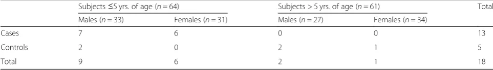
A total of 125 stool samples were tested in this study. Premier™Rotaclone® kit detected rotavirus in 11.2% (14/ 125) while Seeplex® Diarrhea-V ACE Detection kit de-tected rotavirus in 16.0% (20/125). Seeplex® Diarrhea-V ACE Detection kit detected rotavirus in 25.4%, (16/63) of cases and 6.5% (4/62) of the controls. On the other hand, Premier™ Rotaclone® kit detected rotavirus in 22.2% (14/63) of cases and in none of the controls as shown in Tables1and2. Discordant samples noted were from 4 controls and 2 cases, resolution by qRT-PCR re-vealed that the 2 cases and 2 of the controls were true positives while the other 2 controls turned out to be false positives. No cross-reactivity was observed in any of the assays. Sensitivity and specificity were calculated after resolution of discordant samples by qRT-PCR.
The sensitivity and specificity of the Seeplex® Diarrhea-V ACE detection were 100% (95% confidence interval 81.5 to 100.00%) and 98.1% (95% confidence interval 93.41 to 99.77%) respectively, and those of the
Table 1Distribution of Rotavirus among study participants
Subjects≤5 yrs. of age (n= 64) Subjects > 5 yrs. of age (n= 61) Total
Males (n= 33) Females (n= 31) Males (n= 27) Females (n= 34)
Cases 7 6 0 0 13
Controls 2 0 2 1 5
Premier™ Rotaclone® were 77.8% (95% confidence inter-val 52.36 to 93.59%), and 100% (95% confidence interinter-val 96.61 to 100.00%) respectively as shown in Table3
Turnaround time (TAT)
The mean TAT per run of 10 samples was 93 min and 359 min for the Premier™ Rotaclone® and the Seeplex® Diarrhea-V ACE Detection respectively (Table 3). The difference in mean TATs was 266 min (95% confidence interval from 262 to 270 min). The Seeplex® Diarrhea-V ACE Detection kit takes more than threefold the amount of time it takes to run the Premier™Rotaclone®.
Cost
The average cost of kits/reagents used for testing one sample was 13.33USD and 32.94USD for the Premier™ Rotaclone® and the Seeplex® Diarrhea-V ACE Detection assay respectively (Table 4). The Premier™ Rotaclone® only required one kit, a pipette, pipette tips, and sterile tubes while the RT-PCR assay required 2 additional kits (an RNA extraction and a reverse transcription kit), as well as Thermo Scientific™NanoDrop™2000 Specrtophot-ometer~ 9700USD, a thermocycler (Applied Biosystems Veriti 96 well thermocycler ~7000USD), high-speed cen-trifuge (Eppendorf Microcencen-trifuge ~3000USD), an elec-trophoresis power supplier (Thermo Scientific Owl EC-200 XL Hi Current Power supply~ EC-2000USD), an electro-phoresis chamber (Thermo Scientific Owl A1 Large Gel System~650USD), and a gel imaging system (Alpha Inno-tech AlphaImager HP~ 2500 USD). Unlike consumable reagents and test kits that are bought frequently and have predictable prices, laboratory equipment are not bought frequently and vary greatly in prices depending on their capabilities and vendors. For these reasons, cost of
equipment used in this study are only listed for readers’ consideration but not included alongside consumable re-agents and kits in calculation of the average cost per sam-ple represented in Table4.
Additional findings
Due to the multiplex nature of the Seeplex® Diarrhea-V ACE Detection kit, we detected a variety of enteric vi-ruses including a 1 subject who had a rotavirus/noro-virus co-infection (Table5).
Discussion
In this study, the Seeplex® Diarrhea-V ACE Detection kit showed a higher detection frequency and was more sen-sitive than the Premier™ Rotaclone®. The two kits had detection frequencies of 25.4 and 22.2% respectively among cases, all of these were children below 5 years of age. These resulted closely reflects prevalence of 14.5 to 31% that were reported in recent epidemiologic studies done in Kenya among children below 5 years of age hos-pitalized with diarrhea [10,12]. The higher detection fre-quency and sensitivity of the RT-PCR assay could be attributed to its lower detection limit documented by the manufacturer (100 copies/3μl DNA) that enables it to detect trace amounts of viral nucleic acids excreted in stool samples. The observation that rotavirus was de-tected in asymptomatic controls by RT-PCR could be at-tributed to the fact that the virus’nucleic acid is likely to remain detectable for a longer period after the subjects has recovered. In contrast, the EIA targets antigens that are rarely detected more than 1 week after onset of ill-ness [34]. Asymptomatic controls have also tested posi-tive by RT-PCR in other studies including one recently done in Niger [27]. The higher sensitivity (100%) of the Seeplex® Diarrhea-V ACE Detection kit was observed in an evaluation and verification study done in Canada [24]. The higher specificity (100%) exhibited by Premier™ Rotaclone® in this study is also in agreement with previ-ous studies comparing EIA and RT-PCR detection assays U.S.A [31].
The mean TAT per run of 10 samples for Seeplex® Diarrhea-V ACE Detection kit was more than three-fold longer than the TAT for Premier™ Rotaclone®. This was due to several steps involved in the RT-PCR assay that
Table 2Rotavirus detection frequency and descriptive metrics for the two assays
Detection frequency (%)
Seeplex® Diarrhea-V ACE Detection(n= 125)
Premier™Rotaclone® (n = 125)
Casesn= 63 16/63 (25.4%) 14/63 (22.22%)
Controlsn= 62 4/62 (6.4%) 0/62 (0)
Total = 125 20/125 (16.0%) 14/125 (11.2%)
Table 3Comparison of the sensitivity and specificity of the Seeplex® Diarrhea-V ACE Detection and Premier™Rotaclone® assays
Seeplex® Diarrhea-V ACE Detection (n = 125) Premier™Rotaclone® (n = 125)
True positives 18 14
False positives 2 0
True negatives 105 107
False negatives 0 4
Sensitivity 100% (95% CI: 81.5 to100%) 77.8% (95% CI: 52.36 to 93.59%)
include RNA extraction, reverse transcription, and amp-lification. This difference in mean TATs is an important consideration in clinical settings where shorter TATs are of great importance for patient treatment and care [18]. It is important to note that the Seeplex® Diarrhea-V ACE Detection kit is able to detect five different diarrhea causing viruses (rotavirus, adenovirus, astrovirus, noro-virus GI and GII) within the time noted whereas the Premier™ Rotaclone® can only detected rotavirus. See-plex® Diarrhea-V ACE Detection kit was able to detect norovirus GII in 12 samples (8 from children below 5 years and 4 from individual above 5 years), adenovirus in 6 samples (5 from children under 5 years and 1 from a 6 years old child), 1 astrovirus from a 5 years old child and one rotavirus/norovirus GII co infection from a 1 year and 4 months old child. This gives a hint of the heavy disease burden imposed especially on children below 5 years old by enteric viruses circulating in Kenya. As ob-served by Goldenberg et al. 2015, the ability to detect multiple pathogens is an important consideration to make when identifying platforms to use for future clin-ical as well as outbreak investigations and surveillance work [35].
The average cost of testing one sample by Seeplex® Diarrhea-V ACE Detection was more than 2-fold higher than that of testing by the Premier™ Rotaclone®. The higher cost can be attributed to the fact that the RT-PCR assay required an RNA extraction, reverse tran-scription as well as amplification kits whereas the Prem-ier™ Rotaclone® procedure only required one kit. Seeplex® Diarrhea-V ACE Detection also utilized more laboratory equipment: a spectrophotometer, thermocy-cler, a high-speed centrifuge, an electrophoresis power
supplier, an electrophoresis chamber, and a gel imaging system making its operational cost much higher than the Premier™ Rotaclone®. The Premier™ Rotaclone® re-quired fewer supplies due to fewer steps involved in its procedure. The RT-PCR platform had longer steps with both RNA extraction and agarose gel electrophoresis that required more supplies. This made the Seeplex® Diarrhea-V ACE Detection assay more labor intensive and time consuming than the EIA platform [36].
Accurate diagnosis and surveillance of rotavirus and other enteric viruses causing diarrhea in Kenya is critical for the rapid identification of infected patients who are potential sources of infection to others. Having this cap-ability at the point-of-care or as close as possible would enhance patient management, reduce the spread of en-teric pathogens, and improve the management of disease burden, especially among children [37]. Evaluation stud-ies like this will ensure quality and improvement of en-teric viruses’ testing assays, implementation of better testing platform, improved knowledge on disease dy-namics and better patient care. These outcomes will have a beneficial effect especially on the health of chil-dren in Kenya and other developing countries that bear the biggest disease burden associated with enteric vi-ruses. This will lead to better growth, development and general well being of such children.
Conclusion
In this study, the Seeplex® Diarrhea-V ACE Detection kit (Seegene, Seoul, Republic of Korea), a RT-PCR, showed a higher detection frequency and sensitivity compared to the established EIA, Premier™ Rotaclone® kit(Meridian Bioscience, INC, Cincinnati Ohio, U.S.A). However, the Premier™ Rotaclone® kit had a higher specificity, lower average cost, and shorter TAT.
With a higher detection frequency, sensitivity, and ability to detect 5 viruses, the Seeplex® Diarrhea-V ACE Detection kit is an ideal tool for outbreak investigations and surveillance of enteric viruses causing diarrhea in Kenya. Its higher cost, complexity, and longer TAT make it unsuitable for routine clinical testing especially in aus-tere setups within Kenya and other developing nations.
Table 4Summary of TAT and average reagent and kits cost per assay
Seeplex® Diarrhea-V ACE Detection
Premier™Rotaclone®
Mean TAT
(per run of 10 samples)
359 93
Average cost (per sample)
$32.94 $13.33
Table 5Enteric viruses detected by the Seeplex® Diarrhea-V ACE Detection Assay
Subjects≤5 yrs. of age (n= 64) Subjects > 5 yrs. of age (n= 61) Total
Males Females Males Females
Rotavirus 9a 6 2 3 20
Adenovirus 3 2 0 1 6
Astrovirus 1 0 0 0 1
NoVGI 0 0 0 0 0
NoVGI1 3a 5 2 2 12
Total 16 23 4 6 39
a
However, if validated and approved by the relevant au-thorities in Kenya, it can be a useful tool in well-funded clinical laboratories within the country. This platform can be easily implemented in well-funded research insti-tutions in Kenya and can play an important role in eluci-dating the incidences, prevalence, distribution, and disease burden associated with enteric viruses circulating in the country. The longer TAT and higher cost of the Seeplex® Diarrhea-V ACE assay may be justifiable by the number of viruses it can detect in each run.
The Premier™Rotaclone® is suitable for routine clinical testing of rotavirus especially in austere settings in Kenya. It has a shorter turnaround time, is 100% spe-cific, cheaper and can be easily implemented even in re-mote clinics that have limited resources and financial constraints that inhibit their ability to implement com-plex platforms like RT-PCR.
Further evaluation and validation of newer multiplex assays that can detect several enteric pathogens includ-ing parasites and bacteria circulatinclud-ing in Kenya should be encouraged, as this will elucidate their applicability and make scientists and policy makers aware of the most suitable platforms to implement in routine clinical and research work in Kenya. The additional data that multi-plex assays are able to provide can enhance surveillance of etiological agents causing diarrheal illness and en-hance public health measures and policy decisions on diarrheal diseases. Other than rotavirus, there is very limited information on the prevalence and trends of other enteric viruses circulating in Kenya. As rotavirus studies continue, more effort should be put on studying the other enteric viruses as well. This should be done by the most suitable testing platforms that can be imple-mented efficiently to generate the most accurate data possible.
Limitations of the study
Limitations of this study include the fact that we did not look at severity of illness and focused only on 5 enteric viruses thus we cannot provide information on other pathogens including bacteria and parasites that are the other likely causes diarrhea in the cases that were nega-tive for the enteric viruses tested in this study.
Abbreviations
CAP:College of American Pathologists; dNTPs: Deoxyribonucleotide triphosphate; EIA: Enzyme immunoassay; EM: Electron microscopy; EM: Electron microscopy; IRB: Institutional Review Board; KEMRI: Kenya Medical Research Institute; MHK: Microbiology Hub Kericho; MoH: Ministry of Health; mRT-PCR: Multiplex reverse transcription polymerase chain reaction; PCR: Polymerase chain reaction; qRT-PCR: Real-time reverse transcription Polymerase chain reaction; TAT: Turn around time; UKNEQAS: United Kingdom National External Quality Assessment Service; USAMRD-A: United States Army Medical Research Directorate-Africa; WHO: World Health Organization; WRP: Walter Reed Project
Acknowledgements
We acknowledge the role of United States Army Medical Research Directorate-Africa, the Microbiology Hub–Kericho, and Kenya Medical Research Institute for facilitating this study. Ethics committees from both WRAIR and KEMRI-SERU played a critical role in ensuring that this study is feasible and meets the required standards. We also acknowledge the role played by Dr. Alexander Flynn for his support during the critical stage of submission for publication. Special thanks go to Dr. Lilian Musila from USAMRD-K/KEMRI, Rukia Kibaya from KEMRI, and Teresa Soderberg from WRAIR for invaluable assistance during ethical and scientific review. We also acknowledge and appreciate the critical role played by both WRAIR and KEMRI IRBS during review and approval of this study. Special thanks to Leelgo Kimeto, Michael Obonyo and Judith Bosuben from the Walter Reed Project-Kericho Regulatory office for their guidance and support.
Authors’contributions
CP and EK contributed to study design, laboratory testing, data analysis, and drafting of the manuscript. BS, EM, BD, SB and CH contributed in the study design and manuscript review. RK, MK, EO, NK, AO, JN and MK contributed in data analysis and manuscript review. All authors read and approved the final manuscript.
Authors’information
Cliff Philip, Erick Kipkirui, Ronald Kirera, Mary Kirui, Elizabeth Odundo, Nancy Kipkemoi, Abigael Ombogo, Janet Ndonye and Margaret Koech are all employees at United States Army Medical Research Directorate-Africa’s/ Microbiology Hub Kericho. Brett Swierczewski is a senior research scientist at Walter Reed Army Institute of Research (WRAIR), USA. Esther Magiri is a Professor at Jomo Kenyatta University of Agriculture and Technology, Kenya. Brook Danboise was affiliated to USAMRD-A & WRAIR during study conception and is now a student at the Michigan State University College of Human Medicine. Christine Hulseberg was previously affiliated to USAMRD-A and WRAIR and is now a student at the University of Virginia, School of Medicine. Stacey Bateman was previously affiliated to USAMRD-A and WRAIR but is currently affiliated to Madigan Army Medical Center. Alexander Flynn is the current laboratory director of the Microbiology Hub Kericho.
Funding
Funding for this study was provided through AFHSB-GEIS program through support of Enteric Pathogen Surveillance program and Protocol #1549. This study was a sub-study of WRAIR/KEMRI # 1549.
Availability of data and materials
All data and material used to generate information published in this paper shall be made available to publisher or any relevant party that may need the information.
Ethics approval and consent to participate
The investigators have adhered to the policies for protection of human subjects as prescribed in AR 70–25. This study was reviewed and approved by both WRAIR and KEMRI IRBS. This study was designated WRAIR #2443 and KEMRI/SERU/CCR/0052/3384 and approved as a sub-study of WRAIR#1549 by both WRAIR and KEMRI SERU. Specimen collection and laboratory analysis was conducted under human-use protocol WRAIR #1549; Enteric Pathogen Surveillance study approved by WRAIR and KEMRI SERU. It was determined that it did not involve interaction with human subjects and that consent to participate was adequately covered by protocol WRAIR#1549.
Consent for publication
Consent for publication was granted from all authors, institutional review boards, and funding agency. Material has been reviewed by the Walter Reed Army Institute of Research. There is no objection to its presentation and/or publication. The opinions or assertions contained herein are the private views of the author, and are not to be construed as official, or as reflecting true views of the Department of the Army or the Department of Defense.
Competing interests
Author details
1United States Army Medical Research Directorate-Africa, Nairobi, Kenya. 2University of Michigan Medical School, Arbor, USA.3Center for Genome
Sciences, US Army Medical Research Institute of Infectious Diseases, Frederick, Maryland, USA.4Madigan Army Medical Center, Washington, USA. 5Walter Reed Army Institute of Research, Silver Spring, USA.6Jomo Kenyatta
University of Agriculture and Technology, Juja, Kenya.
Received: 25 January 2019 Accepted: 21 June 2019
References
1. McAuliffe GN, Anderson TP, Stevens M, Adams J, Coleman R,
Mahagamasekera P, et al. Systematic application of multiplex PCR enhances the detection of bacteria, parasites, and viruses in stool samples. J Infect. 2013;67(2):122–129. [cited 2014 mar 13] Available from:http://www.ncbi. nlm.nih.gov/pubmed/23603249.
2. Curry JA, Riddle MS, Gormley RP, Tribble DR, Porter CK. The epidemiology of infectious gastroenteritis related reactive arthritis in U.S. military personnel: a case-control study. BMC Infect Dis. 2010;10:266.
3. Anteneh ZA, Andargie K, Tarekegn M. Prevalence and determinants of acute diarrhea among children younger than five years old in Jabithennan District, Northwest Ethiopia, 2014. BMC Public Health. 2017. Available from: https://doi.org/10.1186/s12889-017-4021-5.
4. Kotloff KL, Blackwelder WC, Nasrin D, Nataro JP, Farag TH, Van A, et al. The Global Enteric Multicenter Study ( GEMS ) of Diarrheal Disease in Infants and Young Children in Developing Countries : Epidemiologic and Clinical Methods of the Case / Control Study. 2012;55(Suppl 4):S232–45.
5. Riddle MS, Smoak BL, Thornton SA, Bresee JS, Faix DJ, Putnam SD. Epidemic infectious gastrointestinal illness aboard U.S. Navy ships deployed to the Middle East during peacetime operations--2000-2001. BMC Gastroenterol. 2006;6:9.
6. Guerra AH, Stockmann C, Pavia AT, Hersh AL, Thorell EA, Weng HY, et al. Laboratory-Confirmed Rotavirus Disease in Utah Children: Clinical and Economic Impact of Rotavirus Vaccination. J Pediatric Infect Dis Soc. 2012; 1(4):268–77 Available from:http://www.pubmedcentral.nih.gov/articlerender. fcgi?artid=3656544&tool=pmcentrez&rendertype=abstract.
7. Parashar UD, Hummelman EG, Bresee JS, Miller MA, Glass RI. Global Illness and Deaths Caused by Rotavirus Disease in Children. Emerg Infect Dis. 2003; 9(5):565–72.
8. Gutiérrez-Aguirre I, Steyer A, Boben J, Gruden K, Poljšak-Prijatelj M, Ravnikar M. Sensitive detection of multiple rotavirus genotypes with a single reverse transcription-real-time quantitative PCR assay. J Clin Microbiol. 2008;46(8): 2547–54.
9. Muendo C, Laving A, Kumar R, Osano B, Egondi T, Njuguna P. Prevalence of rotavirus infection among children with acute diarrhoea after rotavirus vaccine introduction in Kenya, a hospital cross-sectional study. [cited 2018 Dec 17]; Available from:https://doi.org/10.1186/s12887-018-1291-8. 10. Bitek A. Caccination era: a cross-sectional study. 2017;8688:1–12. 11. Mohan VR, Karthikeyan R, Babji S, Mcgrath M, Shrestha S, Shrestha J, et al.
Rotavirus Infection and Disease in a Multisite Birth Cohort : Results From the MAL-ED Study. J Infect Dis. 2017;216(February 2014):305–16.
12. Muendo C, Laving A, Kumar R, Osano B, Egondi T, Njuguna P. Prevalence of rotavirus infection among children with acute diarrhoea after rotavirus vaccine introduction in Kenya, a hospital cross-sectional study; 2018. p. 1–9. 13. Sk R, Nyangao J, Kombich J, Sang C, Gikonyo J, Ongus JR. Original article
human rotavirus group a serotypes causing gastroenteritis in children less than 5 years and hiv-infected adults in viwandani slum, Nairobi. p. 2. 14. Junaid SA, Umeh C, Olabode AO, Banda JM. Incidence of rotavirus infection
in children with gastroenteritis attending Jos university teaching hospital, Nigeria. Virol J. 2011;8(1):233 [cited 2015 Jan 14]. Available from:http:// www.pubmedcentral.nih.gov/articlerender.fcgi?artid=3107812&tool= pmcentrez&rendertype=abstract
15. Liu Y, Xu ZQ, Zhang Q, Jin M, Yu JM, Li JS, et al. Simultaneous detection of seven enteric viruses associated with acute gastroenteritis by a multiplexed luminex-based assay. J Clin Microbiol. 2012;50(7):2384–9.
16. Lalani T, Tisdale MD, Liu J, Mitra I, Philip C, Odundo E, et al. Comparison of stool collection and storage on Whatman FTA Elute cards versus frozen stool for enteropathogen detection using the TaqMan Array Card PCR Comparison of stool collection and storage on Whatman FTA Elute cards
versus frozen stool for enteropath. 2018. Available from:https://doi.org/1 0.1371/journal.pone.0202178
17. Swierczewski BE, Odundo EA, Koech MC, Ndonye JN, Kirera RK, Odhiambo CP, et al. Enteric pathogen surveillance in a case-control study of acute diarrhoea in the town of Kisii, Kenya. J Med Microbiol. 2013;62:1774–6. 18. Escherichia ST. Clinical Evaluation of a Real-Time PCR Assay for Identification
of Salmonella, Shigella, Campylobacter (Campylobacter jejuni and Specimens). J Clin Microbiol. 2013;51(12):4001–7.
19. Omore R, Tate JE, Reilly CEO, Ayers T, Williamson J, Moke F, et al. Epidemiology , Seasonality and Factors Associated with Rotavirus Infection among Children with Moderate-to-Severe Diarrhea in Rural Western Kenya, 2008–2012 : The Global Enteric Multicenter Study (GEMS ) 2016;2008–2012. Available from:https://doi.org/10.1371/journal.pone.0160060
20. Gikonyo JN, Nyangao J, Mbae C, Sang C, Njagi E, Ngeranwa J, et al. Molecular characterization of group A rotaviruses in Mukuru slums Kenya: detection of novel strains circulating in children below 5 years of age. BMC Res Notes. 2017;10(1):290 Available from:http://bmcresnotes.biomedcentral. com/articles/10.1186/s13104-017-2611-z.
21. Kiulia NM, Kamenwa R, Irimu G, Nyangao JO, Gatheru Z, Nyachieo A. The Epidemiology of Human Rotavirus Associated with Diarrhoea in Kenyan Children : A Review; 2008. p. 401–5.
22. For C, Control D. Annual report 2014; 2014.
23. Mwenda JM, Ntoto KM, Abebe A, Enweronu-Laryea C, Amina I, Mchomvu J, et al. Burden and epidemiology of rotavirus diarrhea in selected African countries: preliminary results from the African Rotavirus Surveillance Network. J Infect Dis. 2010;202(Suppl 1):S5–11 [cited 2015 Jan 14]. Available from:http://www.ncbi.nlm.nih.gov/pubmed/20684718.
24. Higgins RR, Beniprashad M, Cardona M, Masney S, Low DE, Gubbay JB. Evaluation and verification of the Seeplex Diarrhea-V ACE assay for simultaneous detection of adenovirus, rotavirus, and norovirus genogroups I and II in clinical stool specimens. J Clin Microbiol. 2011;49(9):3154–3162 [cited 2014 Mar 13]. Available from:http://www.pubmedcentral.nih.gov/ articlerender.fcgi?artid=3165607&tool=pmcentrez&rendertype=abstract 25. Swierczewski BE, Odundo EA, Koech MC, Ndonye JN, Kirera RK, Kirera CP, et
al. Surveillance for enteric pathogens in a case-control study of acute diarrhea in Western Kenya. Trans R Soc Trop Med Hyg. 2013;107(September 2009):83–90.
26. Ope M, Nyoka R, Unshur A, Oyier FO, Mowlid SA, Owino B, et al. Evaluation of the Field Performance of ImmunoCard STAT ! ® Rapid Diagnostic Test for Rotavirus in Dadaab Refugee Camp and at the Kenya–Somalia Border. Am J Trop Med Hyg. 2017;96(6):1302–6.
27. Lagare A, Moumouni A, Kaplon J, Langendorf C, Pothier P, Grais RF, et al. Diagnostic accuracy of VIKIA ® Rota - adeno and premier™Rotaclone ® tests for the detection of rotavirus in Niger. BMC Res Notes. 2017:1–5. 28. Xia Z, Johansson ML, Gao Y, Zhang L, Zhan A, Haffner GD, et al.
Conventional versus real - time quantitative PCR for rare species detection. Ecol Evol. 2018;8:11799–807.
29. Zemtsova GE, Montgomery M, Levin ML. Relative Sensitivity of Conventional and Real- Time PCR Assays for Detection of SFG Rickettsia in Blood and Tissue Samples from Laboratory Animals. 2015;1–7. Available from:https:// doi.org/10.1371/journal.pone.0116658.
30. Parikh R, Mathai A, Parikh S, Chandra Sekhar G, Thomas R. Understanding and using sensitivity, specificity and predictive values. Indian J Ophthalmol. 2008;56(1):45–50. [cited 2018 Dec 6]. Available from:http://www.ncbi.nlm. nih.gov/pubmed/18158403.
31. Gautam R, Lyde F, Esona MD, Quaye O, Michael D, Viruses R. Detection of Rotavirus Antigen in Stool Specimens. 2015;58(1):292–4.
32. Goswami B, Singh B, Chawla R, Gupta VK, Mallika V. Turn Around Time ( TAT ) as a Benchmark of Laboratory Performance. Indian J Clin Biochem. 2010;25(4):376–9. 33. Gerlach J, Sequeira M, Alvarado V, Cerpas C, Balmaseda A, Gonzalez A, et al.
Cost analysis of centralized viral load testing for antiretroviral therapy monitoring in Nicaragua, a low-HIV prevalence, low-resource setting. J Int AIDS Soc. 2010;13(1):43.
34. Hagbom M, Franco MA, Greenberg HB, Ryan MO. Rotavirus infection. 2018. 35. Goldenberg SD, Bacelar M, Brazier P, Bisnauthsing K, Edgeworth JD. A cost
benefit analysis of the Luminex xTAG gastrointestinal pathogen panel for detection of infectious gastroenteritis in hospitalised patients. J Infect [Internet]. 2015;70(5):504–11. Available from:https://doi.org/10.1016/j.jinf.2014.11.009. 36. Hoet AE, Chang K, Saif LJ. Comparison of ELISA and RT-PCR versus immune
37. Dennehy PH, Schutzbank TE, Thorne GM. Evaluation of an automated immunodiagnostic assay, VIDAS rotavirus, for detection of rotavirus in fecal specimens. J Clin Microbiol [Internet]. 1994;32(3):825–7. Available from: http://www.pubmedcentral.nih.gov/articlerender.fcgi?artid=263132&tool= pmcentrez&rendertype=abstract.
Publisher’s Note