MINI REVIEW
The Potentials and Applications of Cellulose Acetate in biosensor
technology
Hadi Baharifar1; Elham Honarvarfard2; Mohammad Haji Malek-kheili3; Hassan Maleki4; Mohammad Barkhi5; Ali Ghasemzadeh6; Kamyar Khoshnevisan7,8*
1Department of medical nanotechnology, Applied Biophotonics Research Center, Science and Research Branch,
Islamic Azad University, Tehran, Iran
2Department of Chemistry and Biomolecular Science, Clarkson University, Potsdam, NY 13699-5810, USA
3Departemant of chemistry, Islamic Azad University, Science and Research Branch, Tehran, Iran
4Department of Medical Nanotechnology, School of Advanced Technologies in Medicine, Tehran University of Medical Sciences, Tehran, Iran
5University of Applied Science and Technology (UAST), Zar Center, Karaj, Iran
6School of Chemical Engineering, Iran University of Science and Technology, Narmak 16846-13114, Tehran, Iran
7Biosensor Research Center, Endocrinology and Metabolism Molecular-Cellular Sciences Institute, Tehran University of Medical Sciences, Tehran, Iran
8Endocrinology and Metabolism Research Center, Endocrinology and Metabolism Clinical Sciences Institute, Tehran University of Medical Sciences, Tehran, Iran
*Corresponding Author Email: [email protected]
The interest in cellulose and its derivatives has been exponentially increasing due to its excellent thermal stability, biocompatibility, chemical persistence and biodegradability. Among various cellulose derivatives, cellulose acetate (CA) has been applied in many applications including sensor systems, drug delivery systems, separation membrane, and tissue engineering. Recently, the electrospun nanofibers have been employed and have gotten more attention in the biotechnology and the biomedical applications. In this case, Electrospinning methods widely used to fabricate and generate novel nanomaterials along with the well-aligned structure of electrospun nanofibers. Electrospinning has emerged as a powerful method to produce nanofibrous assemblies from a variety of polymers and composites including CA fibers. These fibers obtained from this method were applied in biomedical applications specially for sensing process in the medical diagnostic kit. In this review article, the recent progress and development of electrospun CA fibers and nanofibers and also their nanocomposites for advanced sensing systems are presented. Several sensors and biosensors including optical/colorimetric, and electrochemical-based on CA are discussed in this study.
ARTICLE INFO
Article History:
Received 02 October 2017 Accepted 20 December 2017 Published 27 December 2017
Keywords: Cellulose acetate Sensing
Optical/Colorimetric Electrochemical
ABSTRACT
How to cite this article
Baharifar H, Honarvarfard E, Haji Malek-kheili M, Maleki H, Barkhi M, Ghasemzadeh A, Khoshnevisan K, The Potentials and Applications of Cellulose Acetate in biosensor technology, Nanomed Res J, 2017; 2(4):216-223. DOI: 10.22034/nmrj.2017.04.002
INTRODUCTION
Biodegradable and biocompatible materials and polymers show significant advantages over other materials for enhancing mats in biomedical applications [1]. These materials, due to their
applications [9]. Among the various techniques of creating CA nanofibers, electrospinning has been extensively used. Electrospinning is a well-organized process for synthesizing of ultrafine fibers with diameters ranging from several micrometers down to a few nanometers, which works with a high voltage across a conductive needle attaching to a polymer liquid-containing reservoir and a collector [10, 11]. Recently, the electrospinning of CA has been extensively studied because of its exceptional thermal stability, chemical resistance, biocompatibility, and biodegradability [12]. electrospun CA nanofibers can be applied to the surface device of diagnostic kits and biosensing materials.
Major sensing approaches include acoustic wave, photoelectric, resistive, optical, amperometric, and other sensors are the main types of these sensors that were employed on the surface of electrospun nanomaterials [13, 14].
The purpose of this article is to investigate recent researches conducted on sensing by using CA fibers involving optical/colorimetric, and electrochemical biosensors.
Methods of Sensing by Using Cellulose Acetate Polymeric composites and polymeric materials have a wide array of applications in the realm of technology. Among these materials, the derivatives of cellulose such as CA are widely employed for biomedical applications. Depending on the processing method employed, CA can be used for high varies of applications (e.g., films, membranes or fibers). In this section, the recent trend in the exploitation of electrospun CA fibers in the nanoscale for sensing applications have been reviewed.
Based on literature review, at least three kinds of sensing approach could be achieved using
substrate or product for the Vis spectrum and determining the best lambda Max and a linear region. It should be noted that CA in colorimetric methods is utilized only as a support membrane for attaching the biological elements (i.e., enzymes, antibodies, and aptamers).
CA usually activated before enzyme immobilization. Modifications of CA could improve the biological element attachment or sensing process via different mechanisms. Among different methods, using cross-linking agents is one of the common methods in CA activation.
CA membrane could also be activated using treatment with sodium periodate solution, ethylenediamine solution, and glutaraldehyde respectively. Then different enzymes such as cholesterol oxidase could be immobilized on the activated CA membrane [16].
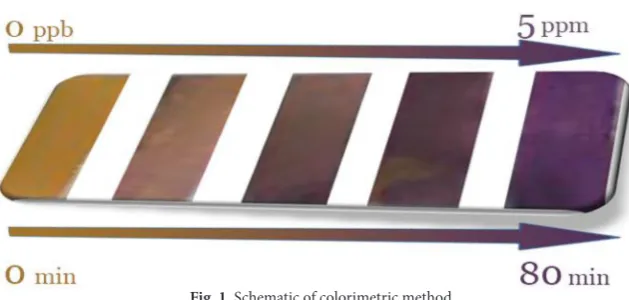
Uranium is one of the important metal ions that exhibits toxic and radioactive effects [17]. A soluble form of uranium is uranyl (UO2 2+). For the detection of uranyl, there are many techniques such as gamma spectrometry, ion chromatography and alpha spectrometry which are expensive. Other methods such as laser fluorimetry and stripping voltammetry are highly sensitive and selective. However, they require complicated procedures [18-24].
with this technique, the quantitative detection for uranyl is achieved [26]. The colorimetric method at different times and various concentration were shown in fig. 1.
Metalloprotein is a generic type of a protein that contains a metal ion cofactor. The solid-state nanofiber-based optical sensors with an anionic fluorescent dendrimer (AFD) via a fluorescence resonance energy transfer (FRET) mechanism were employed to detect the low concentration of metalloproteins [27].
In this method, the AFD has been encapsulated in electrospun CA nanofibers, and for improving the sensing performance, CA is deacetylated to cellulose to generate secondary porous structures which are desirable for enhancing molecular interactions. The protein sensing properties of the fibers were studied by monitoring the quenching behaviors of cytochrome c (cyt c), hemoglobin (Hgb), and bovine serum albumin (BSA) as a function of concentration. The quenching effect is a result of energy/electron transfer processes between iron-containing proteins (i.e., cyt c and Hgb) and the fluorescent core. To achieve the highest fluorescence intensity, five water-soluble fluorescent dendritic compounds (AFD-1, AFD-2, AFD-3, AFD-4, and AFD-5) were synthesized [27]. Except the AFD-3, the others dendrimers commonly demonstrated low visible fluorescent emission. The fluorescence images of the AFD-3-doped cellulose nanofibers before the quenching process indicate the evidence fluorescence emission and the uniform dispersion of fluorophores in cellulose, which is beneficial to sensing performance [27].
CA-based electrochemical Sensors
Amperometric sensors are based on measuring the produced current when a potential is applied between two electrodes. The current is then related to the concertation of the analyte present in the system.
Biosensors that uses current for detection, usually need a transducer. Therefore, CA could not be a support for amperometric sensors alone. Modification of CA for immobilization of enzyme is an important part in the amperometric sensors. CA/Au nanorods composites are employed as supports for designing amperometric sensors to detect glucose. In one study, glucose oxidase (GOx) enzyme was immobilized using glutaraldehyde as a cross-linking agent. The designed biosensors showed high sensitivity (8.4 µA cm-1 mM-2) and acceptable limit of detection (2 × 10⁻⁵ M) [29]. Catalase-based biosensor has been successfully designed using immobilization of enzyme on modified (activated) CA beads. For the preparation of CA beads, the polymer was dissolved in acetone and then dropwise was added to hexane solution under stirring. First, CA beads were activated using Ce(SO4)2 and then were immobilized with catalase. Immobilization of enzyme occurred by entrapment and cross-linking by activated CA beads. Activation of CA beads resulted in oxidizing its OH groups to aldehyde groups in the presence of Ce(SO4)2. After that, spacer arms were composed with the help of bovine serum albumin (BSA), and enzyme entrapped using cross-linking agents. Enzyme activity was evaluated in 10.5 mM of hydrogen peroxidase at 240 nm using a spectrophotometer and demonstrated that enzyme had optimal activity at pH 7.0 and T=35°C [15].
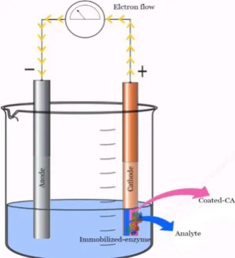
Gilmartin et al. developed a uric acid sensor based on CA fibers as a support. In their work, modified and unmodified cobalt phthalocyanine (CoPc) electrodes coated with CA and immobilized with uricase were used for sensing of uric acid. First, the electrodes were doped with screen printed carbon (SPCEs) and coating after that, the desired area of CoPc-SPCEs was coated with 2% solution of CA. The coated electrodes with CA immersed in different concertation of uricase solution for 5 minutes. Then, the electrodes were dried overnight and rinsed with distilled water to remove unbounded enzymes. Obtained results revealed that in the range of 1×10⁻⁶ to 13×10-6 mol/dm of
uric acid, the amperometric calibrations were linear, and the optimum uricase loading was 1 U [30]. Amperometric method based on CA and immobilized enzyme was shown in Fig. 2.
Constantinos et al. developed a CA-based amperometric sensor for detection of glycolic acid in various complex matrixes such as cosmetics, instant coffee, and urine. Glycolic acid is a constituent of sugar cane juice and has been used widely in the industry (e.g., processing the textile, leather, and food). There are limited number of methods to determine glycolic acid such as gas chromatography/mass spectrometry, HPLC, ion-exchange HPLC, etc. which require derivatization steps, complex isolation, and expensive instrumentation. In their work, they developed an amperometric glycolic acid sensor
based on glycolate oxidase/catalase immobilized into a CA membrane. First, CA membrane was prepared by mixing CA and Polyvinyl Acetate (PVA) with acetone and cyclohexane and was then placed on the platinum surface to eliminate interference from electroactive species. In the next step, the membrane was immersed in a solution of enzymes followed by superimposed with an outer polycarbonate membrane to protect enzymes from leaking and microbial attack. This system demonstrates a linear relationship between the response and the glycolate concentration in the range 0.01-1mM with a correlation coefficient, r = 0.997 and the detection limit of 6µM glycolate [31]. CA could be activated using ionic liquids such as 1-butyl-3-methylimidazolium bis(trifluoromethylsulfonyl)imide (BMI•N(Tf)2) for designing of biosensors. Methyldopa was detected using immobilized laccase on CA/ (BMI•N(Tf)2) support. The Support was prepared by adding CA to acetone in a reaction flask and incubating overnight under the nitrogen atmosphere. Then BMI•N(Tf)2 was added to CA solution and stirred until homogeneous phase appeared. Afterward, homogeneous phase spread on a glass electrode and calcinated at 300°C and triturated to achieve the product. Voltammetric evaluation of methyldopa showed linearity from 34.8 to 370.3µM and detection limit about 5.5µM [32]. In another work, modified electrodes were prepared by immobilizing of
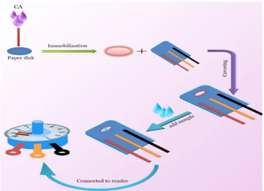
alcohol dehydrogenase on CA for voltammetric detection of ethanol. The CA solution was prepared in different concentrations, and the hydroxyl group of CA converted to imidazoylcarbamate derivatives using 1,1'-carbonyldiimidazole (CDI). After that, the solution was coated on a glassy carbon electrode followed by dropping toluidine blue on the electrodes. Alcohol dehydrogenase solution in phosphate buffer (pH 7.5) was added to the electrodes surface followed by adding glutaraldehyde for cross-linking of the enzyme. Prepared electrode response was linear between 1×10⁻⁵ M and 4 × 10⁻⁴M ethanol and detection limit was about 5× 10⁻⁶ M [33]. Generally, electrochemical method based on CA modified surface was illustrated in fig. 3. In this schematic, CA was applied to immobilize onto paper disk and then after was used for the working electrode. In Tkác et al. work fructose dehydrogenase was immobilized on a modified CA membrane for designing a voltammetric fructose biosensor. Ferrocene embedded CA membrane and ferrocene/Nafion modified CA membrane were prepared by dissolving ferrocene and ferrocene/ Nafion in CA solution and immobilization of enzyme on the electrodes directly. The initial sensitivity of electrodes was about 226 nA/mM, and both modified CA membrane demonstrated high stability [34].
Ammonia is a hazardous alkaline gaseous pollutant which widely used in the chemical
processes, medical diagnosis kits, etc. Ammonia concentration over 55ppm, can be easily identified by smell, however for concentration below 55ppm, we need to apply effective methods for detecting ammonia [35].
There are various sensing techniques for gas sensing such as optical and electrical techniques [33-36]. Fabrication of a sensor coated by a quartz crystal microbalance (QCM) with an electrostatic layer-by-layer (LBL) self-assembly technique is a novel approach to detect the ammonia concentration. In this approach, positively charged polyethylenimine (PEI) and negatively charged graphene oxide (GO) were embedded on the surfaces of negatively charged electrospun CA nanofibers on the QCM electrode. In the gas-sensing tests, the CA/PEI/ GO-based QCM sensor not only exhibited a low detection limit and rapid response, but also performed excellent reversibility and selectivity with respect to ammonia detection [37].
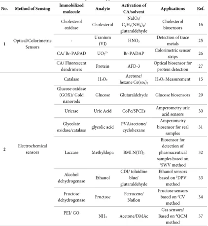
Table 1. describes the various types of CA-based sensors and its potential applications for detection of different analytes such as hydrogen peroxide, glucose, ethanol, and cholesterol as well as the modification and activation of CA substrates for employing in enzymatic and non-enzymatic sensors.
Conclusions and Future Perspective
In this review paper, recent progresses in electrospun CA-based sensors and a comprehensive
overview of fabrication and modification techniques of CA fibers are presented. Furthermore, different types of CA-based sensors including optical/colorimetric, and electrochemical as well as applications of CA in these sensors have been introduced.
High thermal stability, biocompatibility, and biodegradability of cellulose and its derivatives make them a great alternative to common substrate
Considering CA has a great potential for disposable, cost-effective, and biocompatible devices, further researches are needed to be conducted to improve its capabilities. Currently, the majority of the performed CA-based sensors studies are in vitro, and some further investigations devoted to in vivo CA-based sensors are needed. Since the potential applications of CA can be foreseen in a much broader area than those of optical/colorimetric, and electrochemical sensors, more studies are required to resolve current challenges for future biosensors.
CONFLICTS OF INTEREST
The authors declare that there are no conflicts of interest regarding the publication of this manuscript.
REFERENCES
1. Gheibi A, Khoshnevisan K, Ketabchi N, Derakhshan MA, Babadi AA. Application of Electrospun Nanofibrous PHBV Scaffold in Neural Graft and Regeneration: A Mini-Review. Nanomedicine Research Journal, 2016;1 (2):107-111. 2. Mehrabi F, Shamspur T, Mostafavi A, Saljooqi A, Fathirad F.
Synthesis of cellulose acetate nanofibers and its application in the release of some drugs. Nanomedicine Research Journal, 2017;2 (3):199-207.
3. Idris A, Yet LK. The effect of different molecular weight PEG additives on cellulose acetate asymmetric dialysis membrane performance. Journal of Membrane Science, 2006;280 (1):920-927.
4. Lv J, Zhang G, Zhang H, Yang F. Exploration of permeability and antifouling performance on modified cellulose acetate ultrafiltration membrane with cellulose nanocrystals. Carbohydrate Polymers, 2017;174 (Supplement C):190-199. 5. Voicu SI, Condruz RM, Mitran V, Cimpean A, Miculescu F,
Andronescu C, Miculescu M, Thakur VK. Sericin Covalent Immobilization onto Cellulose Acetate Membrane for Biomedical Applications. ACS Sustainable Chemistry & Engineering, 2016;4 (3):1765-1774.
6. Konwarh R, Karak N, Misra M. Electrospun cellulose acetate nanofibers: the present status and gamut of biotechnological applications. Biotechnology advances, 2013;31 (4):421-437. 7. Wang X, Kim Y-G, Drew C, Ku B-C, Kumar J, Samuelson
LA. Electrostatic assembly of conjugated polymer thin layers on electrospun nanofibrous membranes for biosensors. Nano Letters, 2004;4 (2):331-334.
8. Schiffman JD, Schauer CL. A review: electrospinning of biopolymer nanofibers and their applications. Polymer reviews, 2008;48 (2):317-352.
9. Rezaei A, Nasirpour A, Fathi M. Application of cellulosic
nanofibers in food science using electrospinning and its potential risk. Comprehensive Reviews in Food Science and Food Safety, 2015;14 (3):269-284.
10.Greiner A, Wendorff JH. Electrospinning: a fascinating method for the preparation of ultrathin fibers. Angewandte Chemie International Edition, 2007;46 (30):5670-5703. 11.Li D, Xia Y. Electrospinning of nanofibers: reinventing the
wheel? Advanced materials, 2004;16 (14):1151-1170. 12.Rodríguez K, Gatenholm P, Renneckar S. Electrospinning
cellulosic nanofibers for biomedical applications: structure and in vitro biocompatibility. Cellulose, 2012;19 (5):1583-1598.
13.Ding B, Wang M, Wang X, Yu J, Sun G. Electrospun nanomaterials for ultrasensitive sensors. Materials Today, 2010;13 (11):16-27.
14.Sapountzi E, Braiek M, Chateaux J-F, Jaffrezic-Renault N, Lagarde F. Recent Advances in Electrospun Nanofiber Interfaces for Biosensing Devices. Sensors, 2017;17 (8):1887. 15.Yildiz H, Akyilmaz E, Dinçkaya E. Catalase immobilization
in cellulose acetate beads and determination of its hydrogen peroxide decomposition level by using a catalase biosensor. Artificial cells, blood substitutes, and biotechnology, 2004;32 (3):443-452.
16.Wang S, Li S, Yu Y. Immobilization of cholesterol oxidase on cellulose acetate membrane for free cholesterol biosensor development. Artificial cells, blood substitutes, and biotechnology, 2004;32 (3):413-425.
17.Elabd AA, Zidan WI, Abo-Aly MM, Bakier E, Attia MS. Uranyl ions adsorption by novel metal hydroxides loaded Amberlite IR120. Journal of Environmental Radioactivity, 2014;134 (Supplement C):99-108.
18.Sundar U, Ramamurthy V, Buche V, Rao DN, Sivadasan PC, Yadav RB. Rapid measurements of concentrations of natural uranium in process stream samples via gamma spectrometry at an extraction facility. Talanta, 2007;73 (3):476-482. 19.Brunel B, Philippini V, Mendes M, Aupiais J. Actinide
oxalate complexes formation as a function of temperature by capillary electrophoresis coupled with inductively coupled plasma mass spectrometry. Radiochimica Acta, 103;2015:27-37.
20.Shaw MJ, Hill SJ, Jones P, Nesterenko PN. Determination of uranium in environmental matrices by chelation ion chromatography using a high performance substrate dynamically modified with 2,6-pyridinedicarboxylic acid. Chromatographia, 2000;51 (11):695-700.
21.Benedik L, Vasile M, Spasova Y, Wätjen U. Sequential determination of 210Po and uranium radioisotopes in drinking water by alpha-particle spectrometry. Applied Radiation and Isotopes, 2009;67 (5):770-775.
determination of trace amount of uranium (VI) in different aqueous and organic streams of nuclear fuel processing using 2-(5-bromo-2-pyridylazo-5-diethylaminophenol). Journal of Radioanalytical and Nuclear Chemistry, 2010;285 (3):675-681.
26.Hu L, Yan X-W, Li Q, Zhang X-J, Shan D. Br-PADAP embedded in cellulose acetate electrospun nanofibers: Colorimetric sensor strips for visual uranyl recognition. Journal of Hazardous Materials, 2017;329 (Supplement C):205-210.
27.Davis BW, Niamnont N, Hare CD, Sukwattanasinitt M, Cheng Q. Nanofibers Doped with Dendritic Fluorophores for Protein Detection. ACS Applied Materials & Interfaces, 2010;2 (7):1798-1803.
28.Niamnont N, Siripornnoppakhun W, Rashatasakhon P, Sukwattanasinitt M. A Polyanionic Dendritic Fluorophore for Selective Detection of Hg2+ in Triton X-100 Aqueous Media. Organic Letters, 2009;11 (13):2768-2771.
29.Ren X, Chen D, Meng X, Tang F, Du A, Zhang L. Amperometric glucose biosensor based on a gold nanorods/ cellulose acetate composite film as immobilization matrix. Colloids and Surfaces B: Biointerfaces, 2009;72 (2):188-192.
for methyldopa detection. Biosensors and Bioelectronics, 2011;26 (8):3549-3554.
33.Alpat Ş, Telefoncu A. Development of an alcohol dehydrogenase biosensor for ethanol determination with toluidine blue O covalently attached to a cellulose acetate modified electrode. Sensors, 2010;10 (1):748-764.
34.Tkáč J, Voštiar I, Gemeiner P, Šturdık E. Stabilization of ferrocene leakage by physical retention in a cellulose acetate membrane. The fructose biosensor. Bioelectrochemistry, 2002;55 (1):149-151.
35.Bendahan M, Lauque P, Lambert-Mauriat C, Carchano H, Seguin JL. Sputtered thin films of CuBr for ammonia microsensors: morphology, composition and ageing. Sensors and Actuators B: Chemical, 2002;84 (1):6-11.
36.Mader HS, Wolfbeis OS. Optical Ammonia Sensor Based on Upconverting Luminescent Nanoparticles. Analytical Chemistry, 2010;82 (12):5002-5004.