Review
Causes and consequences of spatial within-host viral
spread
Molly E. Gallagher1, Christopher B. Brooke2,3, Ruian Ke4, Katia Koelle1,∗ 1. Department of Biology, Emory University, Atlanta, GA 30322.
2. Department of Microbiology, University of Illinois at Urbana-Champaign, IL 61801.
3. Carl R. Woese Institute for Genomic Biology, University of Illinois at Urbana-Champaign, IL 61801 4. T-6, Theoretical Biology and Biophysics, Los Alamos National Laboratory, NM 87545
1
2
3
4
5
6
7
8
9
10
11
12
13
14
15
16
17
18
* Correspondence:[email protected];Tel.:+1-404-727-8996
Abstract: The spread of viral pathogens both between and within hosts is inherently a spatial process. While the spatial aspects of viral spread at the epidemiological level have been increasingly
well characterized, the spatial aspects of viral spread within infected hosts are still understudied.
Recent experimental studies, however, have started to shed more light on the mechanisms and
spatial dynamics of viral spread within hosts. Here, we review these experimental studies as
well as the limited number of computational modeling efforts that have begun to integrate spatial
considerations for understanding within-host viral spread. We limit our review to influenza
virus to highlight key mechanisms affecting spatial aspects of viral spread for pathogens of the
respiratory tract. There is considerable empirical evidence for highly spatial within-host spread of
influenza virus, yet few computational modeling studies that shed light on possible factors that
structure the dynamics of this spatial spread. In existing modeling studies, there is also a striking
absence of theoretical expectations of how spatial dynamics may impact the dynamics of viral
populations. To mitigate this, we turn to the extensive ecological and evolutionary literature to
provide informed theoretical expectations for what viral and host factors may impact the spatial
patterns of within-host viral dynamics and for how spatial spread will affect the genetic composition
of within-host viral populations. We end by discussing current knowledge gaps related to the
spatial component of within-host influenza virus spread and the potential for within-host spatial
considerations to inform the development of disease control strategies.
Keywords: Influenza virus; within-host viral dynamics; spatial spread; within-host evolution 19
1. Introduction 20
More often than not, viral populations are spatially structured. At the between-host level, this 21
spatial structure is evident for endemic pathogens from observed patterns of genetic differences 22
across space, such as those observed for measles virus at large geographic scales [1] and dengue virus
23
even at intracity scales [2]. In the case of epidemic pathogens, both surveillance data and viral genetic
24
data often point to the occurrence of spatial spread, for example, in seasonal epidemics of influenza 25
viruses in the U.S. [3,4]. In recent years, the processes driving these spatial dynamics have been
26
increasingly well characterized, and include mobility patterns [3,5,6] and activity patterns of hosts
27
and vectors [7–9], among other factors. Characterizing these spatial dynamics and understanding the
28
factors driving them are important for anticipating local timing of disease incidence and for guiding 29
more informed control strategies. 30
At the within-host level, many viral populations also exhibit spatial structure. For chronic viral 31
infections such as cytomegalovirus and SIV/HIV, evidence for this structure comes from genetic 32
compartmentalization [10–14]. In acute or slowly progressing chronic infections, spatial spread 33
has been documented through spatially-explicit ‘surveys’ of viral populations, for example for 34
influenza [15] and hepatitis C virus [16,17]. The specific processes driving these spatial dynamics
35
have also been increasingly well characterized at the within-host level, through empirical studies 36
focused on elucidating factors that influence viral dissemination and cell/tissue tropism [18–20]. A
37
better understanding of the spatial patterns of viral spread within infected hosts is important for 38
anticipating the timing of infection in specific tissues and for guiding more informed control strategies 39
at the individual level. 40
From an ecological perspective, populations are regulated by what are known as bottom-up 41
and top-down processes [21]. Bottom-up processes determine the extent of resources available to
42
a population, while top-down processes primarily determine the death rates of individuals in a 43
population. One can also adopt this perspective to examine the ecology of within-host viral infections 44
through bottom-up processes such as the availability of susceptible target cells and top-down 45
processes such as viral clearance by immune cells. For all populations, including viruses, these 46
bottom-up and top-down processes occur at characteristic spatial scales that determine the spatial 47
dynamics of organismal spread and further impact their evolutionary dynamics. 48
Here, we first review patterns and mechanisms of within-host viral spread from this 49
bottom-up/top-down perspective. We then turn to the computational modeling literature to review 50
insights gained from modeling studies as to how bottom-up and top-down processes, acting at 51
characteristic spatial scales, can drive patterns of within-host viral spread. We limit our reviews 52
of the empirical and modeling studies to human influenza A viruses (IAVs) as a representative and 53
well-studied acute infection of the respiratory tract. For this virus, we surprisingly find only a very 54
limited number of computational studies that explicitly consider the causes and consequences of 55
spatial within-host spread. This is worrisome, given the extensive number of studies in the ecological 56
and evolutionary literature that underscore the importance of space in regulating the dynamics of 57
populations and in shaping their genetic composition. We thus then turn to this more extensive 58
theoretical literature to shed light on what characteristics of viral populations are likely to impact 59
patterns of spatial viral spread and the evolutionary consequences of this spread. While much of 60
our review focuses on influenza viruses, these theoretical insights should be applicable to other viral 61
systems undergoing spatial within-host spread. 62
2. Experimental studies point towards spatial within-host influenza virus spread 63
The within-host spatial dynamics of influenza virus infection have been increasingly well 64
characterized over the last decade. Early work relied on immunohistochemistry and in situ
65
hybridization approaches to determine the extent of spatial heterogeneity in viral presence/absence 66
across infected host tissues. For example, by examining lung tissue blocks from several human 67
patients who had fatal influenza infections, Guarner and colleagues found evidence for focal 68
influenza infection in the epithelium of large bronchi in a subset of patients [22]. Interestingly, they
69
found that viral antigen was only found within a fraction of the lung tissue blocks they examined, 70
thus providing one of the first lines of evidence that influenza infections have a spatial dimension. 71
While this study may have been biased based on the exclusive focus on fatal influenza infections, 72
bronchoscopy of patients with nonfatal influenza infections also indicated that influenza virus spread 73
was highly spatial, with significant variation in the degrees of inflammation and epithelial damage 74
between bronchi within individual hosts [23]. Immunohistochemistry-based analysis of infected
75
ferrets of influenza revealed that different subtypes of influenza virus all exhibited spatial viral 76
spread, with notable differences in the spatial (and temporal) signatures of viral infection across the 77
subtypes examined [15].
78
Recent advances in the development of recombinant viruses expressing fluorescent reporters 79
have greatly advanced our ability to understand the spatial aspects of within-host influenza 80
and cellular tropism of the virus in mouse lungs [24]. Imaging of excised lungs four days post 82
infection showed focal areas of viral infection (Figure1A), similar to what was observed in human
83
tissue samples. Fukuyama et al. engineered four distinct influenza viruses that stably encode 84
different fluorescent reporter proteins [25]. By infecting mice with a mixture of these four ‘Color-flu’
85
viruses and tracking them independently, they observed clusters of the same fluorescent color 86
within bronchial epithelial cells at 2 days post-infection. By day five post-infection, infected
87
alveolar cells showed expression of single fluorescent proteins. Both of these observations point 88
to local (and possibly occasional long-distance) dispersal of virions. The authors also found that 89
approximately 20% of bronchial epithelial cells were infected with more than one Color-flu virus 90
at day 2 post-infection, suggesting the frequent occurrence of cellular coinfection during influenza 91
infection. The frequency of coinfection may have significant consequences for the spatial dynamics 92
of infection (more below). 93
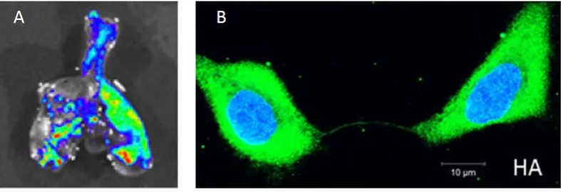
Figure 1. Experimental findings of within-host influenza virus spread. (a) Influenza virus spread visualized through bioluminescent imaging. Figure shows fluorescence from excised lungs of infected mice. Figure reproduced from [24]. (b) The genome and proteins of influenza virus can be transferred between cells via intercellular pathways called tunneling nanotubes. Figure reproduced from [26].
The development of viruses that stably express luciferase has allowed the visualization of 94
longitudinal infection dynamics and spatial distribution within live hosts [27–31]. In mice, multiple
95
studies have demonstrated the existence of clear viral foci in the lungs that spread in spatial extent 96
before recovery from viral infection [27–29]. Subsequent studies have demonstrated the utility of
97
these luciferase reporter viruses in tracking the spatio-temporal dynamics of infection in ferrets and 98
in the context of pre-existing immunity [30,31].
99
Altogether, there is an increasing body of experimental work in systems ranging from mice to 100
humans that indicates that influenza infections are highly spatially structured. Along with this, recent 101
studies are also providing a better understanding of the specific mechanisms that may be responsible 102
for the establishment of this spatial structure. One clear factor is the spatial heterogeneity of cells with 103
certain receptor distributions that mediate efficient viral attachment and therewith modulate cellular 104
tropism. To elaborate, IAV primarily binds host cells through interactions with the galactose-sialic 105
acid (SA) linkages present on the termini of complex glycan structures. The SA structures used by 106
influenza viruses are structurally diverse, but are typically classified as eitherα2,3 orα2,6 based on
107
the orientation of the bond between the galactose and sialic acid moieties [32]. The specificity of
108
HA forα2,3 orα2,6 linkages is thought be a key determinant of species and cellular tropism, with
109
avian strains primarily bindingα2,3, and human strains binding α2,6 [32]. Multiple studies have
110
demonstrated a correlation between the presence of α2,6 linked SA receptors on the cell surface
111
and virion binding and infection in human airway and nasal epithelial cultures, as well as within 112
sections of human respiratory tissue [20,33–36]. Consistent with this, deep sequencing different
113
anatomical sub-compartments within the ferret respiratory tract revealed clear compartmentalization 114
of viral variants based on the distribution of receptor structures [37]. Importantly, the ubiquity of
SA receptors throughout the mammalian respiratory tract lumen may limit the spatial spread of 116
virions. Release and efficient spread of newly produced virions depends upon the ability of the 117
viral neuraminidase protein (NA) to efficiently cleave SA receptors from the surface of the infected 118
cell [38,39]. In addition, the airway lumen contains an abundance of heavily sialylated host factors
119
such as pentraxins and mucins that can bind virions and restrict their free diffusion [40,41]. Thus,
120
both cellular and cell-free SA may act to restrict or structure the spatial spread of the virus by limiting 121
free diffusion. This effect may be counteracted to varying degrees by the activity of the viral NA, 122
which can differ between different viral strains [42].
123
Recent studies have suggested that influenza virus may also be able to spread spatially via an 124
entirely separate mechanism that does not depend on diffusion of extracellular virions. Specifically, 125
Roberts et al. showed that viral proteins can spread between adjacent cells via intercellular
126
actin pathways (“tunneling nanotubes”) without going through the standard budding and release 127
process [43]. These proteins include the viral replicase machinery (nucleoprotein and polymerase
128
proteins), as well as NS1. Subsequently, Kumar and colleagues showed that the genomes of influenza 129
viruses can also spread between cells via these nanotubes (Figure1B) [26].
130
Thus, influenza viruses may use at least two modes of transmission between cells: (1) the 131
textbook process of extracellular spread by virions, and (2) the intercellular spread of viral genomes 132
and proteins between neighboring cells. Infection therefore likely occurs at two characteristic spatial 133
scales: a scale with the possibility of long-distance dispersal (with cell-free virions having a small 134
possibility to infect cells at appreciable distances from the cells from which they budded) and a scale 135
that involves highly localized spread (with intercellular pathways having the ability to form only 136
between adjacent target cells). To our knowledge, the actual distances traveled by cell-free virions 137
have not been measured experimentally, but given the spread of cell-free virions between hosts, it 138
is likely that cell-free virions also have some degree of long-distance dispersal capabilities within a 139
host. The relative importance of these two modes of spread may differ between infected hosts. For 140
example, hosts with pre-existing anti-influenza immunity may clear cell-free virions more rapidly 141
than naïve hosts, resulting in a more dominant role for intercellular viral spread in these individuals. 142
These two modes of viral spread can be considered bottom-up processes in that they impact 143
the rate of viral spread via access to the resources necessary for replication. The spatial aspects of 144
top-down processes that control viral spread, such as the activities of immune cells and cytokines in 145
neutralizing virions, clearing infected cells, and rendering cells refractory to infection, have not been 146
extensively studied to our knowledge. Questions that need to be addressed are thus how locally 147
the immune system acts and the degree of spatial heterogeneity in the immune response across 148
the respiratory tract. Further empirical work is needed to understand the potential for top-down 149
regulation of viruses. However, because the respiratory tract is not a highly immune-privileged 150
site, and numerous soluble components of the anti-viral immune response such as antibodies and 151
interferon are thought to act at the tissue-wide or systemic level, we expect the spatial dynamics of 152
influenza virus spread to be regulated more strongly by the bottom-up process of local target cell 153
availability than by the top-down process of immune-mediated viral clearance. 154
In sum, the experimental findings we reviewed here indicate that human influenza viruses 155
exhibit strong patterns of within-host spatial spread, driven predominantly by local movement of 156
cell-free virions and the intercellular spread of viral genomes and proteins. In the next section, we 157
review computational models that address the role that certain viral and host factors may play in 158
shaping the spatial aspect of within-host influenza virus spread. 159
3. Computational models of spatial within-host influenza virus spread 160
The overwhelming majority of computational within-host influenza models do not incorporate 161
a spatial aspect to viral spread. Despite this, they have provided insights into the processes
162
regulating within-host virus dynamics. These dynamics are typically characterized by exponential 163
approximately 106TCID50/mL [44]. Virus generally becomes undetectable within 5-6 days following 165
infection [45] . The decline in viral load is often biphasic, with an initial rapid decline, followed by a
166
longer, slower decline in viral load [46,47]. Figure2A shows these characteristic viral load dynamics
167
in a human subject experimentally infected with the H1N1 influenza A subtype. 168
Several non-spatial models have been able to reproduce these characteristic infection dynamics. 169
The most basic versions of these models consider only target cells and virus, yet can reproduce 170
the exponential growth of the viral population within a host, followed by an exponential viral 171
decline once target cells have been depleted (Figure 2A) [44]. These models, however, fail to
172
reproduce observed biphasic viral declines. More complex models have incorporated the host
173
response, typically by considering the role of interferon and cells of the innate and adaptive immune 174
response [46–49]. These models have been able to reproduce many (and in some cases, all observed)
175
patterns of viral growth and decline without unrealistic target cell depletion. The importance of the 176
host immune response in regulating within-host influenza dynamics, identified by these non-spatial 177
models, lay the groundwork for more complex models that explicitly account for spatial structure. 178
Figure 2. (a) Fit of a non-spatial, target-cell limited within-host influenza model to viral load data from a human subject experimentally infected with influenza A subtype H1N1. The data are shown as black points. The black lines show model fits of viral load data. The blue lines show model-predicted declines in the number of target cells. Solid lines show the fits of the basic model; dashed lines show the fits of a more complex model with an eclipse phase before infected cells produce virus. Figure is reproduced from [44]. (b) Cellular automata model of within-host influenza infection under assumptions of local cell regeneration and localized recruitment of immune cells. Simulations reproduce the appearance of infected foci. Figure reproduced from [50].
Incorporating spatial structure into within-host disease models can have important 179
consequences. Most notably, parameter estimates inferred for spatial within-host models are often 180
biologically more reasonable than those inferred for non-spatial models. This has been shown for 181
models of influenza virus [51], as well for models of other pathogens [52]. This finding indicates that
182
spatial aspects of viral spread might inappropriately bias inferred parameter values of non-spatial 183
models. Spatial models also lead to qualitative predictions of viral dynamics that may be more 184
biologically reasonable [53]. For example, under certain parameter regimes, one might expect a
185
viral infection to become chronic or for viral load to equilibrate steadily; spatial models for chronic 186
have a greater tendency for viral infections to be stochastically cleared or to exhibit dampening 188
oscillatory dynamics [53].
189
For influenza virus, several spatially explicit within-host models have been developed in the last 190
10-15 years [54–56]. These models all allow virus to spread locally from infected cells to nearby
191
susceptible cells. In some of these models, other processes, such as host cell regeneration and 192
immune cell recruitment, are also spatially structured. The most common approach to explicitly 193
model spatial aspects of within-host influenza virus spread has been through the implementation of 194
agent-based models. These generally consist of a 2-dimensional grid of cells and a defined set of 195
rules that determine viral kinetics and cell death kinetics, among other kinetics such as those of the 196
host immune response. Beauchemin and colleagues showed that an agent-based model of this sort 197
could successfully reproduce certain features of acute influenza infections, including the timing of 198
peak viral load and the 5-7 day duration of infection [54] . However, in their simulations, the number
199
of infected cells appears to grow linearly until viral load peaks; this stands in contrast to observed 200
patterns of exponential viral growth over the first few days of influenza infection. More work needs to 201
be done to determine whether and under what scenarios one would expect exponential viral growth 202
in spatially structured influenza infections. 203
In a second study, Beauchemin considered the dynamical effect of factors occurring at different 204
spatial scales [50]. Specifically, this study considered the regeneration dynamics of epithelial cells
205
to occur either globally or locally. Which of these assumptions was adopted clearly would affect 206
resource availability for the virus, and thereby shed light on the importance of this bottom-up 207
process’s spatial scale in shaping the spatial distribution of the viral population. The study further 208
considered the recruitment dynamics of immune cells to occur either at random or preferentially 209
at infection sites. Considering these alternative assumptions allowed Beauchemin to evaluate the 210
importance of spatial scale in the immune response’s top-down control of the viral population. 211
Finally, this study considered different possible dispersal distances for the virus. Overall, the study 212
found that local cell regeneration and short viral dispersal distances reproduced observed empirical 213
patterns, including foci of infected cells, better than other combinations (Figure 2B). Whether
214
recruitment of immune cells was at random or localized at infected sites did not have an appreciable 215
effect as long as cell regeneration was localized. Better experimental data are still needed to quantify 216
cellular regeneration, and whether it should be expected to impact influenza virus dynamics over a 217
5-6 day period. 218
Following this work, Levin et al. assessed in more detail the importance of the host immune 219
response in regulating spatial within-host influenza virus spread [57]. In this study, the authors
220
showed that T-cells were unable to control the spread of influenza viruses with high replication rates. 221
This inability of the host immune response to control the viral infection was due to delays in T-cell 222
migration to the infection site. The results of this spatial model sheds light on how the localized 223
interaction between the immune system and the virus could result in some viral strains, but not 224
others, being able to evade top-down control by the host immune response. 225
Despite the increase in their use, agent-based models are still computationally intensive and 226
frequently do not allow for effective interfacing with data or analytical insight. Fortunately, several 227
alternative approaches exist for modeling spatial aspects of within-host viral spread that do not rely 228
on agent-based model simulations. One such approach is to compartmentalize the respiratory tract 229
into several distinct ‘patches’, with low levels of viral transmission between one another. Within 230
a patch, virus is assumed to have equal access to all cells and is similarly targeted equally by all 231
host immune responses. A study by Reperant et al. provides an example of this type of approach, 232
in which viral dynamics are considered across three tissue compartments: the trachea/bronchi, 233
the bronchioles, and the alveoli [58]. These compartments captured spatial heterogeneity in host
234
cell types across tissues by differing in their initial number of susceptible target cells, in their 235
viral clearance rates, and in their immunoglobulin distributions. By simulating this multi-patch 236
tissue differences lead to strain-specific variation in viral localization along the respiratory tract, and 238
therewith differences in the onward transmission potential of different influenza subtypes. A second 239
alternative approach makes use of partial differential equations (PDEs), which can deterministically 240
simulate the dynamics of a viral population over both space and time. With PDEs, certain processes 241
can occur locally while others can occur over more extensive spatial scales. For within-host influenza 242
dynamics, for example, virus production, infection and death of cells, and immune activation can all 243
occur locally, while viruses, cytokines, and certain cells can diffuse or migrate over more extensive 244
ranges across space. 245
A third alternative approach to agent-based models is to consider space implicitly, rather than 246
explicitly. This can be done by including saturating (instead of mass-action) terms in non-spatial 247
mathematical within-host models [49]. While mass-action terms are often used in within-host models
248
to describe virus infection of target cells, using a saturating term (such as a Michaelis-Menton term) to 249
describe the infection process would allow for deviation from a well-mixed assumption. The rationale 250
for using such a term is that when a virus is produced from infected cells, it cannot reach all target cells 251
in a host. Instead, there are only a small number of target cells that are available to the virus to infect. 252
Thus, the rate at which susceptible cells become infected can rapidly saturate even while many target 253
cells remain susceptible. A final approach for modeling space implicitly is to allow for overdispersion 254
of virus among target cells, by assuming, for example, a negative binomial distribution for viral 255
particles across host cells rather than a Poisson distribution [59]. With overdispersion, a small number
256
of target cells are infected with a large number of virions, while a large number of target cells might 257
still be uninfected. Overdispersion can thus capture expected viral distribution patterns under the 258
assumption of spatial viral spread. 259
While spatial within-host models have helped us understand how influenza virus infections 260
spread within a host, the current literature has not addressed many open questions that seem 261
particularly important in the context of spatial viral spread. One question is how the eclipse phase 262
of infected cells impacts viral population growth and spatial spread dynamics. A second question is 263
how cellular coinfection impacts the rate of viral spread. In a spatially structured infection, we expect 264
substantially more cellular coinfection than in a non-spatial setting, where virus is spread more evenly 265
over an entire population of cells. As such, the effect that cellular coinfection has on the rate of viral 266
production will be critical to determining how quickly the viral population will spatially expand. 267
Higher levels of cellular coinfection in a spatially structured setting will also impact viral reassortment 268
rates [60], and thereby also impact the adaptive potential of influenza viruses. Intriguingly, these
269
questions, among others, have already been addressed, albeit not in the specific context of within-host 270
viral dynamics. Indeed, there is a rich ecological literature that can be mined to inform us of answers 271
to these questions, provided that we make effective analogies between processes identified in this 272
literature and those acting on within-host viral populations. 273
4. Ecological factors driving patterns of spatial spread 274
One of the most straightforward questions we can ask about within-host disease spread is 275
how fast it will progress. On this question, the ecological literature has shown that spatially
276
unstructured populations grow faster than their spatially structured counterparts when starting from 277
small population sizes [61,62]. When population sizes are small, spatially unstructured populations
278
are expected to grow exponentially, whereas spatially structured populations are expected to grow 279
slower than exponentially (that is, sub-exponentially). This reduced growth rate is due to the lower 280
relative availability of resources in spatially structured populations: resources are scarce in the centers 281
of expanding populations, and resources are only abundant for those individuals at the very front of 282
the expanding population wave. In the context of within-host viral spread, this means that infections 283
that are highly spatially structured have constrained growth rates relative to those infections that are 284
in the rate of influenza virus diffusion reduces the growth rate of the virus and its overall population 286
size [50].
287
While spatial structure is known to slow overall population growth, the ecological literature 288
has also delved more specifically into what factors impact the rate of spatial population spread. In 289
general, a population expanding outward from its point of origin is theoretically expected to spread 290
as a “traveling wave”, that is, at a constant rate and with a wavefront shape that is maintained 291
over time [63,64]. This traveling wave dynamic is expected when a population is expanding in a
292
single dimension along a line (Figure3A) or in 2-dimensional space (Figure3B), the latter of which
293
would be more relevant for the within-host spread of influenza virus populations. If a population is 294
expanding in a single dimension, the amount of occupied area is expected to grow linearly in time 295
[64]. Alternatively, if a population is expanding in two dimensions, the square root of occupied area
296
is expected to grow linearly in time [64] (Figure3C).
297
Figure 3. Patterns and dynamics of spatial spread from the ecological literature. (a) Populations expand as a “traveling wave” in a single spatial dimension. (b) Populations expand as a “traveling wave” in two-dimensional space. Figures (a) and (b) reproduced from [63]. (c) When populations expand in two spatial dimensions, the square root of the area that is inhabited is expected to grow linearly in time. Figure reproduced from [64]. (d) Types of density-dependence. Negative density-dependence occurs when per capita growth rates decrease with increases in local population densities. Allee effects occurs when per capita growth rates first increase, and then decrease, with increases in local population densities. Figure reproduced from [65].
One important factor that affects the velocity of the traveling wave is the type of 298
“density-dependence” that the population is subject to, where density-dependence refers to the 299
relationship between local population density and individual (per capita) growth rates. Populations 300
are said to undergo density-independent growth when an individual’s growth rate is not influenced 301
by local population density. Negative density-dependence is said to occur when an individual’s 302
growth rate decreases with increases in local population density, such that the maximum per capita 303
growth rate occurs at small population sizes (Figure3D). Positive density-dependence is said to occur
304
when an individual’s growth rate increases with increases in local population density. Systems can 305
be subject to multiple forms of density-dependence. For example, in populations with an Allee effect, 306
there is a transition from positive to negative density-dependence with increases in local population 307
density (Figure 3D). In populations that are strictly subject to negative density-dependence, the
308
(asymptotic) velocity of the traveling wave is given by p4f‘(u)D, where D is the dispersal rate,
309
measured in units of dispersal distance2/time, and f‘(u) is the individual growth rate at low
310
population density [63]. In populations with an Allee effect, the (asymptotic) velocity of the traveling
311
wave is lower than in similar populations without an Allee effect [66–68]. Thus, the type of
312
While a spatially-expanding population is generally expected to exhibit traveling wave 314
dynamics, it may initially exhibit transient dynamics that differ from its asymptotic, long-term 315
behavior. In many cases , these transient dynamics are expected to have a slower velocity than 316
those of the asymptotic traveling wave [63,69,70], with the rate of spread expected to accelerate
317
once the local population has reached a threshold density [71]. This expected increase in the rate of
318
spatial population spread is an important theoretical finding that, if ignored, could lead to dramatic 319
underestimation of the rate at which a population will ultimately spread and the total distance that it 320
will ultimately travel. In the case of an Allee effect, some population expansions may even fail due to 321
local populations failing to exceed certain threshold densities [68].
322
Unfortunately, little is known about density-dependence in viral populations specifically. Within 323
a host, the characterization of density-dependence in a viral population would require determining 324
how the number of viral progeny from a given intracellular viral particle depends on the multiplicity 325
of infection of the cell the viral particle resides in. For some viral pathogens, host cell machinery 326
may be the primary limiting factor. In this case, the population would be strictly subject to negative 327
density-dependence. In influenza, there is some indication that an Allee effect may be at play. This 328
expectation derives from a study that showed that over 90% of the time, singularly infected cells 329
fail to produce viral progeny [72]. This failure to produce viral progeny stems from the failure of
330
one or more of influenza’s eight gene segments to be delivered to the nucleus. The existence of 331
these “semi-infectious particles” [73] that can produce viral progeny through complementation can
332
therefore be thought of as bringing about positive density-dependence at low cellular multiplicities 333
of infection (MOIs). With host cell machinery ultimately limiting viral production at high cellular 334
MOIs, IAV growth may therefore be characterized by an Allee effect. As such, we may expect some 335
infections to fail due to threshold population sizes not being reached, and we may expect the rate of 336
viral spread to be slower for strains of IAV that have higher proportions of semi-infectious particles. 337
A second ecological factor that is known to impact the rate of spatial population spread is the 338
frequency of long-distance dispersal events [74–76]. The primary ecological effect of long-distance
339
dispersal events is an increase in the rate at which populations expand spatially [77]. This increase
340
in the rate of spatial spread further leads to an overall increase in population growth rates because 341
dispersed individuals have access to more resources than they would otherwise have had. In a study 342
that compared two different modes of range expansion (exclusively short-range diffusion versus a 343
combination of short-range diffusion and long-distance dispersal), populations were found to invade 344
more quickly when long-distance dispersal occurred, even if these events occurred only rarely [75].
345
Increases in the rate of spatial population spread with higher frequencies of long-distance dispersal 346
events is consistent with the findings that asymptotic velocity of a traveling wave increases with the 347
dispersal rateD(see equation above).
348
Given the importance of long-distance dispersal events on the dynamics of spatially structured 349
populations, knowledge of how frequently virions disperse at these long distances within infected 350
hosts appears critical. To the best of our knowledge, the frequencies of these events have not 351
been quantified, eitherin vivoorin vitro. Clearly, transmission of influenza particles between hosts
352
constitutes a long-distance dispersal event. While we know that influenza virions generally infect 353
nearby cells, and can even be transmitted between cells directly via intercellular actin pathways, the 354
extent to which virions travel long distances within a host is unknown. Intriguingly, the observed 355
exponential growth of the viral population for the first 2-3 days following infection may be an 356
indication that long-distance within-host dispersal occurs; in its absence, we would expect a pattern of 357
subexponential viral growth. As mentioned above, long-distance dispersal mitigates to some extent 358
the growth-slowing depletion of local resource (target cells, in the case of viruses), and thereby brings 359
the rate of viral population growth closer to an exponential form. The frequency of long-distance 360
viral dispersal in IAV infections should be investigated further, given the evidence in the ecological 361
Spatial heterogeneity is a third key factor that can impact the rate of spatial population spread. 363
Spatially heterogeneous environments might be caused by irregularities in the landscape such as 364
unevenly distributed resources or barrier zones. While we normally expect populations to expand 365
through space as a traveling wave, there is evidence that spatial heterogeneity can result in much 366
more complex patterns. For example, Keeling and colleagues showed that heterogeneity across the 367
landscape in resource distribution and quality helped to explain why a disease outbreak traveled 368
irregularly and was difficult to predict [77]. Another important example can be found in a study by
369
Sharov and Liebhold, who used empirical data and a spatially heterogeneous model to show that a 370
single “barrier zone” could greatly reduce that rate of population spread [78]. Similar results have
371
been found in other studies on the importance of barrier zones, which can serve to reduce the rate of 372
spatial expansion or even halt it entirely [79].
373
These effects of spatial heterogeneity are an important consideration for within-host viral spread. 374
We know that flu infections occur in a spatially heterogeneous environment. Across the length of the 375
respiratory tract, the ‘landscape’ is heterogeneous in terms of cell densities and receptor structures. 376
Progressing from the upper to the lower respiratory tract, we see an increase in the number of 377
α2,3 SA binding receptors relative to α2,6 receptors, meaning there are fewer appropriate target 378
cells for human influenza viruses to bind to [80,81]. This change in resource distribution could
379
affect the pattern and rate of viral spread, helping to explain why many human influenza infections 380
are confined to the upper respiratory tract. Furthermore, we can expect that within-host patterns 381
of immune response would also add heterogeneity to the environment, in the form of interferon 382
diffusing as it is released from infected cells and immune cells moving through the system. This is 383
an active area of study, and recent advances in within-host imaging techniques will no doubt greatly 384
advance our understanding of within-host spatial heterogeneity and its effects on viral spread. 385
Finally, the presence of other species can strongly affect the ability of a species to invade. 386
Competitors can act to reduce the availability of resources or alter the environment in other ways 387
that make it more difficult for a species to disperse and survive. Unsurprisingly, most models 388
suggest that the presence of a competitor will act to slow down the rate of spatial spread [82]. The
389
competitor can still have this effect even if it is less fit than the focal species. This is especially 390
true if the competitor is already established in the new location before the focal species arrives [76],
391
but this is not a requirement. Similarly, predators can also slow down the rate at which a species 392
can invade a new territory, and, depending on their distribution in the landscape and their time of 393
release, they may even make it such that the invasion dynamics of the prey species can no longer be 394
characterized by a traveling wave [63]. In the context of influenza, while the virus may not explicitly
395
be subject to interspecific interactions, we could perhaps think of components of the immune response 396
as either competitors or predators. In particular, exposure to interferon-α is known to make cells
397
refractory to viral infection, thereby reducing the number of susceptible target cells available to a 398
virus. Interferon-αcould therefore potentially be considered as an asymmetrical competitor of IAV
399
within a host. The depletion of susceptible target cells by interferon-α would act to slow down
400
the rate of viral spread within a host. The cellular and humoral immune responses of hosts could 401
instead be thought of predators of the within-host IAV population, by neutralizing free virus or 402
killing infected cells. Viruses infecting hosts with pre-existing immunity would thereby experience 403
top-down, predator-like control from the immune system. This dynamic would lead to a slower rate 404
of viral spatial spread, and potentially the abrogation of a traveling wave form. 405
In sum, our understanding of ecological dynamics can help us to better understand within-host 406
viral dynamics, and to fill in some of the gaps in our knowledge about factors that may impact rates 407
of viral spread. To consider how these spatial aspects of viral spread will in turn impact the genetic 408
5. The consequences of spatial spread on population evolution 410
The evolutionary literature provides insight into how spatial spread impacts patterns of 411
population genetic diversity, how it impacts the processes of purifying and positive selection, 412
and how spatially-distinct selection pressures may shape population phenotypes. Here, given the 413
intrinsically spatial aspect of influenza virus spread within hosts, we review this literature and again 414
make ties to observations from the flu field where possible. 415
An important effect of spatial population expansion is a significant reduction in population 416
genetic diversity. This effect is one of the more robust effects of spatial spread, with a large number 417
of studies showing that genetic diversity is rapidly eroded when population expansion occurs 418
locally, as with a range expansion [83–87]. In the case of spatial expansion in two dimensions, this
419
reduction of genetic diversity from stochastic founder effects results in sectors that are genetically 420
homogeneous [85] (Figure4A). Intriguingly, these patterns are consistent with a recent analysis of
421
within-host viral populations in individuals experiencing acute influenza infections [88]. Specifically,
422
McCrone and colleagues found that stochastic effects dominated in the structuring of the within-host 423
flu populations, and that, despite high viral titers, only 57% of single nucleotide variants from an 424
early sample were still present in a later sample from the same individual when samples were taken 425
one or more days apart. These results are consistent with the phenomenon of spatial spread, where 426
rapid drops in standing genetic variation would be expected further into the range expansion due to 427
genetic drift at the wavefront. Rapid losses of genetic diversity were also evident in a mouse model for 428
influenza infection, where the authors found, using four distinct colors of fluorescently labeled viral 429
proteins, that the majority of individual alveoli only showed the presence of a single color [25]. This
430
indicates that at the furthest extent of within-host viral spread, spatial founder effects and bottlenecks 431
appear to be at play. 432
Several factors have been identified in the population genetic literature that will modulate the 433
extent to which the genetic diversity of a spatially expanding population will be eroded. In most 434
cases, these factors have clear analogues for within-host viral populations. First, the ‘dispersal kernel’ 435
is known to affect the rate at which populations will lose genetic diversity, where the dispersal kernel 436
quantifies the distribution of distances individuals in a population will seed their progeny. Intuitively, 437
one might think that higher levels of long-distance dispersal will always mitigate the loss of genetic 438
diversity. However, Bialozyt and colleagues showed instead that increases in the number of long 439
distance dispersal events will counterintuitively first have the effect of reducing genetic diversity [89].
440
Further increases in the number of long-distance dispersal events will then act to increase levels of 441
genetic diversity again. This pattern results in the minimum level of population genetic diversity 442
being present at some level of long-distance dispersal. This pattern results from what has been termed 443
an ‘embolism’ effect (Figure4B), where rare long distance dispersal events lead to single individual
444
founders with substantial replication resources surrounding them. The rapid expansion of these 445
single individual founders leads to dramatic reductions in the overall population’s genetic diversity. 446
The dispersal kernel, as one might expect, will also impact the genetic ‘patchiness’ of the population 447
across space [90]. In light of the two possible modes by which flu viruses infect target cells, the
448
relative roles of cell entry through receptor binding by free virus versus cell entry through tunneling 449
nanotubes will likely be important in understanding patterns of genetic diversity in within-host flu 450
populations. If TNTs are a major source of cellular infection, as they may be in previously infected 451
individuals with strong antibody responses, then dispersal is expected to be more highly localized, 452
and long-distance dispersal events will be fewer. However, whether this will lead to higher or lower 453
levels of genetic diversity relative to a case with higher levels of cell entry via receptor binding by free 454
virus is unclear, given that the relationship between genetic diversity and the number of long-distance 455
dispersal events is non-monotonic [89].
456
A second factor affecting the rate at which population genetic diversity will be lost in a spatially 457
expanding population are the life history characteristics of the population. For example, it has been 458
in populations [91]. This is because a juvenile stage slows down the colonization process and allows 460
for more genetic diversity to accumulate at the wavefront. An ‘eclipse’ phase in viral populations is 461
analogous to this juvenile stage: infected cells are not productive immediately following infection; 462
rather it can take several hours for viral progeny to be produced. For influenza, the duration of this 463
eclipse phase has been quantified experimentally, with most recent estimates being on the order of 464
2-4 hours [92].
465
Figure 4. The effects of spatial spread on a population’s evolutionary dynamics. (a) Local movement in 2-dimensional space leads to the generation of genetically homogeneous ‘sectors’. Figure reproduced from [93]. (b) Intermediate levels of long distance dispersal result in major reductions in genetic diversity, as described by the ‘embolism effect’. Figure reproduced from [89]. (c) Mutations can ‘surf’ to high frequencies, regardless of whether they are deleterious (left), beneficial (right), or neutral (not shown). Figure reproduced from [84]. (d) Spatially expanding populations can select for cooperative phenotypes at the leading edge. Figure reproduced from [94].
A third factor affecting the extent to which genetic diversity will be eroded is how an individual’s 466
reproductive rate depends on nearby population density. For example, theoretical studies have 467
shown that Allee effects have the potential to maintain genetic diversity in a spatially expanding 468
system [85,86]. A higher level of genetic diversity is maintained in populations with an Allee
469
effect because in these cases it is not only the furthest members of a population that contribute to 470
the expanding population. As discussed in the previous section, Allee effects may be at play in 471
within-host viral populations that require complementation, including influenza. 472
A fourth factor affecting levels of genetic diversity in spatially expanding systems is the 473
extent of spatial heterogeneity. Specifically, Wegmann and coauthors showed that environmental 474
heterogeneity leads to loss of genetic variation within similar regions and further leads to greater 475
genetic differences between regions [95]. This finding may be applicable to within-host viral
476
populations that exist across different regions of different cell types. For example, within-host 477
regions of cells having predominantly α2,3 versus α2,6 sialic acid receptors might result is less
478
genetic variation within each region and greater levels of population genetic differentiation between 479
distribution of viral sequence variants between tissue compartments within ferrets that differ in 481
receptor distribution [37].
482
Beyond impacts on population-level patterns of genetic diversity, populations that are spatially 483
expanding are known to be subjected to a phenomenon called “surfing” [84,85,96,97], whereby
484
genetic variants present on the wavefront of an expanding population may rapidly rise to high 485
frequencies due to the dominance of genetic drift in the small wavefront populations. With the 486
process of genetic drift (over selection) dominating at the wavefront of an expanding population, 487
de novo mutations(whether beneficial, deleterious, or neutral) that occur at the right place at the right 488
time can rise to high frequencies and even fix in populations (Figure4C). Since, in many systems,
489
the majority of mutations appear to be deleterious, this surfing phenomenon results in deleterious 490
mutations fixing at considerably higher rates in spatially expanding populations than in populations 491
that are growing in the absence of a spatial dimension [84,98]. As such, these spatially-extended
492
systems are expected to carry an “expansion load” [99,100], defined as the deleterious mutation
493
load a population carries that is due to spatial founder effects from small populations at the 494
wavefront. While there is no evidence yet for within-host viral populations being subject to the 495
surfing phenomenon and to expansion loads, one should theoretically expect this to be the case. This 496
is because most RNA virus mutations are known to be deleterious, with recent experimental findings 497
providing evidence for this specifically for influenza virus [101].
498
While this surfing phenomenon is also relevant to beneficial mutations, the consequences of 499
genetic drift dominating at the wavefront results in lower rates of beneficial mutation accumulation in 500
a spatially expanding population than would be anticipated in population expanding in the absence 501
of a spatial dimension. This is for two reasons: first, beneficial mutations are rare, so, relative 502
to deleterious mutations, de novo mutations are unlikely to be beneficial. Second, if a beneficial
503
mutation does arrive in the right place at the right time, it is unlikely for it to be brought to high 504
frequencies through selection because of the dominance of genetic drift at the wavefront. This surfing 505
phenomenon is thus known to slow the rate of adaptation of spatially expanding populations, and 506
could even lead to fixation of deleterious mutations within hosts. Spatial within-host dynamics may 507
therefore provide a mechanism to explain why RNA viruses, including influenza, appear to carry 508
deleterious mutation loads [102–104].
509
Finally, spatially expanding populations may select for different phenotypes than ones that do 510
not have a spatial dimension. This would occur, for example, if individuals residing at the wavefront 511
experience different selection pressures from the ones residing at the interior of the population 512
range. Rather than this being an unlikely case, different selection pressures at different points in 513
the population range are theoretically expected in many situations. At the wavefront, resources are 514
relatively abundant, and individuals with a high intrinsic growth rate (“r”) are known to outcompete 515
others. In contrast, in the interior of a population range, resources are limiting, and individuals 516
with more efficient resource use do best (i.e., those individuals with higher basic reproduction 517
numbers,R0). This difference inrversusR0selection pressures has been considered in the context of
518
infectious diseases, and is at the core of why epidemic pathogens (with abundant host resources) are 519
expected to evolve to higher virulence compare to endemic pathogens (with scarce resources) [105].
520
Analogously, one would expect more virulent viruses to be selected for at the wavefront of an 521
expanding population, compared to the interior [61], regardless of whether we are considering the
522
viral population to be expanding within hosts or across the globe. In the context of within-host flu 523
dynamics, spatial spread may therefore select for phenotypes that kill infected cells more rapidly but 524
have higher rates of viral production. Another within-host phenotype that may be at least partly 525
under viral genetic control is the viral dispersal kernel. Given theoretical findings that the evolution 526
of long-distance dispersal is favored during an expansion process [106], perhaps one might even
527
expect influenza virus to evolve a preference for cell entry via budding over cell entry via TNTs. 528
Finally, a recent study intriguingly found that cooperative phenotypes have a selective advantage 529
along the wavefront of expanding populations [94] (Figure4D). This theoretical finding is particularly
relevant to recent work examining the evolution of viral cooperation, collective interactions, and more 531
generally, the budding research area of “sociovirology” [107].
532
In sum, the spatial aspect of within-host viral spread will generally reduce viral genetic diversity, 533
slow the rate of viral adaptation, more easily enable the fixation of deleterious mutations, and result in 534
the evolution of viral phenotypes that may be advantageous for only a subset of the viral population. 535
Discussion 536
We have reviewed current understanding and open questions regarding patterns and 537
mechanisms of within-host viral spread from both empirical and computational perspectives. Our 538
ability to visualize within-host spatial structure has improved greatly thanks to advances in imaging 539
techniques, particularly the use of fluorescent reporters and luciferase expressing viruses. The 540
recently discovered ability of viruses to spread directly from cell to cell via ‘tunneling nanotubes’ is 541
an exciting development, but the feasibility and frequency of long-range dispersal remains unknown. 542
Spatially explicit and non-spatial models have been applied to viral data; non-spatial models are far 543
more common, and allow one to interpret data on viral load kinetics. However, ignoring spatial 544
structure in the infection processes can lead to biased or incorrect estimates of parameter values. 545
Non-spatial models are also less useful in understanding the roles that viral infection processes 546
such as cellular multiplicity of infection and viral reassortment play in regulating viral dynamics. 547
They are also less relevant to understanding the factors that govern viral population dynamics and 548
evolutionary dynamics, such as patterns of genetic diversity and viral mutation loads. Further, 549
ignoring spatial heterogeneity and its consequence on viral population structure may prevent us 550
from interpreting experimental data beyond viral load measurements and make result in imprecise 551
predictions about the impact of therapeutic interventions. 552
We then turned to the ecological and evolutionary literature to provide theoretical insight 553
into the population dynamics and genetics of spatial within-host viral spread. Ecological and
554
evolutionary studies indicate that the within-host spread of a virus should be strongly influenced 555
by its own dispersal patterns and life history characteristics, as well as the spatial heterogeneity 556
in the host environment. The ecology literature has also given us insight into critical gaps in 557
our knowledge about within-host viral spread. Specifically, we need more empirical data on viral 558
density-dependence and the extent to which Allee effects are present in the system. Studies to 559
determine the distance that viruses disperse would also greatly improve our understanding of what 560
regulates the rate of within-host spread. Long-range dispersal greatly increases the speed of invasion, 561
even if that dispersal is rare; but the extent to which long-range dispersal occurs in flu is currently 562
unknown. The evolutionary literature has provided us with theoretical expectations for how the 563
genetics of the viral population will change over time in a flu infection, and the effect that space may 564
have on the ability of the viral population to adapt. These predictions should be tested empirically, 565
using available imaging and sequencing techniques. 566
Perhaps most importantly, the ecology and evolution literature has the potential to inform the 567
development of control strategies. Current control strategies focus on treatment and prevention of 568
infection using drug therapies and vaccination. These interventions introduce antibodies or antivirals 569
into the system, both of which are functionally similar to predators from the standpoint of a virus 570
in a host. They can be very effective under the right circumstances, but vaccines are notoriously 571
difficult to formulate due to the rapid evolution of seasonal flu strains, and antiviral resistance is not 572
uncommon. In order to control an infection, the host must be able to contain the virus and prevent 573
its ongoing spread. In vivo, this seems to be possible because the immune system responds quickly
574
to the location of infection, and the virus ultimately runs out of local susceptible cells to infect. Early 575
intervention is likely to be most effective, not only because there are fewer total virions and infected 576
cells, but because influenza may be subject to strong Allee effects. Control efforts should be focused 577
on reducing the maximum intrinsic growth rate of a population, not the transient initial rate [69].
Studies of spatial heterogeneity suggest that introducing a barrier zone can be a very effective 579
control strategy [77]. In wildlife populations, an artificial barrier has been successfully introduced
580
at times to prevent the spread of rabies, by depositing vaccine-laden food items [108]. While it is
581
likely not possible to introduce a physical barrier within the host’s respiratory tract, the concept of a 582
barrier is somewhat analogous to the local action of the immune system to “immunize” susceptible 583
cells that are close to the site of infection. Different cell types and tissues in the respiratory tract may 584
also function as a kind of barrier, because the virus is not equally able to infect each of these. 585
Finally, influenza infection could potentially be controlled by introducing defective interfering 586
particles (DIPs) into the system. DIPs are naturally occurring during infections, and they essentially 587
parasitize wild-type virus, reducing the amount of infectious offspring that is produced from cells 588
coinfected with DIPs and wild-type virus. The ability of DIPs to interfere with wild-type virus 589
depends on the local cellular MOI, because in the absence of co-infection with a wild-type (“helper”) 590
virus, DIPs cannot replicate [109]. Studies in both mice and ferrets have shown that DIPs can modify
591
within-host influenza virus dynamics, decreasing peak viral loads and delaying its timing [110].
592
Further, the administration of DIPs can reduce influenza symptoms and virulence [110].
593
While we have focused here on influenza virus, insight from the ecological and evolutionary 594
literature is also applicable to a broad range of other viral infections. Accounting for the ecological 595
and evolutionary dynamics of within-host spatial spread will deepen our understanding of the 596
behavior and outcomes of a wide variety of viral infections and potentially lead to new conceptual 597
advances in infection control strategies. 598
Acknowledgments: MG, CB, RK, and KK were funded by DARPA INTERCEPT W911NF-17-2-0034. MG and 599
KK were further supported by funding from MIDAS CIDID Center of Excellence (U54-GM111274) for this work. 600
Conflicts of Interest:The authors declare no conflict of interest. 601
Bibliography 602
1. Furuse, Y.; Oshitani, H. Global Transmission Dynamics of Measles in the Measles Elimination Era.Viruses 603
2017,9. 604
2. Salje, H.; Lessler, J.; Berry, I.M.; Melendrez, M.C.; Endy, T.; Kalayanarooj, S.; A-Nuegoonpipat, 605
A.; Chanama, S.; Sangkijporn, S.; Klungthong, C.; Thaisomboonsuk, B.; Nisalak, A.; Gibbons, R.V.; 606
Iamsirithaworn, S.; Macareo, L.R.; Yoon, I.K.; Sangarsang, A.; Jarman, R.G.; Cummings, D.A.T. Dengue 607
diversity across spatial and temporal scales: Local structure and the effect of host population size.Science 608
2017,355, 1302–1306. 609
3. Viboud, C.; Bjornstad, O.N.; Smith, D.L.; Simonsen, L.; Miller, M.A.; Grenfell, B.T. Synchrony, Waves, and 610
Spatial Hierarchies in the Spread of Influenza. Science2006,312, 447–451. 611
4. Bozick, B.A.; Real, L.A. The Role of Human Transportation Networks in Mediating the Genetic Structure 612
of Seasonal Influenza in the United States.PLOS Pathogens2015,11, e1004898. 613
5. Charu, V.; Zeger, S.; Gog, J.; Bjørnstad, O.N.; Kissler, S.; Simonsen, L.; Grenfell, B.T.; Viboud, C. Human 614
mobility and the spatial transmission of influenza in the United States.PLOS Computational Biology2017, 615
13, e1005382. 616
6. Bajardi, P.; Poletto, C.; Ramasco, J.J.; Tizzoni, M.; Colizza, V.; Vespignani, A. Human Mobility Networks, 617
Travel Restrictions, and the Global Spread of 2009 H1N1 Pandemic. PLoS ONE2011,6, e16591. 618
7. Stoddard, S.T.; Forshey, B.M.; Morrison, A.C.; Paz-Soldan, V.A.; Vazquez-Prokopec, G.M.; Astete, H.; 619
Reiner, R.C.; Vilcarromero, S.; Elder, J.P.; Halsey, E.S.; Kochel, T.J.; Kitron, U.; Scott, T.W. House-to-house 620
human movement drives dengue virus transmission. Proceedings of the National Academy of Sciences2012, 621
110, 994–999. 622
8. Kraemer, M.U.G.; Perkins, T.A.; Cummings, D.A.T.; Zakar, R.; Hay, S.I.; Smith, D.L.; Reiner, R.C. Big 623
city, small world: density, contact rates, and transmission of dengue across Pakistan. Journal of The Royal 624
Society Interface2015,12, 20150468. 625
9. Messina, J.P.; Kraemer, M.U.; Brady, O.J.; Pigott, D.M.; Shearer, F.M.; Weiss, D.J.; Golding, N.; 626
C.J.; Marinho, F.; Scott, T.W.; Hay, S.I. Mapping global environmental suitability for Zika virus.eLife2016, 628
5. 629
10. Renzette, N.; Pokalyuk, C.; Gibson, L.; Bhattacharjee, B.; Schleiss, M.R.; Hamprecht, K.; Yamamoto, A.Y.; 630
Mussi-Pinhata, M.M.; Britt, W.J.; Jensen, J.D.; Kowalik, T.F. Limits and patterns of cytomegalovirus 631
genomic diversity in humans. Proceedings of the National Academy of Sciences2015,112, E4120–E4128. 632
11. Renzette, N.; Gibson, L.; Bhattacharjee, B.; Fisher, D.; Schleiss, M.R.; Jensen, J.D.; Kowalik, T.F. Rapid 633
Intrahost Evolution of Human Cytomegalovirus Is Shaped by Demography and Positive Selection. PLoS 634
Genetics2013,9, e1003735. 635
12. Ball, J.K.; Holmes, E.C.; Whitwell, H.; Desselberger, U. Genomic variation of human immunodeficiency 636
virus type 1 (HIV-1): molecular analyses of HIV-1 in sequential blood samples and various organs 637
obtained at autopsy. Journal of General Virology1994,75, 867–879. 638
13. Korber, B.; Kunstman, K.J.; Patterson, B.K.; Furtado, M.; McEvilly, M.M.; Levy, R.; Wolinsky, S.M. 639
Genetic differences between blood-and brain-derived viral sequences from human immunodeficiency 640
virus type 1-infected patients: evidence of conserved elements in the V3 region of the envelope protein of 641
brain-derived sequences.Journal of virology1994,68, 7467–7481. 642
14. Law, K.M.; Komarova, N.L.; Yewdall, A.W.; Lee, R.K.; Herrera, O.L.; Wodarz, D.; Chen, B.K. In vivo 643
HIV-1 cell-to-cell transmission promotes multicopy micro-compartmentalized infection.Cell reports2016, 644
15, 2771–2783. 645
15. van den Brand, J.M.A.; Stittelaar, K.J.; van Amerongen, G.; Reperant, L.; de Waal, L.; Osterhaus, A.D.M.E.; 646
Kuiken, T. Comparison of Temporal and Spatial Dynamics of Seasonal H3N2, Pandemic H1N1 and 647
Highly Pathogenic Avian Influenza H5N1 Virus Infections in Ferrets. PLoS ONE2012,7, e42343. 648
16. Graw, F.; Balagopal, A.; Kandathil, A.J.; Ray, S.C.; Thomas, D.L.; Ribeiro, R.M.; Perelson, A.S. 649
Inferring Viral Dynamics in Chronically HCV Infected Patients from the Spatial Distribution of Infected 650
Hepatocytes.PLoS Computational Biology2014,10, e1003934. 651
17. Wieland, S.; Makowska, Z.; Campana, B.; Calabrese, D.; Dill, M.T.; Chung, J.; Chisari, F.V.; Heim, M.H. 652
Simultaneous detection of hepatitis C virus and interferon stimulated gene expression in infected human 653
liver.Hepatology2014,59, 2121–2130. 654
18. Sanjuán, R. Collective Infectious Units in Viruses. Trends in Microbiology2017. 655
19. Balsitis, S.J.; Coloma, J.; Castro, G.; Alava, A.; Flores, D.; McKerrow, J.H.; Beatty, P.R.; Harris, E. Tropism 656
of dengue virus in mice and humans defined by viral nonstructural protein 3-specific immunostaining. 657
The American journal of tropical medicine and hygiene2009,80, 416–424. 658
20. Matrosovich, M.N.; Matrosovich, T.Y.; Gray, T.; Roberts, N.A.; Klenk, H.D. Human and avian influenza 659
viruses target different cell types in cultures of human airway epithelium. Proceedings of the National 660
Academy of Sciences2004,101, 4620–4624. 661
21. Shurin, J.B. Top-Down and Bottom-Up Regulation of Communities, 2012. 662
22. Guarner, J.; Shieh, W.J.; Dawson, J.; Subbarao, K.; Shaw, M.; Ferebee, T.; Morken, T.; Nolte, K.B.; Freifeld, 663
A.; Cox, N.; Zaki, S.R. Immunohistochemical and In Situ Hybridization Studies of Influenza A Virus 664
Infection in Human Lungs. American Journal of Clinical Pathology2000,114, 227–233. 665
23. Walsh, J.J.; Dietlein, L.F.; Low, F.N.; Burch, G.E.; Mogabgab, W.J. Bronchotracheal response in human 666
influenza: type A, Asian strain, as studied by light and electron microscopic examination of bronchoscopic 667
biopsies. Archives of internal medicine1961,108, 376–388. 668
24. Manicassamy, B.; Manicassamy, S.; Belicha-Villanueva, A.; Pisanelli, G.; Pulendran, B.; Garcia-Sastre, A. 669
Analysis of in vivo dynamics of influenza virus infection in mice using a GFP reporter virus. Proceedings 670
of the National Academy of Sciences2010,107, 11531–11536. 671
25. Fukuyama, S.; Katsura, H.; Zhao, D.; Ozawa, M.; Ando, T.; Shoemaker, J.E.; Ishikawa, I.; Yamada, S.; 672
Neumann, G.; Watanabe, S.; Kitano, H.; Kawaoka, Y. Multi-spectral fluorescent reporter influenza viruses 673
(Color-flu) as powerful tools for in vivo studies. Nature Communications2015,6. 674
26. Kumar, A.; Kim, J.H.; Ranjan, P.; Metcalfe, M.G.; Cao, W.; Mishina, M.; Gangappa, S.; Guo, Z.; Boyden, 675
E.S.; Zaki, S.; York, I.; García-Sastre, A.; Shaw, M.; Sambhara, S. Influenza virus exploits tunneling 676
nanotubes for cell-to-cell spread.Scientific Reports2017,7. 677
27. Pan, W.; Dong, Z.; Li, F.; Meng, W.; Feng, L.; Niu, X.; Li, C.; Luo, Q.; Li, Z.; Sun, C.; Chen, L. Visualizing 678


![Figure 4.The effects of spatial spread on a population’s evolutionary dynamics.(a) Localmovement in 2-dimensional space leads to the generation of genetically homogeneous ‘sectors’.Figure reproduced from[93].(b) Intermediate levels of long distance dispers](https://thumb-us.123doks.com/thumbv2/123dok_us/1055437.1605860/12.595.107.496.177.442/population-evolutionary-localmovement-dimensional-generation-genetically-homogeneous-intermediate.webp)
