Original Research Article
Incidence of vitamin D deficiency in pediatric neurology outpatient
department of a tertiary care hospital
Purva Keni Karnavat*, Anaita Udwadia Hegde, Fazal Nabi
INTRODUCTION
There is widespread prevalence of varying degrees (50- 90%) of vitamin D deficiency with low dietary calcium intake in Indian population according to numerous studies published earlier.1 Pediatric neurology outpatient population are a vulnerable subgroup due to obvious reasons like non-ambulatory states, chronic use of anti-epileptics, frequent falls, low exposure to sunlight etc. Studies done in the West have revealed high prevalence of deficiency in children on anti-epileptics while studies on vitamin D deficiency in neurologically ill children in India were very few.2 We therefore decided to do a study to understand the prevalence, current spectrum and risk factors of the vitamin D deficiency in pediatric neurology
outpatient department attending a tertiary care specialized clinic.
METHODS
Patient selection
This was a cross sectional study conducted in a tertiary care unit in Mumbai on an outpatient department basis approved by the local Ethics and Scientific Committee. A total of 300 randomly selected children attending the pediatric neurology OPD were screened during the study period from June 2011 to June 2012. Written consent was obtained from the parents or guardians of children involved in the study. They were interviewed as per the
ABSTRACT
Background: Pediatric neurology outpatient populations are a vulnerable subgroup for vitamin D deficiency. The aim was to study the incidence of vitamin D deficiency in pediatric neurology patients by studying relevant biochemical profile and to assess the contributory factors for the same.
Methods: A hospital based cross sectional study was conducted at a tertiary care setup in Mumbai between 2011 and 2012. A total of 284 children aged 0.5-18 years, attending pediatric neurology OPD were enrolled. Statistical analysis was carried out with SPSS version 17.0 and included descriptive statistics; Chi squared and unpaired t-tests to investigate significance of proposed predictors for vitamin D status.
Results: 89.1% of the children were deficient in vitamin D levels (≤30 ng/ml); out of these 11% were severely deficient. Factors significantly associated with vitamin D deficiency were increasing age (p=0.034), absence of intake of calcium supplements (p=0.00) and upper socio-economic class (p=0.001). Low serum calcium levels (p=0.01) and high PTH levels (p=0.00) were associated significantly. Prevalence was similar in children with higher BMI as compared to lower BMI. Duration of AEDs rather than number of drugs was more associated with deficiency.
Conclusions: A high prevalence of vitamin D inadequacy in noted in pediatric neurology patients.
Keywords: Pediatric neurology, Vitamin D deficiency
Department ofPediatrics, Jaslok Hospital and Research Centre, Mumbai, Maharashtra, India
Received: 27 January 2018
Revised: 06 March 2018
Accepted: 07 March 2018
*Correspondence:
Dr. Purva Keni Karnavat, E-mail: [email protected]
Copyright: © the author(s), publisher and licensee Medip Academy. This is an open-access article distributed under the terms of the Creative Commons Attribution Non-Commercial License, which permits unrestricted non-commercial use, distribution, and reproduction in any medium, provided the original work is properly cited.
proforma. Out of these 300, 16 children were excluded as per the exclusion criteria. The remaining 284 children were studied for the various parameters as per the proforma. The prevalence of vitamin D was assessed in the selected cohort. Each of the predictor was then individually analyzed for any significant correlation with their vitamin D status. For the study, vitamin D deficiency was defined as serum values ≤30 ng/ml.3 Subsequently, treatment was offered to these children with vitamin D deficiency.
5 ml of whole blood was collected from a peripheral vein and serum was separated. 25–OH vitamin D was measured using an anti- fluorescein monoclonal mouse antibody covalently bound to paramagnetic particles (PMP), an anti- 25 (OH) vitamin D monoclonal mouse antibody labeled with acridium ester and a vitamin D analog labeled with fluorescein. Intact PTH assay was measured using chemiluscent immunoassay (CMIA) for the quantitative determination in human serum on the system. Photometric color test using Ca- Arsenazo III complex was used for the quantitative determination of total calcium, photometric UV test for the determination of inorganic phosphorus and kinetic color test for the quantitative determination of alkaline phosphatase in serum on AU Beckman Coulter analyzers. All assays
were performed according to the manufacturers' instructions.
Statistics
Statistical analysis was carried out with SPSS version 17.0 and included descriptive statistics; Chi squared and unpaired t-tests to investigate significance of proposed predictors for vitamin D status. Mean and standard deviation were calculated for variables studied in biochemistry.
RESULTS
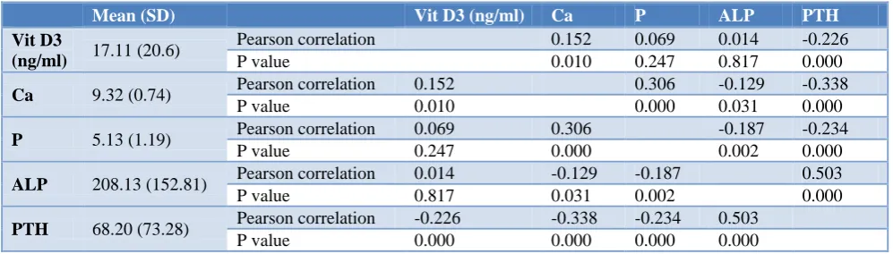
89.1% of the children were deficient in vitamin D levels with 11% being severely deficient. Factors significantly associated with vitamin D deficiency were increasing age (p=0.034), absence of intake of calcium supplements (p=0.00) and upper socio-economic class (p=0.001). Incidence was similar in children with higher BMI when compared to lower BMI. Duration of AEDs rather than number of drugs was more associated with deficiency though not significantly. Children with motor disability had a higher incidence of deficiency. Low serum calcium levels (p=0.01) and high PTH levels (p=0.00) were associated significantly (Table 1).
Table 1: Biochemical profile and its relation.
Mean (SD) Vit D3 (ng/ml) Ca P ALP PTH
Vit D3
(ng/ml) 17.11 (20.6)
Pearson correlation 0.152 0.069 0.014 -0.226
P value 0.010 0.247 0.817 0.000
Ca 9.32 (0.74) Pearson correlation 0.152 0.306 -0.129 -0.338
P value 0.010 0.000 0.031 0.000
P 5.13 (1.19) Pearson correlation 0.069 0.306 -0.187 -0.234
P value 0.247 0.000 0.002 0.000
ALP 208.13 (152.81) Pearson correlation 0.014 -0.129 -0.187 0.503
P value 0.817 0.031 0.002 0.000
PTH 68.20 (73.28) Pearson correlation -0.226 -0.338 -0.234 0.503 P value 0.000 0.000 0.000 0.000
DISCUSSION
This was a pediatric population based, cross sectional study, evaluating vitamin D status in children with a wide age range (0.5–18 years) in a selected cohort of neurologically ill children from the western regions of India. In our study 89.1% of children had vitamin D levels less than 30 ng/ml. In this category about 10.9% were severely deficient (SD), 70.8% were deficient (D) and 7.4% were insufficient (I) in vitamin D. A study by Shellhaas4 showed only 25% of children to have vitamin D levels in sufficient range in children with epilepsy. Our study revealed a prevalence of 89% which may be higher as compared to studies on neurological illnesses internationally. However, when compared with the general prevalence in pediatric population in India it still is comparable.1,5 The mean level of vitamin D was like studies from the South.6
Our study group comprised of 69% males and 31% females with no difference in the vitamin D status between girls and boys (p=0.682). Previous studies have reported a gender bias and attributed this finding to behavioral practices of the subcontinent.7,8 Shellhaas et al demonstrated female gender to be significantly associated with vitamin D deficiency in their study on epileptic children which was attributed to biological/ behavioral differences.4
14 years in females. Increased confinement to indoors in children with neurological illness as they grow associated with increase in screen time thereby decreasing effective sunlight exposure in these children besides poor supplementation could be cited as some of the reasons behind this observation.
Analysis of the dietary habits revealed no influence of diet over the vitamin D status in these children (p=0.765). Around 70% of children were inadequate in dietary calcium intake. However, 86% children with adequate intake of calcium in their diet also reported deficiency of vitamin D. Children who were taking vitamin D supplements had higher overall vitamin D levels. Mona et al studied the dietary intake of vitamin D across different populations of the world and revealed that though Japan had no food fortification programme their overall vitamin D intake was better due to heavy consumption of fish in the country.10 Though a higher percentage (58%) of our study population consumed a mixed diet, many children taking a mixed diet would consume meat or fish only on certain days of the week. This showed that dairy products in terms of calcium intake though important are not the only entity required to maintain adequate vitamin D levels in blood. A Great Britain population based study showed comparable results with supplementation of calcium and vitamin D.9 The mean duration and extent of exposure did not reveal any significance in our study. 94.8% exposed for more than 3.5 hours/week were found to be deficient in vitamin D with no significance. A study by Goswami and et al showed similar findings.11 Other studies have clearly shown that the 25(OH)D levels were directly proportional to the duration of exposure to sunlight.11,12
No significance was seen between BMI and vitamin D deficiency status. It however showed a high prevalence among children in over nourished category. Numerous studies have showed obesity to be associated with decreased vitamin D status.13-17 It would therefore be advisable to take necessary dietary precautions in these children to prevent obesity and thus reduce the chances of vitamin D deficiency.
91% of normally nourished children were seen to be vitamin D deficient. However, there was no significance seen in the class of malnutrition and vitamin D deficient status (p=0.108). Studies around Asia have demonstrated co-existence of severe malnutrition and vitamin D deficiency. Socioeconomic status was assigned as per Kuppuswamy’s grading showed that better socioeconomic status had a high number of vitamin D deficiency (p 0.001).18 Though the number of children were low in grade 1, all of them were seen to be vitamin D deficient. Similar study from India revealed a high prevalence of vitamin D deficiency even in upper strata in India.19 In contrast, another study from Delhi has shown a higher prevalence of vitamin D deficiency in lower socio-economic strata (LSES) as compared to upper socio-economic strata (USES).8 It is seen that with
improving socio-economic conditions, many children get confined to indoors resulting in decreased exposure to the sun.20,21
It was seen that 88.5% of disabled children were found to be deficient as against 90% of non-disabled children (p 0.846). While among children with motor disabilities, deficiency was noted in 90%, it was still higher among children with non-motor disabilities (92%). In a similar study from Australia, the percentage prevalence of hypovitaminosis D was high in both chronically ill and physically/intellectually disabled children with no difference attributable to intellectual versus physical disability.22 Sullivan and et al reported that in children with severe motor disabilities, reduced physical activity and decreased muscle mass and strength are obvious risk factors for secondary osteoporosis along with oral-motor difficulties and dysphagia, predisposing to feeding problems and suboptimal energy, calcium, and vitamin D intakes.23
Epilepsy has been associated with vitamin D deficiency owing to the role of anti-epileptic drugs (AEDs) on renal and hepatic metabolism. The incidence of vitamin D deficiency in epileptic group was found to be 88% while in those without epilepsy it was 92%. Duration of AEDs rather than number of drugs was associated more with vitamin D deficiency. Children on longer duration had a higher incidence of vitamin D deficiency (91%) as compared to those taking the drugs for less than a year (87%). A prospective case control study showed development of hypovitaminosis D in ambulatory children on various anti-epileptic drugs.24 Certain studies have concluded that persons taking long-term AEDs should have routine examination of BMD by using DXA, particularly if prescribed an enzyme-inducing AED.25
Biochemical profile
The blood level of calcium was found to be significant (p=0.010) and co related positively with vitamin D status (Table 1). A randomized controlled trial done on children in India with nutritional rickets showed comparable results.26 The study by Krishnamurthy and colleagues suggested that simultaneous co-administration of calcium and 25-OHD in RDA dosage is beneficial in limiting the changes in calcium and vitamin D metabolism in patients on AEDs in all ages.27 Similarly, significant association was seen in serum PTH levels and vitamin D status and had a negative co relation.
Steve and et al evaluated the relationships among vitamin D status, PTH, and calcium absorption in mid-pubertal boys and girls.28 They found comparable results and thus suggested that in adolescents, especially in the presence of vitamin D insufficiency, PTH secretion increases to adapt to higher rates of bone formation associated with growth resulting in higher serum 1,25(OH)2D
and phosphorus levels. Similar observations were made in a study on healthy school children in Lebanon.29 Another studyobserved that alkaline phosphatase could not be used as a screening test for vitamin D deficiency as there was no positive correlation seen.30 There was a significant positive correlation of mean serum alkaline phosphatase with PTH in an Indian study.8 We too noted a significant correlation between alkaline phosphatase and PTH levels (p=0.000).
CONCLUSION
The prevalence of vitamin D deficiency in children with neurological illness is comparable to apparently normal Indian children and cannot be neglected. Growing neurologically ill children, especially late childhood and adolescent should be adequately supplemented with vitamin D and calcium. Children on drugs for a long time should therefore be screened periodically for vitamin D deficiency.
Limitations
The size of the cohort is relatively small and larger population-based studies are required to arrive at important risk factors. Certain risk factors like sunlight exposure and dietary intake could not be studied.
ACKNOWLEDGEMENTS
We thank all children and their care-givers for the participation.
Funding: No funding sources Conflict of interest: None declared
Ethical approval: The study was approved by the Institutional Ethics Committee
REFERENCES
1. Harinarayan CV, Joshi SR. Vitamin D status in India-Its implications and Remedial Measures. J Assoc Physicians India 2009;57:40-8.
2. Dietary guidelines for Indian a manual drafted by NIN 2011.
3. Hollick MF. Vitamin D deficiency. N Engl J Med 2007; 357:266-81.
4. Borkar VV, Devidayal, Verma S, Bhalla AK. Low levels of vitamin D in North Indian children with newly diagnosed type 1 diabetes. Pediatr Diabetes. 2010;11(5):345-50.
5. Shellhas RA, Joshi SM. Vitamin D and bone health among children with epilepsy. Paediatric neurology 2010;42(6):385-9.
6. Harinarayan. Prevalence of VDD in children - Tirupati, South India. IJMR. 2008.
7. Sahu M, Bhatia V, Aggarwal A, Rawat V, Saxena P, Pandey A, et al. Vitamin D deficiency in rural girls and pregnant women despite abundant sunshine in
northern India. Clin Endocrinol (Oxf) 2009;70(5):680–4.
8. Marwaha RK, Tandon N, Reddy DR, Aggarwal R, Singh R, Sawhney RC, et al. Vitamin D and bone mineral density status of healthy schoolchildren in northern India. Am J Clin Nutr. 2005;82(2);477-82. 9. Absoud M, Cummins C, Lim MJ, Wassmer E, Shaw
N. Prevalence and Predictors of Vitamin D Insufficiency in Children: A Great Britain Population Based Study. PLoS ONE. 2011;6(7):e22179.
10. Calvo MS. Vitamin D Intake: A Global Perspective of Current Status. J Nutr. 2005;135:310–6.
11. Goswami R, Kochupillai N, Gupta N, Goswami D, Singh N, Dudha A. Presence of 25(OH) D deficiency in a rural North Indian village despite abundant sunshine. J Assoc Physicians India. 2008;56:755-7.
12. Arya V, Bhambri R, Godbole MM, Mithal A. Vitamin D status and its relationship with bone mineral density in healthy Asian Indians. Osteoporos Int. 2004;15:56-61.
13. Gilbert-Diamond D, Baylin A, Mora-Plazas M, Marin C, Arsenault JE, Hughes MD, et al. Vitamin D deficiency and anthropometric indicators of adiposity in school-age children: A prospective study. Am J Clin Nutr. 2010;92:1446.
14. Khor GL, Chee WS, Shariff ZM, Poh BK, Arumugam M, Rahman JA, et al. High prevalence of vitamin D insufficiency and its association with BMI-for-age among primary school children in Kuala Lumpur, Malaysia. BMC Public Health. 2011;11:95.
15. Gordon CM, DePeter KC, Feldman HA, Grace E, Emans SJ. Prevalence of vitamin D deficiency among healthy adolescents. Arch Pediatr Adolesc Med. 2004;158:531-7.
16. Rockell JE, Green TJ, Skeaff CM, Whiting SJ, Taylor RW, Williams SM, et al. Season and ethnicity are determinants of serum 25-hydroxyvitamin D concentrations in New Zealand children aged 5-14 y. J Nutr. 2005;135:2602-8. 17. Holick, Michael F. Vitamin D deficiency in obesity
and health consequences Current Opinion in Endocrinology & Diabetes. 2006;13(5):412-8. 18. Kumar N, Gupta N, Kishore J. Kuppuswamy's
socioeconomic scale: Updating income ranges for the year 2012. Indian J Public Health. 2012;56:103-4.
19. Chittari HV, Joshi S. Vitamin D Status of Upper Socioeconomic Status Subjects of South Indians Living in High Altitude (Bengaluru) Endocr Rev, 2012.
20. Spinks A, Macpherson A, Bain C, McClure R. Determinants of sufficient daily activity in Australian primary school children. J Paediatr Child Health. 2006;42:674–9.
Melbourne, Victoria, 1985–2001. Australian New Zealand J Public Health. 2005;29(4):337–42. 22. Greenway A, Zacharin M. Vitamin D status of
chronically ill or disabled children in Victoria. J Paediatr Child Health. 2003;39(7):543-7.
23. Sullivan PB, Lambert B, Rose M, Ford-Adams M, Johnson A, Griffiths P. Prevalence and severity of feeding and nutritional problems in children with neurological impairment: Oxford feeding study. Dev Med Child Neurol. 2000;42:674-80.
24. Nicolaidou P, Georgouli H, Kotsalis H, Matsinos Y, Papadopoulou A, Fretzayas A, et al. Effects of Anticonvulsant Therapy on Vitamin D Status in Children: Prospective Monitoring Study J Child Neurol. 2006;21:205-10.
25. Alison M. Pack. The Association Between Antiepilepticc Drugs and Bone Disease Epilepsy Curr. 2003;3(3):91–5.
26. Aggarwal V, Seth A, Marwaha RK, Sharma B, Sonkar P, Singh S, et al. Management of Nutritional Rickets in Indian Children: A Randomized Controlled Trial. J Trop Pediatr. 2013;59(2):127-33. 27. Murthy J. Antiepileptic drugs and bone health: Dietary calcium and vitamin D-the confounding factors. Neurol India. 2010;58:175-6.
28. Steven A. Abrams, Ian J. Griffin, Keli M. Hawthorne, Sheila K. Gunn, Caren M. Gundberg, Thomas O. Carpenter Relationships among Vitamin D Levels, Parathyroid Hormone, and Calcium Absorption in Young Adolescents J Clin Endocrinol Metab. 2005;90(10):5576–81.
29. Ghada El-Hajj Fuleihan, Mona Nabulsi, Mahmoud Choucair,Mariana Salamoun, Carmen Hajj Shahine, Aline Kizirian, and Raja Tannous Hypovitaminosis D in Healthy Schoolchildren Pediatrics. 2001;107:4-53.
30. Shaheen S, Noor SS, Barakzai Q. Serum alkaline phosphatase screening for vitamin D deficiency states. J Coll Physicians Surg Pak. 2012;22(7):424-7.