Original Research Article
Prognostic subtyping based on estrogen receptor, progesterone
receptor, and human epidermal growth factor receptor2 expression in
breast cancer cases among north Indian females
Kouser Sideeq Lone
1*, Shaheena Parveena
2INTRODUCTION
Breast cancer is the most common cancer of women globally.1 In India, breast cancer is second to cancer of the cervix among women, but is considered as the leading cancer in many parts of the country like the metro cities of Mumbai and Bangalore and also has recently become the leading cancer among women in Kashmir.2,3 It is estimated that 12% breast cancers occur between 20-34 years.1 Breast cancer is strongly related to age, with only 5% of all breast cancers occurring in women under 40 years old.4 There are several pathological features having
prognostic significance in breast carcinoma which includes histologic subtypes, grade, lymph node status, estrogen receptor (ER)/progesterone receptor (PR) status, growth factor and its receptors, proliferation activity and DNA content with tumor suppressor genes and oncogenes.4 Worse Prognosis of breast carcinoma is with the features of higher grade, tumor subtypes like medullary and lobular carcinoma, lymph node metastasis, positivity for Her-e/neu, negativity for ER, PR, and also presence of BRCA gene.4 The receptor status like ER, PR and Her-2/neu immunostaining are a very useful tool for rapid diagnosis, treatment as well as for prognosis of the
ABSTRACT
Background: Cancer of breast has recently become the leading cancer among women in Kashmir. Worse prognosis of this disease is with the features of higher grade, positivity for Her-2/neu, negativity for ER, PR, and presence of BRCA gene. Thus the receptor status like are a very useful tool for rapid diagnosis, treatment as well as for prognosis of the disease.The objective of the study was to analyze the pattern of expression of hormone receptors which include ER, PR and Her-2/neu in breast cancer cases among Kashmiri females and to find their association with various clinicopathological parameters.
Methods: A cross sectional hospital based study done in a tertiary health care centre of Kashmir valley. We selected 102 consecutive breast cancer cases for the period of one year. Cases were sent for biopsy of the tumour lesion and for receptor status analysis.
Results: In about 50.9%of cases tumour had spread to T2 stage. There was a significant difference in the tumor stage and the age groups at presentation. The HER 2 status was positive in 85.5% of cases. About 65% were ER positive and 51.9 were PR positive. Only 8 were triple negative and 42 were triple positive for receptors. We did not found any significant relationship between grade, stage of disease and age when compared for receptor status.
Conclusions: We found a high percentage being receptor positive tumours which has been found to be a good prognostic sign.
Keywords: Breast cancer, Receptor status, North India 1
NIRRH, ICMR, Mumbai, Maharashtra, India 2
GMC, Srinagar, Jammu and Kashmir, India
Received: 04 June 2018
Accepted: 06 July 2018
*Correspondence:
Dr. Kouser Sideeq Lone, E-mail: [email protected]
Copyright: © the author(s), publisher and licensee Medip Academy. This is an open-access article distributed under the terms of the Creative Commons Attribution Non-Commercial License, which permits unrestricted non-commercial use, distribution, and reproduction in any medium, provided the original work is properly cited.
disease. It is the standard practice to determine status of these receptors on biopsy specimens prior to therapeutic intervention.2 Currently, the research suggest that the level of PR determines the tumor response to endocrine treatment, PR- breast tumors have less sensitivity to tamoxifen than PR+ tumors in ER+ breast cancer.5 So it there are differences in approach to treatment in triple negative and triple positive tumors so the protocols are focusing on separating these two types for better treatment and prognosis.
With this background knowledge, the present study was done to analyze the pattern of expression of hormone receptor ER, PR and Her-2/neu in invasive breast carcinoma among Kashmiri females and to find their association with various clinicopathological parameters.
METHODS
This cross sectional hospital based study was done in one of the two tertiary health care centres of Kashmir valley, the SHMS hospital under the department of community medicine involving the departments of radiation and oncology and department of surgery of the same hospital. We selected consecutive breast cancer cases for the period of one year (2013-2014) from the departments of radiation oncology and department of surgery. All cases of breast cancer coming to these departments during this period were selected. Data was collected by using a predesigned semi structured questionnaire information was collected regarding the demographic variables, clinical history of the patient, and the medical records of the patients were also analysed. All the cases were sent for biopsy of the tumour lesion to the department of pathology while as for receptor status of ER and PR and her 2 nucleotide the samples were sent to private pathology labs as non-availability of this facility in the hospital. Around 102 cases were selected in the study but only 83 had their receptor status done during the study period. Those cases whose receptor status was awaiting or not sent due to any reason at the first visit were visited again and receptor status was asserted and if after 2nd visit still the receptor status was not available those cases were excluded from the receptor status analysis and association but all 102 cases were analysed for other aspects like age and histopathology.
RESULTS
A total of 102 cases of breast cancer were selected in this study but only 83 cases had their receptor status done. The presentation and association of receptor status with other variables was thus only done for these 83 cases. Majority of the participants belonged to the age group of 31 to 50 years with mean age of 47.87±13.6 years (Table 1). Ductal carcinoma was the histopathological diagnosis in majority of the cases (88.2% cases) (Figure 1). In about 50.9%of cases tumour had spread to T2 stage, among 21.5% it was at T1 stage and in 5.88% cases it had gone to T4 level (Figure 2). About 35%of cases had no
nodal involvement and 33.3% had nodal involvement to the level of N1 and only 7% had involvement to the level of N2. In approximately 13% of the cases there was distant metastasis and in about 34% of cases metastasis could not have been assessed at the time when data was taken from them. There was a significant difference in the tumor stage and the age groups at presentation (Figure 2) but not in nodal involvement and age groups. Stage II was the stage at presentation in maximum patients and grade 3 was the grade at presentation in 51.5% of patients (Table 2).
Table 1: Age distribution of patients.
Age group (in years) Number (%)
21-30 9 (8.82)
31-40 28 (27.45)
41-50 32 (29.41)
51-60 18 (18.62)
61-70 9 (9.80)
≥71 6 (5.88)
Total 102 (100)
Figure 1: Histopathology of breast cancer lesion among the patients.
Figure 2: Tumour staging of the breast cancer lesion in three age groups of the study participants
(significantly different with p<0.001).
88.2 4.9 1.9 0.9 0.9 0.9 0.9 0.9
0 50 100
Invasive Ductal Carcinoma(NOS) Insitu Ductal Carcinoma Invasive Ductal Carcinoma With
neuroendocrine Features Metaplastic Carcinoma Invasive Ductal carcinimaWith
Pagets
Lobular Carcinoma Malignat Phylloid Tumour Medullary Carcinoma Percentage H isto pa thholo g y o f the ca ncer 0 10 20 30 40 50 60 70 80
T1 T2 T3 T4
age <30
age 31-50
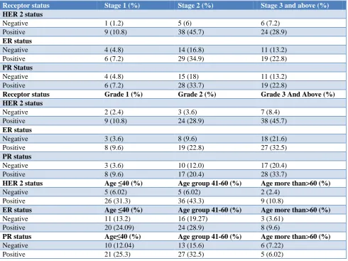
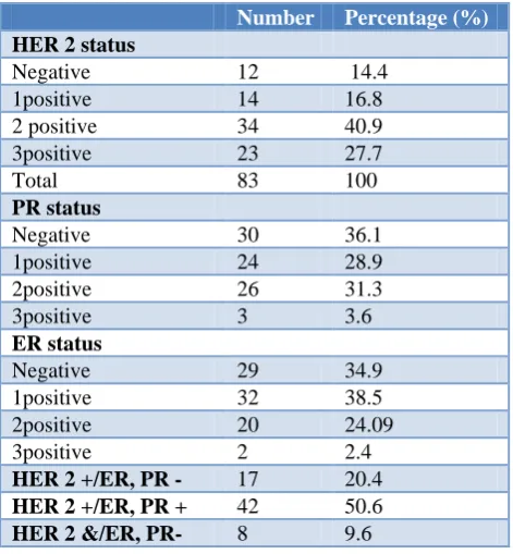
Out of total 102 patients only 83 had their receptor status done. The human epidermal growth factor (HER) 2 status was negative among 14.4% of patients and among them maximum patients (7.2%) were diagnosed in stage 3 of the disease (Table 2). This receptor status was positive in majority (85.5%) of the cases (2+ in 47.8% and 3+ in 32.3% of cases) and among them maximum were diagnosed in stage 2 of the disease (Table 2). Estrogen receptor status was positive in majority of (64.9%) (Table 2) cases and among them it was 1+ in 59%, 2+in 37.7%of cases and 3+ in only 3% of cases (Table 3). The ER receptor was negative in 34.8% of cases. Among both of the types of cases majority were diagnosed in stage 2 of the disease with 16.2% having ER receptor negative and 34.9% having ER receptor positive status in the stage 2 of disease (Table 2). Progesterone receptor (PR) status was also positive in majority of the cases (63.7%) and negative only in 36% of cases (Table 2). Among the PR positive cases about 23.5% and 25.5% were 1+ and 2+ respectively (Table 3). Maximum cases of PR receptor positive as well as negative cases (33.7% and 22.8% respectively out of total 83 cases) were diagnosed in stage 2nd of the disease (Table 2). Total 50% of cases out of all PR negative cases were diagnosed in stage 2nd of the disease and 52% of all PR positive cases were diagnosed in stage 2nd of the disease (Table 2). Among
the 12 cases with HER 2 receptor negative status, majority (7) were diagnosed at grade 3 or above and also among the HER 2 receptor positive cases majority were diagnosed in grade 3 or above of the disease (Table 2). Among ER status negative cases 18 (62%) out of total 29 cases were diagnosed in grade 3 or above of the disease and among the ER positive cases also majority were diagnosed at grade 3 or above of the disease (50% out of total 54 cases) (Table 2). Among patients with PR negative status again the majority (17 out of 30 patients) of the patients were diagnose in grade 3 or above of the disease and a total of 28 out of 53 among PR positive cases were again diagnosed in grade 3rd or above(Table 2). Majority of the patients (36 out of total 71 cases) with HER receptor status belonged to the age group of 41-60 years. Equal number of patients (5 out of 12) among HER 2 status positive belonged to each age groups of ≤40 and 41-60. Majority of the patients among both ER status negative as well as positive cases belonged to 41 to 60 years of age (Table 2). Similar results were obtained for PR receptor negative as well as positive cases. Out of 83 cases only 8 were triple negative and 42 were triple positive for receptors (Table 3). We did not found any significant relationship between grade and stage of disease when compared for receptor status, same results were found while comparing age with the receptor status.
Table 2: Comparison of receptor status with stage and grade of the disease.
Receptor status Stage 1 (%) Stage 2 (%) Stage 3 and above (%)
HER 2 status
Negative 1 (1.2) 5 (6) 6 (7.2)
Positive 9 (10.8) 38 (45.7) 24 (28.9)
ER status
Negative 4 (4.8) 14 (16.8) 11 (13.2)
Positive 6 (7.2) 29 (34.9) 19 (22.8)
PR Status
Negative 4 (4.8) 15 (18) 11 (13.2)
Positive 6 (7.2) 28 (33.7) 19 (22.8)
Receptor status Grade 1 (%) Grade 2 (%) Grade 3 And Above (%)
HER 2 status
Negative 2 (2.4) 3 (3.6) 7 (8.4)
Positive 9 (10.8) 24 (28.9) 38 (45.7)
ER status
Negative 3 (3.6) 8 (9.6) 18 (21.6)
Positive 8 (9.6) 19 (22.8) 27 (32.5)
PR status
Negative 3 (3.6) 10 (12.0) 17 (20.4)
Positive 8 (9.6) 17 (20.4) 28 (33.7)
HER 2 status Age ≤40 (%) Age group 41-60 (%) Age more than>60 (%)
Negative 5 (6.02) 5 (6.02) 2 (2.4)
Positive 26 (31.3) 36 (43.3) 9 (10.8)
ER status Age ≤40 (%) Age group 41-60 (%) Age more than>60 (%)
Negative 11 (13.2) 16 (19.27) 3 (3.61)
Positive 20 (24.09) 24 (28.9) 8 (9.6)
PR status Age≤40 (%) Age group 41-60 (%) Age more than>60 (%)
Negative 10 (12.04) 13 (15.6) 6 (7.22)
Table 3: Receptor status of breast cancer in our study population.
Number Percentage (%)
HER 2 status
Negative 12 14.4
1positive 14 16.8
2 positive 34 40.9
3positive 23 27.7
Total 83 100
PR status
Negative 30 36.1
1positive 24 28.9
2positive 26 31.3
3positive 3 3.6
ER status
Negative 29 34.9
1positive 32 38.5
2positive 20 24.09
3positive 2 2.4
HER 2 +/ER, PR - 17 20.4
HER 2 +/ER, PR + 42 50.6
HER 2 &/ER, PR- 8 9.6
DISCUSSION
Out of total 102 patients only 83 had their receptor status done. It is well established now that assessing the receptor status is very important step to assess the treatment modalities as well as the prognosis of breast cancer but still this facility is not available at a tertiary care hospital in our state, this need to be looked at. Human epidermal growth factor 2 status was positive in 85.5% of cases and many other similar studies have shown much similar results.6 HER 2 status of a primary breast carcinoma carries clinical utility in determining patient treatment options and overall prognosis. Estrogen receptor status was positive in 65% of cases and Progesterone receptor status was positive in 51.9%of cases. The prevalence of ER and PR expression has been found similar in some Indian studies but in western literature studies have found much higher prevalence as compared to the studies in India.6-9 This difference could be because of different genetic pool and ethnic differences. We found about 9.6% of cases being triple negative for receptors in our study population. This prevalence of triple negative breast cancer in our study is comparable to other international studies.6 Magdalena et al did a study by Analysing 38,813 cases from the national cancer database in United Kingdom and they found that the incidence of TNBC varied by region from 10.8% in New England to 15.8% in the east south central US.10 Triple negative breast cancers in Asian population are found to be associated with younger age of onset, increasing tumour size, increased prevalence of axillary lymph nodal involvement, higher histological grade of tumor and poor prognosis though our study did not found any significant relationship when we compared the
receptor status with grade, stage of disease and age groups.11 This difference in results could be because of the increased prevalence of higher grade tumours and late diagnosis in our population leading to more cases without ER/PR positivity.
CONCLUSION
In view of the increasing cases of breast cancer in Kashmir and being the most common cancer among females the facilities for receptor status assessment should be available at all the institutes taking care of cancer patients in the valley. We found a high percentage being ER and PR receptor positive tumours which has been found to be a good prognostic sign and thus if the cancer is diagnosed in early stage and receptor status found at early stage better treatment and prognostic value could be achieved.
Funding: No funding sources Conflict of interest: None declared
Ethical approval: The study was approved by the Institutional Ethics Committee
REFERENCES
1. Torre L, Rebecca Siegel AJ. Global Cancer Facts & Figures 3rd Edition. American Cancer Society; 2015.
2. Indian Council of Medical Research. Consensus Document for Management of Breast Cancer. 2014: 1–40.
3. Wani MA, Jan FA, Khan NA, Pandita KK,
Khurshid R, Khan SH. Cancer trends in Kashmir; common types, site incidence and demographic profiles: National Cancer Registry 2000-2012. Indian J Cancer. 2014;51(2):133–7.
4. Laamiri FZ, Bouayad A, Hasswane N, Ahid S. Risk Factors for Breast Cancer of Different Age Groups : Moroccan Data ? J Obstet Gynecol. 2015;5:79–87. 5. Hormone Therapy for Breast Cancer. American
Cancer Society. 2016. Available at: https://www. cancer.org/cancer/breast-cancer/treatment/hormone-therapy-for-breast-cancer.html. Accesssed on 24 February 2018.
6. Onitilo AA, Engel JM, Greenlee RT, Mukesh BN. Breast cancer subtypes based on ER/PR and Her2 expression: Comparison of clinicopathologic features and survival. Clin Med Res. 2009;7(1–2):4– 13.
7. Biswal P, Behera S, Kar A, Pradhan D. Correlation of Hormonal Receptors Estrogen Receptor, Progesterone Receptor and Her-2 / Neu with Tumor Characteristics in Breast Carcinoma : Study of 100 Consecutive Cases. Int J Clin Med. 2015;12:961–6. 8. Rajan G, Culas TB, Jayalakshmy P. Estrogen and
9. Kinsella MD, Nassar A, Siddiqui MT, Cohen C. Estrogen receptor (ER), progesterone receptor (PR), and HER2 expression pre- and post-neoadjuvant chemotherapy in primary breast carcinoma: A single institutional experience. Int J Clin Exp Pathol. 2012;5(6):530–6.
10. Plasilova ML, Hayse B, Killelea BK, Horowitz NR, Chagpar AB, Lannin DR. Features of triple-negative
breast cancer. Medicine (Baltimore).
2016;95(35):e4614.
11. Ma KK, Chau WW, Wong CHN, Wong K, Fung N,
Lee JTA, et al. Triple Negative Status is a Poor
Prognostic Indicator in Chinese Women with Breast Cancer: a Ten Year Review. Asian Pacific J Cancer Prev. 2012;13(5):2109–14.