Original Research Article
Clinico-epidemiological features of Japanese encephalitis patients
hospitalized in a tertiary care center
Abu Hasan Sarkar
1*, Bishnu Ram Das
2INTRODUCTION
Human brain is an important product of evolution which has placed human beings above all other animals on Earth. Any infection that causes inflammation of human brain or encephalon is therefore a reason of concern. The term encephalitis is used to define an inflammation of the brain parenchyma.1 Encephalitis, in simple terms, is the inflammation of brain matter which may subside and return to normalcy with or without treatment or result in confusion, coma or death. Recovery from encephalitis may be full or with neurological sequelae.2,3 It is a disease that causes high mortality and disability in the survivors.4 Thus encephalitis can cause a great loss to public health, productivity and economy of a country.
Japanese encephalitis (JE) is a zoonotic disease caused by
Flavivirus belonging to Group B Arbovirus and affects human and animals. Humans are incidental or dead-end hosts, because they usually do not develop high enough concentrations of JE virus in their bloodstreams to infect feeding mosquitoes.5,6 JE was first detected in Japan in 1871 and it affects more than half of the countries across the globe, India being one.7
In India, JE was identified as a public health problem since 1952 while in Assam since 1978 and since then there have been a number of outbreaks.8 Based on endemicity, epidemics, isolation of virus from patients and vectors and general serological surveys, the virus has been shown to be widely prevalent in most parts of south, central, northern and northeast states of India.9 Globally
ABSTRACT
Background: Japanese encephalitis (JE) is of particular interest as it has a high morbidity and mortality. Neurological sequale is the most dreaded damage caused by JE. It is a preventable disease with specific interventions. The objective of the study was to study the demography, clinical profile and outcome of patients with Japanese Encephalitis admitted to the wards of Internal Medicine and Pediatrics at Jorhat Medical College Hospital.
Methods: Hospital based observational study for one year in Jorhat Medical College, Jorhat, Assam.
Results: The mean age for JE was 32.25±27 years for male, 27.47±22 years for female and 29.94±24 years overall. Assessment of clinical signs and symptoms showed that fever and change in mental status were present in 100% of JE cases followed by neck rigidity in 79.3% and headache in 68.9%. 44.8% of JE cases had history of seizure, 37.9% had vomiting, 34.5% had irritability, 13.8% were unconscious. The peak of JE incidence occurred in the month of July (77.6%). Complete recovery was seen in 39.2%, followed by death in 32.6% and recovery with neurological sequalae in 28.2% at the time of discharge.
Conclusions: Vigorous awareness activities should be carried out to sensitize people on prevention of JE.
Keywords: Japanese encephalitis, Clinical profile, Demography, Neurological sequale, Outcome
1District Surveillance Unit, Integrated Disease Surveillance Program, Barpeta, Assam, India 2Department ofCommunity Medicine, Jorhat Medical College, Jorhat, Assam, India
Received: 08 August 2018
Accepted: 07 September 2018
*Correspondence:
Dr. Abu Hasan Sarkar,
E-mail: [email protected]
Copyright: © the author(s), publisher and licensee Medip Academy. This is an open-access article distributed under the terms of the Creative Commons Attribution Non-Commercial License, which permits unrestricted non-commercial use, distribution, and reproduction in any medium, provided the original work is properly cited.
every year around 50,000 people suffer from JE as per WHO reports. According to the reports published by National Vector Borne Disease Control Programme for the period from 1st Jan 2015 to 31st Dec 2015, the figures for JE are 1731 and 614 cases in India and Assam respectively.10
Geographically and culturally Assam has all the hitherto anthropogenic and mosquitogenic factors in abundance required for JE transmission. There is agricultural practice which is dependent on rain based irrigation. In some parts of Assam, the culture of pig rearing is
practiced. Also the vector (Culicine mosquito)
responsible for transmission of JE is present. Earlier JE was thought to be a disease affecting the people living in Upper Assam mostly however this trend is changing and reports have shown a rise in JE cases in entire Assam. Further, the earlier view of JE as a disease affecting children below 15 years of age is changing.11
JE is of particular interest as it has a high morbidity and mortality affecting both pediatric and adult age group in present context. Neurological sequale is the most dreaded damage caused by JE. There is no antiviral treatment. Understanding of epidemiological features and diagnosis of JE on clinical grounds is extremely important to manage the encephalitis cases individually and to contain the epidemic in community. Hence the present study was conducted with the following objective.
Objectives
To study the clinico-epidemiological features of JE patients admitted in Jorhat Medical College Hospital.
METHODS
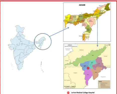
The present hospital based observational prospective study was conducted at Jorhat Medical College Hospital (26.7426° N, 94.1951° E) from 1st July 2015 to 30th June 2016 (one year).
Figure 1: Map showing the study area.
Inclusion criteria
Inclusion criteria were patients of acute encephalitis syndrome who tested JE specific IgM antibody positive in serum or CSF and were admitted under the departments of Internal Medicine and Pediatrics of Jorhat Medical College Hospital; participants or caregivers (in case of cognitive orientation compromised patients or children) who were willing to participate and gave consent.
Exclusion criteria
Patients or guardians (in case of cognitive orientation compromised patients or minors) who were not willing to give consent for the study.
Sample size and sampling technique
All the consecutive cases of acute encephalitis syndrome admitted under Internal Medicine and Pediatrics department from 1st July 2015 to 30th June 2016 who fulfilled the inclusion criteria.
Instrument for data collection
WHO AES case investigation format was used for collection of data. Additional data were collected viz. socio-economic status, Glasgow Coma Scale score etc.
Informed consent
A written informed consent was obtained from all the patients or their guardians individually after explaining the purpose and the scope of the study. They were provided with a participant information sheet which detailed the nature and all other information pertaining to the research study. Assent was obtained from adolescents. Surrogate consent was taken from immediate relative (spouse>parents>sibling), in case the patient was not able to give consent. Later the patient was informed about the study in details and consent was taken if he/she became conscious and oriented. Informed consent forms were used in understandable language of the participants or their guardian. The patient or relatives were requested to sign or give left hand thumb impression whichever was applicable and comfortable to the patients or relatives.
Ethics clearance
Ethical clearance was obtained from the Institutional Ethics Committee (Human), Jorhat Medical College and Hospital, Jorhat.
Data collection technique
WHO AES case definition and laboratory results of JE virus specific of IgM antibody Mac-ELISA were noted from bed head tickets of the patients. Demographic, clinical and epidemiological data were collected from each JE positive patient at the time of case investigation.
Statistical analysis
The data collected were entered in Microsoft Excel 2010 and analyzed using Epi info version 7 for Windows which was downloaded free from the website of Centre for Diseases Control and Prevention (CDC), Atlanta. Statistical significance was considered at 95% confidence interval that is when p value was less than 0.05.
Operational definitions
Clinical signs of JE are indistinguishable from other causes of Acute Encephalitis Syndrome. Laboratory confirmed cases were therefore considered as cases of JE. Laboratory diagnosis of JE can be done in any one of the following ways.12
Presence of JE virus-specific IgM antibody in a single sample of cerebrospinal fluid (CSF) or serum or both, as detected by an IgM-capture ELISA.
Detection of JE virus antigens in tissue by
immunohistochemistry;
Detection of JE virus genome in serum, plasma, blood, CSF or tissue by reverse transcriptase polymerase chain reaction (PCR) or an equally sensitive and specific nucleic acid amplification test;
Isolation of JE virus in serum, plasma, blood, CSF or tissue;
Detection of a four-fold or greater rise in JE virus-specific antibody as measured by haemagglutination inhibition (HI) or plaque reduction neutralization assay (PRNT) in serum collected during the acute and convalescent phase of illness. The two specimens for IgG should be collected at least 14 days apart. The IgG test should be performed in parallel with other confirmatory tests to eliminate the possibility of cross-reactivity.
Laboratory criteria considered in this study for diagnosis of JE was criterion 1 which states that presence of JE virus-specific IgM antibody in a single sample of cerebrospinal fluid (CSF) or serum or both, as detected by an IgM-capture ELISA.
Children/pediatric age
Patient admitted with JE who belonged to age group of less than or equal to 15 years (inclusive) of age.
Adult
Patient admitted with JE who belonged to age group of more than 15 years (exclusive) of age.
Outcome
Outcome was considered in three categories viz. complete recovery, recovery with neurological sequelae and death. The outcome for individual patient was recorded at the time of discharge or death.
Neurological sequale
Neurological sequelae was defined by the presence of one or more of the following at the time of discharge;
impaired consciousness, weakness (monoparesis,
hemiparesis, and quadriparesis), focal or generalized abnormal limb tone (hypertonia and hypotonia), focal or generalized abnormal limb reflexes (hyperreflexia and hyporeflexia), diagnosis of new onset or recurrent seizures, or new or recurrent extra pyramidal movement disorders.13
RESULTS
In the present study out of 169 AES cases, 58 (34.3%) cases were found as having positive laboratory result for JE virus specific IgM antibody in CSF or serum or both. Hence there were slightly more than one-third JE cases amongst all the AES cases admitted.
Table 1: Socio-demographic profile of JE cases.
Age group (in years)
Number of JE cases (N=58)
Total (%)
Male Female
Urban Rural Total (%) Urban Rural Total (%)
<1 0 0 0 (0) 0 0 0 (0) 0 (0)
1-5 0 3 3 (10) 0 2 2 (7.1) 5 (8.6)
5-15 0 10 10 (33.3) 0 12 12 (42.8) 22 (37.9)
15-30 1 2 3 (10) 1 2 3 (10.7) 6 (10.3)
30-60 1 6 7 (23.3) 2 5 7 (25) 14 (24.1)
>60 2 5 7 (23.3) 0 4 4 (14.3) 11 (19)
Figure 2: Clinical signs and symptoms of JE cases.
Figure 3: Distribution of JE cases district wise.
Socio-demographic profile of JE cases
In socio-demographic profile, it was observed that out of the 58 laboratory confirmed JE cases, 52% were male and 48% were female. Among the total JE cases maximum 53.4% cases occurred in adults (>15 years of age) while 46.6% were pediatric age groups (≤15 years). Among children the mostly affected age group was 5-15 years (Table 1).
The mean age for JE was 32.25±27 years for male, 27.47±22 years for female and 29.94±24 years overall. The difference in mean ages for male and female was not
found to be statistically significant. JE cases were seen in patients from 3 years of age to 85 years of age.
It was observed that majority (88%) of the laboratory confirmed JE cases belonged to rural areas and only 12% belonged to urban areas (Table 1).
Clinical profile of JE cases
In the current study assessment of clinical signs and symptoms showed that fever and change in mental status were present in 100% of JE cases followed by neck rigidity in 79.3% and headache in 68.9%. We recorded
0% 10% 20% 30% 40% 50% 60% 70% 80% 90% 100%
100.0% 100.0%
13.8%
44.8%
34.5%
3.4%
79.3%
68.9%
5.2%
37.9%
history of seizure in 44.8% of JE cases while 37.9% had vomiting, 34.5% had irritability and 13.8% were unconscious (Figure 2).
It was also seen that 53.4% of JE cases had a GCS score of more than 8 and in 46.6% GCS score was less than or equal to 8.
Geographical distribution of JE cases
It was seen in the present study that out of the 58 laboratory confirmed JE cases, 55.17% were from Jorhat, 36.21% from Golaghat, 6.9% from Sivasagar and 1.72% were from Karbi Anglong district. The maximum of the laboratory confirmed JE cases were from Jorhat district followed by Golaghat (Figure 3).
Month wise occurrence of JE cases
On arranging the JE cases according to their month of occurrence it was seen that JE transmission mainly occurred during the rainy season starting from the month of June to September. The peak of JE incidence (77.6%) occurred in the month of July (Figure 4).
Figure 4: Showing the month wise occurrence of JE cases.
Vaccination status among the JE cases
Vaccination history of the JE patients with SA 14-14-2 was enquired and it was found that out of total 58 laboratory confirmed JE cases, 56.9% of them were not vaccinated and 6.9% were vaccinated. The immunization status of 36.2% could not be ascertained in the present study due to non-availability of records. The information on vaccination status was based on recall memory as none of the patient could produce the immunization record at the time of enquiry.
Outcome of JE cases
In the present study it was found that out of total 58 JE positive cases, 2 were referred to higher centres for better case management and 10 cases had Left the hospital
Against Medical Advice (LAMA). So outcome analysis was done for 46 JE confirmed cases. Of these, complete recovery was seen in 39.2%, followed by death in 32.6% and recovery with neurological sequalae in 28.2% patients at the time of discharge from hospital.
Figure 5: Vaccination status of JE cases.
DISCUSSION
Sociodemographic profile of JE cases
Socio-demographic profile of JE cases showed that 52% were male and majority of them were from rural areas (Table 1). Our findings corroborate the earlier studies.14-16 It may be explained by the fact that most male members from rural areas primarily depend on agriculture for their livelihood. The JE vector breeds in water logged irrigated rice fields. Also, male remain less covered by clothes as compared to female. For these reasons risk of man-mosquito contact increases resulting increased prevalence of JE in male.
It was observed that according to age group distribution mostly affected age group was adults (>15 years of age) and in the pediatric (≤15 years) age goup 5-15 years was affected most (Table 1). This may be due to the fact that in recent time newer districts are affected which were earlier non-endemic. In non-endemic areas people are immunologically virgin and does not have IgG antibody in their blood. Therefore all age group are affected, mainly those who are more prone to infected mosquito bite.
Clinical profile of JE cases
In our study, assessment of clinical signs and symptoms showed that fever and change in mental status was present in all the JE cases followed by neck rigidity and headache (Figure 2). This finding was similar to earlier studies and is in conformity with our current understanding of the clinical features of JE.14,15
Geographical distribution of JE cases
In the current study it was seen that maximum cases were from Jorhat district followed by Golaghat, Sivasagar and Karbi Anglong districts. Since the institution where the present study was conducted is a tertiary health care
0.0% 0.0% 0.0% 0.0% 0.0% 5.2% 77.6%
15.5%
1.7% 0.0% 0.0% 0.0% 0%
10% 20% 30% 40% 50% 60% 70% 80% 90%
Vaccinated 6.9%
Not vaccinated
56.9%
institution, so there were referral of cases from different neighbouring districts (Figure 3). However, we could assume from the district wise geographic distribution that Jorhat, Sivasagar and Golaghat were more affected districts from severity point of view.
Month wise occurrence of JE cases
On arranging the JE cases according to their month of occurrence it was seen that JE transmission mainly occurred during the rainy season starting from the month of June to September. The peak of JE incidence occurred in the month of July (Table 4) similar to earlier studies.17,18 Our study showed a clear seasonal trend of JE transmission that peaked in the monsoon and post monsoon when JE vector density increases due to rainfall.
Vaccination status among the JE cases
Vaccination history of the JE patients with SA 14-14-2 was enquired and it was found that 56.9% were not vaccinated while only 6.9% had received vaccine. The immunization status of 36.2% could not be ascertained in the present study (Figure 4).13 This finding is similar to earlier conducted studies.14,19 The low level of immunization revealed that people are not yet aware of JE vaccination. It also indirectly reflects the poor social mobilization of target beneficiaries during mass vaccination campaign and in routine immunization drive.
Outcome of JE cases
Our study revealed that complete recovery was seen in 39.2% of JE cases, followed by death in 32.6%. Other studies conducted have reported less deaths and more complete recovery.19 Since Jorhat Medical College Hospital is a newly established institute the facilities for better case management is still lacking in the hospital. This may further be explained that due to prior poor immunity status the mortality might have increased in our study. However, our observation may be confirmed only after doing further in-depth study in this line.
Recovery with neurological deficit was observed in 28.2% of the JE cases in our study. This finding is similar to the observations made by Kakoti et al and Dutta et al.14,19
CONCLUSION
JE is a major public health problem prevalent in this part of the country. The policy maker should initiate appropriate public health intervention to control this dreaded disease of mankind. Improvement in the treatment facilities are needed for better outcome. Awareness has always been the mainstay of prevention. Hence adequate and specific information should be disseminated to the people regarding the determinants and factors of JE occurrence and its prevention.
Limitations of the study
Cases admitted to health institutions other than Jorhat Medical College Hospital were not included in this study and hence there is a possibility of missing those cases.
Patients who were referred to specialized centers and who left against medical advice could not be followed up for outcome. So these cases were excluded from outcome analysis.
Funding: Department of Biotechnology (DBT), Ministry of Science and Technology, Government of India
Conflict of interest: None declared
Ethical approval: The study was approved by the Institutional Ethics Committee
REFERENCES
1. Forbes BA, Sahm DF, Weissfeld AS. Bailey & Scott’s Diagnostic Microbiology.12th ed. Missouri: Mosby Elsevier; 2007: 826.
2. Malakar M, Bose SR. Encephalitis: A Comparative Study in Pediatric and Adult Age Groups. J Pharm Biological Sci. 2016;11(3):24-7.
3. Suryakantha AH. Community Medicine with recent
advances. 3rd ed. New Delhi: Jaypee brothers Medical Publishers; 2014: 449-50.
4. Fischer M, Hills S, Staples E, Johnson B, Yaich M, Solomon T. Japanese encephalitis prevention and control: advances, challenges, and new initiatives. In: Scheld WM, Hammer SM, Hughes JM (eds). Emerging infections 8. Washington: ASM Press; 2008: 93-124.
5. Hills SL, Weber IB, Marc F. Japanese Encephalitis. Centers for disease control and prevention. Available at: http://wwwnc.cdc.gov/travel/yellow
book/2014/chapter-3-infectious-diseases-related-to-travel/japanese-encephalitis. Accessed on 15
February 2015.
6. Transmission of Japanese Encephalitis Virus.
Available from: http://www.cdc.gov/japanese
encephalitis/transmission/index.html. Accessed on 1 February 2015.
7. Nagabhushana RP. Japanese Encephalitis. Indian Pediatr. 2001;38:1252-64.
8. Hazarika NC. Project study on Japanese
Encephalitis vaccination at Gogamukh, Assam. The Indian Acad Pediatr. 1991;28(9):1029-34.
9. Japanese encephalitis. Available at: http://icmr.nic. in/pinstitute/niv/JAPANESE%20ENCEPHALITIS. pdf. Accessed on 1 February 2015.
10. Directorate of National Vector Borne Disease Control Programme- Delhi. Details of AES/JE Cases and Deaths from 2008-2014, National Vector Borne Disease Control Programme. Available from: http://nvbdcp.gov.in/Doc/je-aes-cd-July14.pdf. Accessed on 15 February 2015.
syndrome in India: changing paradigm and
implication for control. J Commun Dis.
2014;46(1):4-11.
12. Guidelines clinical management of Acute
Encephalitis Syndrome including Japanese
Encephalitis. Government of India. Directorate of National Vector Borne Disease Control Programme. Directorate General of Health Services, Ministry of Health & Family Welfare, Delhi. 2009.
13. Malakar M, Mathur A. Distribution of encephalitis in Dhemaji district of Assam. Int J Emerging Tech Innovative Engineering. 2015;3:185-8.
14. Kakoti G, Dutta P, Das BR, Borah J, Mahanta J. Clinical. Profile and outcome of Japanese Encephalitis in children admitted with Acute Encephalitis Syndrome. BioMed Research Int. 2013.
Available at: http://www.hindawi.com/journals/
bmri/2013/152656/. Accessed on 20 February 2015. 15. Mohan DG, Hazarika NK. A clinic-pathological
study and demographic profile of Japanese encephalitis from a tertiary care hospital in Assam, India. Int J Current Microbiol Applied Sci. 2015;4(6):522-9.
16. Chakraborty D, Banerjee S, Maji D, Dey TK, Mondal P, Basu M. A Descriptive Study of Japanese
Encephalitis in West Bengal, India, Based on Surveillance Data: Changing Pattern Observed in Recent Years. Sch J App Med Sci. 2015;3(1):320.
17. Borkotoki U, Borkotoki S, Barua P, Das A,
Rajkhowa P. Japanese encephalitis(JE) among Acute Encephalitic Syndrome (AES) cases- a hospital based study from Upper Assam, India. Int J Health Sci Res. 2016;6(5):72-7.
18. Kakkar M, Rogawski ET, Chaturvedi S, Dhole TN,
Hossain SS, Krishnan SS. Acute Encephalitis Syndrome Surveillance, Kushinagar District, Uttar Pradesh, India, 2011–2012. Emerg Infect Dis. 2013;19(9):1361-7.
19. Dutta A, Konwar M, Kumar PP. A study of clinical
profile of adult patients with acute encephalitis syndrome coming to a tertiary care hospital of North
East India. J Evid Based Med Healthc.
2016;3(46):2299-302.