www.fm.viamedica.pl
Address for correspondence: Dr Mounir N. Ghabriel, The Department of Anatomical Sciences, University of Adelaide, Adelaide, South Australia 5005, tel: +618 83035481, fax: +618 83034398, e-mail: [email protected]
Anatomical variations in the branches
of the human aortic arch: a recent study
of a South Australian population
Kartik Bhatia, Mounir N. Ghabriel, Maciej Henneberg
The Department of Anatomical Sciences, University of Adelaide, South Australia
[Received 31 May 2005; Accepted 4 July 2005]
Variations of the branches of the aortic arch are likely to occur as a result of the altered development of certain branchial arch arteries during the embryonic period of gestation. In the present investigation the pattern of branches of the aortic arch was studied in 81 cadavers from a recent South Australian popula-tion of European descent, who have migrated to (n = 38) or were born and lived in (n = 43) South Australia during the twentieth century. Two principal variations were noted in the present study. Firstly, in 6 cadavers, the left verte-bral artery originated directly from the arch of the aorta, between the left com-mon carotid and the left subclavian arteries. The 6 subjects were acom-mong the subgroup born in South Australia, giving an incidence of 13.95%, which is much higher than in previous reports. The overall incidence of 7.41%, when related to the whole group, is also higher than incidences reported in other populations. The presence of this variation suggests that in some individuals part of the aortic arch is formed from the left 7th inter-segmental artery. Secondly, none of the cadavers examined had the thyroidea ima artery, contrasting with previously reported incidences that varied between 4% and 10%. Since all 6 cadavers with the left vertebral artery variant were born in South Australia, it is suggested that environmental factors may have contributed to this variation. Significant envi-ronmental changes in South Australia around the turn of the twentieth century are discussed. This study represents the first systematic investigation of the branch-es of the aortic arch in a South Australian population and providbranch-es data relevant to the practice of medicine.
Key words: angiogenesis; vertebral artery, thyroidea ima, development
INTRODUCTION
The human arterial system of the head, neck and upper thoracic regions arises from a series of aortic arches that make their appearance in the 4th and 5th
weeks of embryonic development. These arches arise from the ventrally located aortic sac and terminate in the dorsal aortae. In all, 6 pairs of aortic arches
Figure 1. A photograph showing the left vertebral artery (LV) arising
from the arch of the aorta (Ao), between the left common carotid (CC) and left subclavian (LS) arteries; BS — brachiocephalic artery.
documented variations in the branches of the arch of the aorta, and the reported incidences of these variations have differed across studies [6].
The aortic arch has a composite origin as it de-velops from the aortic sac, left 4th
aortic arch, and part of the left dorsal aorta. The aortic arch acquires functional ascendancy during the 4th and 5th weeks
of embryonic development. The branches of the aor-tic arch develop over the 5th and 6th weeks of
gesta-tion. Investigation of the arterial pattern of the head, neck and thorax thus provides insight into the chang-es that occur from the 4th to the 6th weeks of
gesta-tion. In the period from the 6th to the 8th week,
trans-formation of the aortic arch arteries leads to the de-velopment of the adult arterial system [15]. Knowl-edge of the arterial pattern of these regions and any variations are important in the practice of emergency medicine and for cardiothoracic surgical procedures. There are indications that the incidence of varia-tions in other arteries has changed during the twen-tieth century [19]. The present study was carried out to provide data on the incidence of variations in the branches of the aortic arch amongst members of the South Australian population who were born and died in the twentieth century.
MATERIAL AND METHODS
A total of 81 cadavers, 18 females and 63 males, all of European descent, were used in this study. These were donated to the Department of Anatom-ical Sciences, University of Adelaide, by a long es-tablished body donation programme that exists in South Australia. Their years of birth ranged from 1901 to 1945. The cadavers were grouped into birth cohorts by decade of birth (Table 1). The aim of this study was communicated to the families of the do-nors who consented to the study. Demographic data were verified via an investigation of birth, census
and immigration records of South Australia. These included the date of birth, birthplace and date of immigration to South Australia (where applicable). Of the 81 donors 43 were born in South Australia and their years of birth ranged from 1901 to 1934. The remaining donors were born in other Australian states (n = 12), Germany (n = 11), Yugoslavia (n = 1), Austria (n = 2), Italy (n = 1), England (n = 6), Scot-land (n = 2), IreScot-land (n = 2) and France (n = 1). There was no evidence of first or second-degree re-lation between the donors, reducing, but not exclud-ing, the chance of genetic linkage between the sub-jects of the study.
All cadavers were dissected by trained dissectors. The dissections provided a clear view of the arch of the aorta and its branches.
RESULTS
The position of the arch of the aorta was found to be consistent with textbook descriptions in all 81 cadavers. All cadavers had the brachiocephalic trunk, left common carotid and left subclavian arteries as branches of the aortic arch (Fig. 1). However, 6
spec-Table 1. The number of cadavers divided according to
year of birth cohorts, and those showing the variant origin of the left vertebral artery from the arch of the aorta
Birth Number of Number of cohorts dissected cadavers cadavers showing
variation
1901–1910 6 –
1911–1920 28 4
1921–1930 33 1
1931–1940 13 1
imens displayed an additional branch of the aortic arch, the left vertebral artery, arising in these 6 cas-es from the aortic arch between the origins of the left common carotid and left subclavian arteries (Fig. 1). Of the 6 cases, 4 were among the 1911–1920 birth-cohort, 1 was among the 1921–1930 birth-cohort, and 1 was among the 1931–1940 cohort. All 6 subjects were born in South Australia, 5 were male and 1 was female. The overall incidence of this variation in this study was 7.41 ± 2.91%. The incidence among those born in South Australia was 13.95 ± 5.28%. Though this latter is not formally significantly differ-ent from the former, its greater value is of interest. The anatomy of the brachiocephalic trunk was consistent with textbook descriptions in all cases and no variations were found in its origin or branching, as it divided into the right common carotid and right subclavian arteries (Fig. 1). The anatomy of the left common carotid artery was consistent with textbook descriptions in all cases and no variations were found in its origin (Fig. 1). However, in 1 male cadaver, born in England in 1924, the bifurcation of the left and right common carotid arteries into internal and external carotid arteries was found to occur at the level of the 2nd cervical vertebra. No variation was
detected in the left subclavian artery as it arose from the aortic arch (Fig. 1). The thyroidea ima artery was not found in any of the 81 cadavers examined in this study, thus having an incidence of 0%.
DISCUSSION
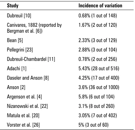
The first notable variation found in this study of the aortic arch branches was a variant origin of the left vertebral artery, which arose directly from the arch of the aorta, between the origins of the left common carotid and left subclavian arteries. This variant occurred in 6 subjects, all born in South Aus-tralia, and the incidence in relation to the whole sam-ple was 7.41%. In addition, the incidence of this vari-ation amongst the subjects born in South Australia was 13.95%. This incidence is in sharp contrast to a previous report that the vertebral arteries tend to be the most constant of the subclavian branches [8]. This variation has been noted in a number of previ-ous studies in other populations but the incidences reported were lower than that found in the present investigation. The results of 12 previous studies are shown in Table 2, and our summarised incidence from these studies indicates the presence of this vari-ation in 116 out of 3,499 persons (3.32%). This is significantly less (c2 = 4.03, df = 1, p < 0.05) than
our finding of 6 amongst 81 cadavers (7.41%). When
the data presented in Table 2 are combined with those of the current study, frequencies increased from 1.12% (3/268) in the nineteenth century to 3.5% (113/3231) in the twentieth century and to 7.41 (6/81) in the twenty-first century. These differences between centuries are significant (c2 = 10.23, df = 4,
p < 0.05).
Bergman et al. [6] discussed several earlier stud-ies and provided a summarised combined incidence of this variation of 1.79%. A larger and more exhaustive literature review conducted by Argenson et al. [4] demonstrated an incidence of 3.9% in 6,475 observations, which is also significantly lower than in our material. The incidences reported in previous studies (Fig. 2) demonstrate a general, significant increase with time. The current incidence of 7.41 in the total sample of the present investigation sup-ports an increasing trend with time (Fig. 2). The much higher incidence of 13.95% reported in the present study, when this variant is related to the subpopula-tion of cadavers born in South Australia, may be re-lated to environmental factors that prevailed in South Australia, different populations, and/or the differ-ent time from previous studies. A recdiffer-ent study from Poland showed this variant in 7 out of 103 speci-mens (6.8%). However, that study was done on spon-taneously aborted foetuses [12], suggesting either maternal or foetal abnormality. The current study was performed on subjects who died of natural caus-es, and is the first to report on a sample of the South
Table 2. Studies on the incidence of variant origin of the
left vertebral artery from the arch of the aorta reported in 12 previous studies
Study Incidence of variation
Dubreuil [10] 0.68% (1 out of 148)
Canivares, 1882 (reported by 1.67% (2 out of 120) Bergman et al. [6])
Bean [5] 2.33% (3 out of 129)
Pellegrini [23] 2.88% (3 out of 104)
Dubreuil-Chambardel [11] 0.78% (2 out of 256)
Adachi [1] 5.43% (28 out of 516)
Daseler and Anson [8] 4.25% (17 out of 400)
Anson [2] 3.6% (36 out of 1000)
Argenson et al. [4] 5.8% (6 out of 104)
Nizanowski et al. [22] 3.1% (8 out of 260)
Matula et al. [20] 3.05% (7 out of 402)
Australian population who died in the last decade of the twentieth century.
The subclavian artery develops from the 7th
cervi-cal inter-segmental artery, which is initially connect-ed to the dorsal aorta caudal to the dorsal attach-ment of the aortic arch [3]. As the neck elongates and the heart descends, this point of origin migrates rostrally along the dorsal aorta and comes to lie op-posite the dorsal end of the 4th aortic arch (Fig. 3).
Since this part of the dorsal aorta on the left side becomes incorporated in the arch of the aorta, the stem of the left 7th inter-segmental artery forms the
origin of the subclavian artery from the arch of the aorta (Fig. 3).
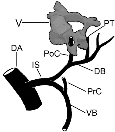
The most common origin of the vertebral artery is from the first part of the subclavian artery. How-ever, it has been known to arise from the arch of the aorta, the left common carotid artery or from the brachiocephalic trunk (right vertebral) [6]. The ver-tebral artery develops from elaboration of the verti-cal anastomosis between the somatic segmen-tal arteries of the dorsal aorta (Fig. 3, 4). The inter-segmental arteries extend from the corresponding dorsal aorta towards the anterolateral aspects of the developing vertebrae, where they give rise to dorsal branches that pass between the vertebral transverse processes to reach the posterior vertebral muscles. The rest of the inter-segmental artery continues lat-erally as the ventral branch. The ventral and dorsal branches of consecutive segments are linked by vertical anastomoses at pre-costal, post-costal and post-transverse positions (Fig. 4). With the
disappear-Figure 2. A graph showing the incidences of the variant origin of
the left vertebral artery from the aortic arch, taken from 12 pre-vious studies (Table 2) and the present study (7.41%). The graph shows a general trend of increase in this variation over time. The incidence of this variation in our sub-sample of subjects born in South Australia (13.95%) is not shown. The correlation coeffi-cient is significantly different from zero (p < 0.05).
Figure 3. A diagram showing the aorta (Ao), pulmonary artery (P)
and aortic arches I–VI joining the dorsal aorta (DA) on each side. Intersegmental arteries 1–7, arise from the dorsal aortae, and communicate via vertical anastomoses, forming the vertebral artery (V) that passes through the foramina transversaria (FT) of the upper 6 cervical vertebrae. The stems of the upper 6 inter-segmental arteries regress and disappear leading to the vertebral artery arising from the dorsal branch (DB) of the 7th intersegmen-tal artery. The stem of the 7th intersegmental artery and its ven-tral branch (VB) normally give rise to the subclavian artery; BC — brachiocephalic artery; TCT — thyrocervical trunk. The arrows indicate the cranial migration of the origin of the subclavian arteries along the dorsal aortae. Vessels and parts of vessels that regress during normal development are lightly shaded. Drawn and adapted after Arey[3], with permission.
Figure 4. Diagram showing the precostal (PrC) vertical
ance of the stems of the first 6 inter-segmental ar-teries from the dorsal aorta (Fig. 3), the post-costal vertical anastomosis forms the part of the vertebral artery located within the foramina transversaria of the upper 6 cervical vertebrae [15]. The vertebral artery, therefore, in most cases appears as a vertical branch from the 7th segmental artery. However, in
the present study, the origin of the vertebral artery from the arch of the aorta may indicate that in this variant the stem of the 7th inter-segmental artery and
part of its ventral branch have been incorporated in the arch of the aorta (Fig. 5). This may not be sur-prising on account of the composite developmental origin of the arch of the aorta, which appears to develop from the aortic sac, the left 4th aortic arch
and part of the left dorsal aorta [3].
The development of blood vessels is associated with local release of certain growth factors such as placental growth factor, fibroblast growth factor-2, angiopoietins and the vascular endothelial growth factor [7, 21]. These factors are a crucial aspect of the tissue development processes, and are released by the developing tissues to ensure that they obtain an adequate blood supply. Thus variation from the normal pattern of arterial morphology may result from premature or late release of these factors. Alternatively, the level of these factors of angiogen-esis may be increased or decreased. This interpreta-tion may be applicable to the aortic arch and its branches. Therefore variations in the arterial
mor-phology of aortic arch branches may be a conse-quence of, or a reflection of, alterations in tissue development. The vertebral arteries provide blood supply to the posterior neck musculature, the rhombencephalon, the mesencephalon, the occipi-tal lobe and part of the temporal lobe of the brain. A direct origin of the vertebral artery from the arch of the aorta may be a reflection of variation in de-mand by these supplied structures.
The increased incidence of aortic origin of the left vertebral artery in this study may be due to both genetic and environmental causes. Environmental perspectives may include changes in maternal nutri-tion, exercise patterns, psychological stressors, health care and a multitude of other environmental factors that have altered over the last century. Social and environmental changes have caused, amongst oth-er things, a tripling in newborn life expectancy, sig-nifying the relaxation of the opportunity for natural selection through differential mortality [17]. Socio-environmental factors, combined with increased geographical mobility over the same period, may result in an increase in the range of variability of anatomical structures [16]. An example of the im-pact that environment can have on developing em-bryonic tissues can be observed in the reduction in the incidence of neural tube defects associated with increased folic acid intake [13]. In relation to the present study, the changes in society and environ-ment may have impacted upon the variability of
ar-Figure 5 A. Depicts the most commonly seen origin of the vertebral artery (V) from the subclavian artery (S). The proximal part of the
vertebral artery, from its origin to its entrance into the 6th cervical foramen transversarium, represents the proximal part of the dorsal branch of the 7th intersegmental artery. The subclavian artery (S) is formed from the stem and the ventral branch of the 7th intersegmen-tal artery. On the left side, segment „A” represents the stem of the 7th intersegmental artery (arising from the aorta, Ao). Segment „B” represents the proximal part of the ventral branch of the 7th intersegmental artery. B. Depicts the variant seen in the present study, where the left vertebral artery (V) arises directly from the arch of the aorta (Ao), between the left common carotid artery (CC) and the left subclavian artery (S). The diagram depicts our hypothesis that in such situation segments „A” and „B” are pulled proximally into the wall of the aorta and contribute to the development of the arch of the aorta.
terial structures. A previous study has reported a progressive increase in the incidence of persistence of the median artery of the forearm [18]. The role of environmental factors in the findings in the present study may be strengthened by the fact that all 6 sub-jects in whom this variant was seen were born in South Australia. Thus it is likely that these subjects have been exposed to similar environmental factors during prenatal development. Social changes and environmental factors present in South Australia at the turn of the twentieth century are discussed be-low. However, the suggested impact of socio-envi-ronmental factors on the findings of the present study is purely speculative, and could only be prov-en through more specific studies of their effects on angiogenesis in the embryo. The fact that there is no evidence of first or second-degree genetic rela-tion between any of the cadavers reduces, but does not exclude, the chance that there is genetic linkage between the cadavers. In addition, genetic studies assessing both genotype and phenotype would be required to assess the role of genetic factors on ar-terial variations.
A second notable result in this study is the ab-sence of the thyroidea ima artery amongst all 81 cadavers. Incidence of the thyroidea ima artery has been reported as being 4–10% [6]. Normally the thyroid gland migrates caudally towards the root of the neck, and receives an adequate blood supply from the superior and inferior thyroid arteries. When present, the thyroidea ima artery arises from the bra-chiocephalic artery, aortic arch, right common ca-rotid artery, subclavian artery or internal thoracic artery. Absence of the thyroidea ima artery from an individual may be a consequence of a delayed mi-gration of the thyroid gland along the path of the thyroglossal duct, in a such way that it lags behind the descending arch of the aorta, leading to attenu-ation and regression of any arterial contribution to the gland from the arch of the aorta. On the other hand, acquisition of a higher functional significance for the inferior thyroid artery may lead to regression of the arterial contribution from the aortic arch to the gland. The inferior thyroid artery is a branch from the thyrocervical trunk, which develops from the vertical pre-costal anastomosis (Fig. 4) from the 7th
inter-segmental artery [15].
Knowledge of variations of the branches of the aortic arch is important in the practice of emergen-cy medicine and cardiothoracic surgery. Furthermore, in certain medical conditions (subclavian steal syn-drome) [9] brain ischaemia is known to occur in
pro-ximal stenosis of the subclavian artery, particularly with ipsilateral limb exercise, as the extra blood sup-ply to the upper limb on the affected side is main-tained at the expense of blood flow to the brain via the vertebral artery. A variant of this syndrome can occur when such a stenosis is combined with a ver-tebral artery of aortic origin [10, 24]. Knowledge of the incidence of the variation of the vertebral artery originating from the aortic arch is relevant in the management of such syndromes.
Discussion of social and environmental factors in South Australia from 1870 to 1930
During the late nineteenth and early twentieth cen-turies the South Australian population consisted of lo-cal Aboriginal peoples, South Australian born people of European descent, and European immigrants [19]. A large influx of German immigrants to South Austra-lia in the late nineteenth and early twentieth centuries is reflected in our sample through the presence of 11 subjects born in Germany. The majority of the South Australian economy and labour force was focused in the fields of agriculture and mining. Prior to World War I most employed women were engaged in do-mestic work. A large proportion of the South Austra-lian population lived in what could be regarded as the lower and middle socio-economic classes [19].
There was a sharp decline in infant mortality rates between 1875 and 1895 and then again between 1900 and 1920 [25]. Prior to 1875 communicable dis-eases, such as typhoid fever, dysentery, diphtheria, whooping cough and measles, were responsible for the majority of infant deaths in the South Australian population; these diseases stemmed from a poor water supply, poverty, deficient sewerage drainage, inadequate disposal of animal wastes and meat, and inadequate health facilities [19]. The installation of a deep drainage system and sewerage farm in Ade-laide in 1881 saw the death toll in AdeAde-laide drop from 23.5/1,000 in 1880 to 14.3/1,000 in 1886, mostly via a reduction in gastrointestinal infections [19]. There was a significant decline in the crude birth rate in South Australia between 1880 and 1900, resulting in an overall reduction in the size of the average South Australian family [25]. The reduction in birth rate eased the financial strain on families, perhaps allowing family members greater access to food and health resourc-es. This plus the improvements in public health and the drop in mortality may have resulted in a relax-ation of natural selection pressures.
drafted into the Allied Forces to fight in World War I (1914–1918). A large number of females entered the labour force and many were employed in factories, which required women to work long hours for a pit-tance in compensation [19]. Laborious work, com-bined with the loss of spousal support and overall reduction in food and financial resources, may have created a high stress environment for pregnant wom-en of the time. Modern studies show that maternal stress and anxiety have a negative impact upon foe-tal development in utero. A recent study [14] showed that anxious or stressed mothers had impaired blood flow through the uterine arteries, which may explain the increased incidence of premature birth and small birth weights in such pregnancies. The study also showed a strong correlation between maternal stress and cortisol levels released in the mother and the foetus, and suggested a direct effect on the devel-opment of the foetal brain [15]. A high-stress ma-ternal environment can have a significant impact on the development of the central nervous system, re-sulting from increased activity of the placental-foet-al neuroendocrine axis in the form of increase in the secretion of placental corticotropin-releasing hor-mone (CRH) [28]. Placental CRH may also have a sig-nificant impact on the development of the vascular system [27]. Because the development of the verte-bral arteries will be influenced by the development of both the central nervous system and the vascular system, it is likely that maternal stress may impact on the development of these arteries in utero.
ACKNOWLEDGEMENT
We would like to thank Wesley Fisk, for assistance in obtaining demographic data of the cadavers, Tavik Morgenstern for photography and Gail Herma-nis for editorial assistance.
REFERENCES
1. Adachi B (1928) Das Arteriensystem der Japaner. Kenkyusha Press, Tokyo, pp. 138–156.
2. Anson BJ (1963) An atlas of human anatomy. WB Saun-ders Company, Philadelphia, p. 467.
3. Arey LB (1965) Developmental anatomy. A textbook and laboratory manual of embryology. W.B. Saunders Company, Philadelphia, pp. 375–395.
4. Argenson C, Francke JP, Sylla S, Dintimille H, Papasian S, di Marino V (1980) The vertebral arteries (Segments V1 and V2). Anat Clin, 2: 29–41.
5. Bean RB (1904) Observations on a study of the subcla-vian artery in man. Johns Hopkins Hosp Bull, 15: 203– –205.
6. Bergman RA, Thompson SA, Afifi AK, Saadeh FA (1988) Compendium of Human Anatomic Variation. Urban & Schwarzenberg, Baltimore, pp. 57–125.
7. Bernanke DH, Velkey JM (2002) Development of the coronary blood supply: changing concepts and cur-rent ideas. Anat Rec, 269: 198–208.
8. Daseler EH, Anson BJ (1959) Surgical anatomy of the subclavian artery and its branches. Surg Gynecol Ob-stet, 108: 149–171.
9. Denkhaus H, Weichert HC, Horn P (1981) Variante des Subclavian Steal Syndrome bei isoliert abgehender Arteria vertebralis sinistra. Radiologe, 21: 77–80. 10. Dubreuil JM (1847) Des Anomalies Arterielles. Bailliere,
Paris, 18–43.
11. Dubreuil-Chambardel L (1926) Variations des Artères Membre Superieur. Masson et Cie, Paris, 127–163. 12. Gielecki JS, Wilk R, Syc B, Musiat-Kopiejka M,
Piwowar-czyk-Nowak A (2004) Digital image analysis of the aor-tic arch’s development and its variations. Folia Mor-phol, 63: 449–454.
13. Giesel J (2003) Folic acid and neural tube defects in pregnancy: a review. J Perinat Neonatal Nurs, 17: 268–279. 14. Glover V (1999) Maternal stress or anxiety during preg-nancy and the development of the baby. Pract Mid-wife, 2: 20–22.
15. Hamilton WJ, Mossman HW (1972) Human embryology. W Heffer & Sons, Cambridge, pp. 228–290.
16. Henneberg M (1992) Continuing human evolution: bodies, brains and the role of variability. Trans Roy Soc S Afr, 48: 159–182.
17. Henneberg M (1997) Human evolution today: which way next? Perspectiv Hum Biol, 3: 1–12.
18. Henneberg M, George BJ (1995) Possible secular trend in the incidence of an anatomical variant: Median ar-tery of the forearm. Am J Phys Anthropol, 96: 329–334. 19. Kwan E (1987) Living in South Australia: A social his-tory. South Australian Government Printer, Adelaide, South Australia.
20. Matula C, Trattnig S, Tschabitscher M, Day JD, Koos WT (1997) The course of the prevertebral segment of the vertebral artery: anatomy and clinical significance. Surg Neurol, 48: 125–131.
21. Nagy JA, Dvorak AM, Dvorak HF (2003) VEGF-A164/165 and PlGF. Roles in angiogenesis and arteriogenesis. Trends Cardiovasc Med, 13: 169–175.
22. Nizanowski C, Noczynski L, Suder E (1982) Variability of the origin of ramifications of the subclavian artery in humans (Studies on the Polish population). Folia Morphol, 41: 281–294.
23. Pellegrini A (1906) Le arteriae subclavia e axllaris nell’uomo studiate col metodo statistico. Arch Ital Anat Embriol, 5: 205–255.
24. Schwarzacher SW, Krammer EB (1989) Complex anom-alies of the human aortic arch system: unique case with both vertebral arteries as additional branches of the aortic arch. Anat Rec, 225: 246–250.
25. Vamplew W, Richards E, Jaensch D, Hancock J (1984) South Australian historical statistics. Historical statistics monograph no. 3, History Project Inc., Kensignton, NSW. 26. Vorster W, Du Plooy PT, Meiring JH (1998) Abnormal origin of internal thoracic and vertebral arteries. Clin Anat, 11: 33–37.
27. Wadhwa PD, Glynn L, Hobel CJ, Garite TJ, Porto M, Chicz-DeMet A, Wiglesworth AK, Sandman CA (2002) Behavioral perinatology: biobehavioral pro-cesses in human fetal development. Regul Pept, 108: 149–157.