R E V I E W
Open Access
Native-mimicking in vitro microenvironment:
an elusive and seductive future for tumor
modeling and tissue engineering
Girdhari Rijal
*and Weimin Li
*Abstract
Human connective tissues are complex physiological microenvironments favorable for optimal survival, function, growth, proliferation, differentiation, migration, and death of tissue cells. Mimicking native tissue microenvironment using various three-dimensional (3D) tissue culture systems in vitro has been explored for decades, with great advances being achieved recently at material, design and application levels. These achievements are based on improved understandings about the functionalities of various tissue cells, the biocompatibility and biodegradability of scaffolding materials, the biologically functional factors within native tissues, and the pathophysiological conditions of native tissue microenvironments. Here we discuss these continuously evolving physical aspects of tissue microenvironment important for human disease modeling, with a focus on tumors, as well as for tissue repair and regeneration. The combined information about human tissue spaces reflects the necessities of considerations when configuring spatial microenvironments in vitro with native fidelity to culture cells and regenerate tissues that are beyond the formats of 2D and 3D cultures. It is important to associate tissue-specific cells with specific tissues and microenvironments therein for a better understanding of human biology and disease conditions and for the development of novel approaches to treat human diseases.
Keywords:ECM, 3D cell culture, Native tissue, Biomaterial, Scaffold, Hydrogel, Microenvironment, Tumor modeling, Tissue engineering, Regeneration
Background
Native microenvironment (NME) of live tissue is a mechanophysiological space provided to tissue cells, which in turn contribute to the overall appearance and function of the tissue. Because of the versatility and het-erogeneity of human tissues and their specific organiza-tions in organs, it is often difficult to precisely define a tissue NME. Thus, NME is rather specified on the basis of physical, physiological, metabolic and other functions of particular tissues or organs. For example, the bone micro-environment is necessary for normal growth and resorp-tion of bone tissues while the heart microenvironment is essential for cardiomyocytes, other heart cells and blood vessels to maintain the heart muscle kinetic functions. Normal NME therefore plays vital roles in maintaining the integrity and functionality of tissues ranging from
growth to resorption and static to kinetic activities, with an exception in regenerative microenvironment (RME), where a reprogrammed tissue growth is involved.
Intracellular, intercellular and extracellular spaces and components comprise the foundation of microenviron-ments under native conditions, which comprehensively include the spatial arrangement and distribution of dif-ferent types of cells as well as their functionally coordin-ating intra- and extra-cellular physical and signaling networks, the structural and mechanical properties of extracellular matrix (ECM), the temperature, the pH, the partial pressure of O2 and CO2 within the interstitial space, etc. Tumor microenvironment (TME) is an ab-normal native physiological condition, where tumor cells and their associated stromal cells undergo uncontrolled growth, proliferation, migration, excessive deposition of certain extracellular proteins and other cancerous cellu-lar activities that result in irregucellu-lar ECM networks and tissue growth [1, 2].With our accumulating knowledge * Correspondence:[email protected];[email protected]
Department of Biomedical Sciences, Elson S. Floyd College of Medicine, Washington State University, Spokane, WA 99210, USA
about ECM, tissue cells and their associated regulating factors under pathophysiological conditions [3, 4], en-couraging advances in the fields of biomedical and bio-engineering research have been achieved by means of the use of various scaffolding materials and techniques for spatial tissue culture as well as for tissue repair and regeneration. These advances have brought about close mimicry of specific tissue microenvironments for more precise modeling of human disease conditions such as breast cancer compared to traditional 2D tissue cultures [5–7]. Importantly, it has been realized that a disease condition within a local tissue microenvironment is the nidus related to a global systemic change [8].
Here we focus on summarizing and discussing the major cells within human connective tissues, the mostly used scaffolding materials to mimic tissue ECMs for spatial cell cultures, certain tissue-associated chemo-kines, growth factors (GFs) and hormones, and physio-logical conditions such as temperature, pH and air gas levels in tissues. The purpose of this review is to better understand the roles of the major factors essential for the maintenance of native microenvironment and to utilize these factors in applications of creating native-like microenvironments in in vitro culture systems for ad-vanced modeling of human diseases and tissues.
Cells of native microenvironment
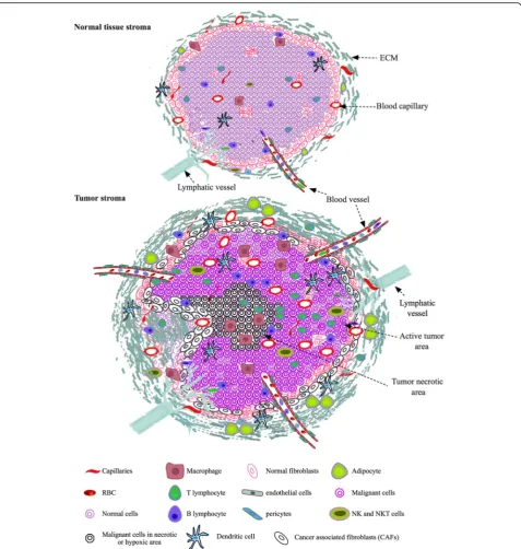
Most of the human connective tissues contain tissue specific cells, cells of vasculature, lymphatic and immune system along with other cells such as migrating stem cells, fibroblasts, pericytes, and tissue associated adipo-cytes (Fig.1). These cells are embedded within the inter-woven fibrillar structures of ECM lattices that are filled with interstitial amorphous ground substance and fluid. Thus, tissue cells live in spatial and interactive microenvironments.
Tissue specific stem cells
Tissue specific stem cells are specified somatic or adult stem cells or mesenchymal cells, which have potentials to differentiate into different types of cells in specific tissues or organs, for example myoepithelial stem cells for glan-dular epithelium [9] and hematopoietic stem cells for vari-ous blood cells [10]. Some tissues or organs have tissue specific stem cells, which are able to regenerate and repair damaged tissues [11]. Breast-specific spindle-shaped myoepithelial cells, which line outside luminal epithelial cells and away from mammary gland ducts, adhere to basement membrane (BM) via hemidesmosomes and to adjacent luminal epithelial and myoepithelial cells by des-mosomes [9, 12]. Cytokeratins (CK) such as CK5, CK14 and CK17 maintain the integrity of myoepithelial cells and support their attachment to BM and adjacent cells [13]. The cytoplasma of myoepithelial cells is filled with
different types of functional proteins such as actin, my-osin, fibronectin, collagen, nidogen, activin and laminin [14,15]. The membranes of myoepithelial cells possess re-ceptors, which include integrins, particularly β4 and α1, and E-cadherins that mediate cell-matrix and cell-cell in-teractions [16]. Furthermore, myoepithelial cells produce BM proteins such as laminin-1, laminin-5, collagen IV, fi-bronectin, and a number of tumor suppressor proteins in-cluding p63, p73, 14–3-3 sigma and maspin. Expression of morphogens and certain GFs in a coordinated manner during morphogenesis of myoepithelial cells helps main-tain the correct polarity of luminal epithelial cells. Myoe-pithelial cells may have hierarchical differentiation pattern among myoepithelial lineages with expression of different types and levels of certain proteins. Together with BM, myoepithelial cells act as a natural barrier with selective permeability to small molecules and tumor suppressors, physically preventing cancer cell invasion and functionally suppressing tumor growth by releasing proteinase and an-giogenic inhibitors [17, 18]. However, myoepithelial cells profoundly contribute to the maintenance of TME for tumor progression through their roles in paracrine signal-ing by expresssignal-ing extracellular proteins, various chemo-kines, angiogenic factors and GFs that remodel BM in favor of the colony expansion of cancer cells. Because of these functionalities, myoepithelial cells are also involved in regulation of the progression of ductal carcinoma in situ (DCIS) to invasive breast cancer [17, 19]. Further-more, myoepithelial cells can be triggered by tumor cells for the expression of invasion-associated molecules such as tenascin to promote tumor invasion and growth [20].
Migrating stem cells
Endothelial cells
Endothelial cells are highly specialized and their functions vary considerably from one type of tissue to another. For example, vascular endothelial cells in blood-brain barrier restrict the passage of most molecules into the brain,
whereas those in fenestrated capillary tuft of kidney glom-erulus filter molecules required by the tissue. Irrespective of certain specific functions, endothelial cells are involved hierarchically in forming blood vessels that transport oxy-gen, nutrients, and various factors throughout the body.
Stability and contractility of large blood vessels are provided by smooth muscle cells (SMCs) that wrap around the endothelial lining, whereas final capillaries are surrounded by pericytes, the perivascular cells that provide structural support to capillary endothelial cells in the microvasculature [28]. Angiogenic factors such as vas-cular endothelial GFs (VEGFs), fibroblast GFs (FGFs), platelet-derived GFs (PDGFs) and chemokines stimulate endothelial cells and pericytes to form new blood vessels and repair damaged vessels to maintain NME in tissues. Abnormal and excessive angiogenic signals either from in-flammatory or malignant cells to the quiescent endothelial cells lead to neovascularization that is needed for TME and tumor growth [29]. The tumor vasculature in TME is abnormal with rapid turnover in its structures and func-tions, including chaotic heterogeneous branching and un-even leaky vessel lumen that increase interstitial fluid pressure and facilitate tumor cell migration [30]. Lymph-atic endothelial cells can also form excessive vessel sprout-ing in TME lymphatic tissues under pathological conditions, such as lymphatic hyperplasia, through ex-pressing high levels of VEGFC or VEGFD and altering im-mune responses to affect cancer progression [31,32].
Fibroblasts
Fibroblasts are the most common but least specialized connective tissue cells that exist in connective tissues throughout the body. They are morphologically hetero-geneous with versatile appearance depending on tissues, organs, and the activities of the cells [33, 34]. In NME, the main function of fibroblasts is to maintain architec-tural integrity of connective tissues by depositing ECM proteins like collagens, glycoproteins, proteoglycans, and laminin. During tissue repairing process in NME, fibro-blasts within the tissue get activated, proliferate, migrate towards the injured site, and produce ECM to heal the injury [35]. After the tissue damage is repair, the activ-ities of the fibroblasts decrease and the cells remain minimally active with normal phenotypes [36].
Fibroblasts in TME, generally known as tumor associ-ated fibroblasts (TAFs), are activassoci-ated fibroblasts that undergo various biological and morphological transition in response to tumor progression. They are one of the major components of tumor stroma with pleiotropic ac-tions on tumors and play important roles in maintaining an optimal TME for cancer cell survival and prolifera-tion [37, 38]. They get activated perpetually without reverting to their normal activity and phenotype, and can withstand severe stress without undergoing apop-tosis that is usually lethal to most of other cells [39]. Tumor is always associated with TAFs that produce dif-ferent biomolecules, support cancer cell transformation, induce local inflammation and angiogenesis to promote tumor growth and metastasis [39,40].
Adipocytes
Adipocytes are stromal cells that normally present in fat-associated connective tissue stroma. Apart from an en-ergy storage, they produce hormones, GFs, chemokines and other cytokines in NME [41]. In tumors, adipocytes form tumor stroma along with cancer cells, fibroblasts and other stromal cells in TME [42]. It was shown that early stage cancer cell growth and invasion occur in close proximity to adipocytes [43]. Adipocytes promote cancer invasion by releasing excessive amount of adipokines, cy-tokines, collagen IV and inducing production of matrix metalloproteinases (MMPs) in TME for cancer cell migra-tion [41,43,44]. In addition, adipocytes provide fatty acids as fuel for the metabolic needs of cancer cells [45].
Immune cells
Immune cells include both granulocytes (neutrophils, eo-sinophils and basophils) and agranulocytes (lymphocytes and macrophages). Neutrophils are granulocytes that account for 50–70% of all leukocytes and responsible for counteracting acute infection. Their maturation depends on various stimulating factors including the colony stimulating factor (G-CSF) and the granulocyte-macrophage-colony stimulating factor (GM-CSF). The release of neutrophils to blood stream from bone marrow depends upon various triggering factors such as IL-23, IL-17, G-CSF and other chemokines [46]. Neutrophils are attracted by various ligands including CXCL1, CXCL2, CXCL5 to tumor site and play central roles as tumor-asso-ciated neutrophil (TAN) in tumor inflammation and devel-opment from initiation to metastasis [47,48].
of regulatory T cells in TME was associated with bad prognosis in some cancers [53, 54], it was suggested to be one of the hallmarks of TME and cancer develop-ment [55]. Both natural killer (NK) and natural killer T (NKT) cells are also lymphocytes that are able to kill transformed cancer cells or viruses. When stimulated, NK and NKT release cytokines such as IL-2, IL-12 and interferon α and β (IFN-α/β) that induce inflammatory responses in tissues and increase cytotoxicities to the assaulting cells. These cytokines are responsible for pro-viding innate immunity and play crucial roles in the con-trol of tumor growth [56]. Macrophages are preeminent mononuclear phagocytes in immune system, killing in-vading pathogens as the first line of defense next to neu-trophils. Apart from serving in defensive system, macrophages are involve in tissue repair, tissue develop-ment, and NME homeostasis [57]. In addition, macro-phages also participate in vasculogenesis, angiogenesis, and maintenance of mammary stem cells [58,59]. Their survival and proliferation are regulated by colony stimu-lating factor 1 (CSF1) [60]. Macrophages can be re-cruited by CSF1 and other tumor chemoattractants such as CCL2, VEGFA, and semaphoring 3A (SEMA3A) in developing TME [61]. Tumor-associated macrophages (TAMs) often interact with adipocytes in tissue stroma for cancer development and progression. TAMs phago-cytose adjacent dead adipocytes and establish inflamma-tory foci known as crown-like structures (CLS) [62,63]. Highly diverse population of macrophages or monocytes, generally known as myeloid cells, are terminally differen-tiated macrophages or dendritic cells (DC). Since these myeloid cells are responsible for suppression of various types of immune response, they are defined as myeloid-derived suppressor cells (MDSCs) [64]. MDSCs usually target T cells through suppressor factors like arginase (ARG1), TGF-β, IL-10, inducible nitric oxide synthase (iNOS), and cyclooxygenase-2 (COX2) [64].
In vitro native microenvironment based on cells
Normal tissue stroma contains different types of cells such as residential specific cells, migrating cells, fibro-blasts, immune cells, adipocytes and endothelial cells as described above (Fig. 1). Their spatial arrangements and communications are tissue-specific. NME transforms to TME in the presence of tumor cells and their associated stromal cells (Fig.1). Mimicking native NME or TME in vitro based on the cell types within specific tissue matri-ces is critical for biologically and clinically relevant can-cer studies and tissue engineering. Hence, it is fundamental to establish in vitro co-culture systems that are able to provide microenvironments highly resem-bling native tissue ECM at structural, mechanical and biochemical levels for different types of cells. Overall, co-culture using two different cell types becomes more
common nowadays than before and contributes to our understanding about intricate cell-cell interactions and signaling mechanisms. For instance, co-culture of pri-mary human mampri-mary fibroblast and breast cancer MCF-7 cells revealed intercellular communications that were only possible to be observed in the presence of the secreted biomolecules such as IL-6, prostaglandin E2 (PGE2), and IL-6sR from the cancer cells [65]. Addition-ally, more complex co-cultures with multiple cell types have been reported with encouraging outcomes. For ex-ample, endothelial cells, fibroblasts and bone marrow stromal cells were used for new bone formation [66], and hepatocytes, fibroblasts and endothelial cells were applied in the study of liver tissue generation containing neocapillaries [67]. However, co-cultures with mixed cell populations in tissue-mimicking environments are bound to face certain technical challenges, such as the requirement of different or specific GFs and additional supplements for different types of cells in a same co-culture system, control of cell orientation and distri-bution, tools used to assess complex cell-cell interac-tions, and the coordination of the different parts of the assembled network for overall functions of the in vitro culture. Cultural conditions need to be formulated in a way to promote optimal survival and growth of all types of cells involved in forging the in vitro stroma of the co-culture, with a control over non-desired stimulation or inhibition of the different types cells in the system.
ECM for native microenvironment
during mammary gland development [75], and TAMs can regulate fibroblasts to produce enzymes and inhibi-tors that mediate ECM degradation in tumors [76]. Furthermore, ECM cross-linkers such as transglutami-nase and lysyl oxidase (LOX) present within TME par-ticipate in tumor ECM modifications by cross-linking the newly generated collagen fibers and other ECM pro-teins. The local tension change in a tumor and its sur-rounding tissues is a key mechanical factor regulating spatial cell migration and cancer progression [77, 78].
The presence of GFs, chemokines, angiogenic molecules, and MMPs in ECM provide additional stimuli to the intracellular and intercellular signaling networks that mediate the process of cancer development. Other pro-teases such as cathepsins and cysteine propro-teases are also highly expressed in TME. They suppress blood clotting mechanism by activating heparanase, thereby aiding angiogenesis and metastasis.
Recent advances in in vivo-mimicking tissue culture systems demonstrated the advantages of applying native
ECM in biomedical research and bioengineering [7, 69, 79–82]. Yet, synthetic materials remain to be a robust source of ECM-mimicry in the field, depending on spe-cific applications of the materials. Overall, both synthetic and native materials should provide not only structural, mechanical, and biochemical supports to cells cultured within the scaffolding materials but also optimally sup-port cell-cell and cell-matrix interactions through native-mimicking signaling events. Hence, ECM is one of the most important components of microenvironment under both in vitro and in vivo conditions for the homeostasis, growth, and repair of tissues [83].
Synthetic ECM
The materials that are bioinert, biodegradable, biocompat-ible, and hydrophilic in nature are used for preparation of synthetic hydrogels. Hydrogels can be further converted to elastic topographical materials, generally known as syn-thetic ECM, after cross-linking of their polymers. Biophys-ical and biochemBiophys-ical cues of hydrogels can be spatially and temporally tuned to mimic native ECM for cellular activities such as adhesion, proliferation, and migration [84]. For example, hydrogels from poly(ethylene glycol) (PEG), poly(2-hydroxyethyl methacrylate) (pHEMA), Poly(ethylene glycol) diacrylate (PEG diacrylate), and poly(vinyl alcohol) (PVA or PVOH) can be prepared to structurally and mechanically mimic the physical aspects of tissue ECM. These synthetic polymeric hydrogels are often used as matrices in bioinks for tissue-mimicking printing. For example, PEG diacrylate and alginate blend serves as robust bioink for fabrication of tissue constructs as demonstrated by Rutz et al. [85] and Hong et al. [86].
PEG is able to polymerize under cytocompatible condi-tions through numerous reaccondi-tions, such as Michael addition, chain polymerization, azide alkyne cycloaddition and thiol-ene. Besides, it can be functionalized by modifying its terminal hydroxyl residue with certain reactive groups, such as alkenes, alkyenes, thiols, N-hydroxysuccinimide (NHS) esters, maleimides and azides [87,88]. Cell adhesion property can be enhanced by introduction of Arg-Gly-Asp (RGD) peptide sequences in PEG with NHS ester on one end and acrylate functional group on the other side [89, 90]. In addition to RGDs, other peptides and different ECM components can be in-corporated into PEG and other biomaterials to maintain self-renewal and differentiation of stem cells. For instance, incorporation of vitronectin-derived heparin-binding pep-tide I (GKKQRFRHRNRKG) into polyacrylamide hydrogel facilitates the interaction of ECM with cell surface glycans, and addition of glycosaminoglycan-binding peptides sup-ports self-renewal of stem cells [91,92]. Likewise, hydro-gels can be fabricated for a specific ECM function. For example, heparin-decorated Hyaluronic acid (HA) hydro-gel was used to release bone morphogenetic protein-2
(BMP-2) for chondrogenic differentiation of murine mes-enchymal stem cells [93]; addition of phosphate functional group into mineralized matrices or PEG hydrogel induced osteogenic differentiation of human mesenchymal stem cells (hMSCs) [94,95] while inclusion of t-butyl moiety in the PEG gel promoted adipogenic differentiation [95]; mineralization of PEG hydrogels modified with varying lengths of anionic pendant side chains terminating with carboxyl groups was used in bone-mimetic composite ma-terial fabrication [96].
In addition to chemical modifications, elasticity and stiffness of hydrogels are key factors that play vital roles in cell differentiation. HMSCs grown on hydrogels at low elasticity (elastic modulus 0.1–1 kPa) undergo neurogenesis, while on stiffer hydrogels (elastic modulus 8–17 kPa) are routed to myogenesis and tougher hydro-gels (elastic modulus 25–40 kPa) to osteogenesis [97]. Interestingly, hMSCs osteogenic differentiation can be triggered by hydrogels with early stiffening and adipo-genic differentiation with late stiffening [97].
Moreover, hMSCs grown on hydrogel with high trac-tion stresses differentiate into osteogenic lineage and undergo adipogenic differentiation on low traction gel [98]. Furthermore, hydrogel hydrophobicity influences cellular organization such as rosette-like clusters that help maintain the cellular morphology and promote differentiation of hMSCs [99]. Certain degree of hydro-phobicity of hydrogel is required to maintain its adhesiveness that is necessary for cell attachment, prolif-eration and migration [99, 100]. A smart surface of hydrogel is therefore critical to allow cell attachment and growth. Hydrogels like poly(N-isopropylacrylamide) (pNIPAm), exhibiting lower critical solution tempera-tures (LCST), are generally used for the preparation of smart surfaces [101]. PNIPAm is hydrophilic at temperature below 37 °C, but becomes hydrophobic at 37 °C that enables nonspecific protein interaction for cell attachment. Some synthetic biomaterials like polycarpo-lactone (PCL) is entirely hydrophobic in nature and diffi-cult to grow cells for tissue engineering and biomedical studies, while some hydrophilic biomaterials such as silk fibroin, aloe vera, and curcumin can be added to PCL to make smart surfaces for cell attachment [102]. PCL can also be coated with cell-laden ECM, alginate or decellu-larized ECM, making it more hydrophilic and native-mimicking for cells [103,104]. Furthermore, PCL is one of the most widely used synthetic ECM in various formats of scaffolds for cancer studies because of its slow degradation kinetics and biocompatibility, support-ing TME that contain cancer cells [105–107].
acid in the presence of water [107,108]. Both byproducts enter into various metabolic pathways in the body under normal physiological conditions. Some other polymers like poly(ethylene) (PE), poly(propylene) (PP), poly(vinyl chlor-ide) (PVC), poly(dimethyl silane) (PDMS), poly(methacry-late) (PMMA), pHEMA, poly(ethylene terephthapoly(methacry-late) (PET, dacron), poly-L-lactic acide (PLLA), poly-D- Lactic acide (PDLA), polydioxanome (PDO), polyether ether ke-tone (PEEK), polyether sulfonate (PES), polyamide nylon, poly(vinlypyrrolidone) (PVP), poly(styrene-b-isobutyle-ne-b-styrene) (SIBS), ultrahigh molecular weight PE (UHMWPE), and polyurethane can be use in tissue engin-eering, suturing and other material engineering applica-tions [109]. Apparently, the versatile synthetic polymers are rich sources for tissue engineering in addition to their applications in mimicking physiological tissue environ-ments such as TME [110].
Native ECM
Chitosan is derived from partial deacetylation of chitin, which contains at least 60% of D-glucosamine residues [111]. Commercial chitosan is usually extracted from the chitin of crustaceans and fungal mycelia. The presence of protonable amino groups in chitosan is a peculiar property of the material. For example, a negatively charged sialic acid provides the mucoadhesion property of chitosan [112] and a positively charged amino acid in chitosan backbone endows its hemostatic activity and interaction with the negatively charged cell membrane, thereby helping reorganization of tight junction and membrane proteins and enhancing chitosan permeability [113]. The antimicrobial, polycationic and biodegradable natures of chitosan support its biomedical applications in different formats like hydrogels, films, sponges and 3D scaffolds [114–117]. It was also shown that chitosan promoted cancer progression in TME by binding to CSCs via CD44 receptors and activating both canonical and non-canonical signaling pathways [118].
Silk fibroin, a macromolecular protein polymer se-creted by silkworm (Bombyx mori) larvae, is biocompat-ible and has been applied for tissue engineering of bone, cartilage, ligament and tendon, skin tissue, blood vessel, liver, spinal cord, trachea, bladder, and ocular tissues. Silk fibroin with or without inducing factors like hy-droxyapatite and bone morphogenic proteins (BMPs) is extensively used for in vivo neobone formation together with different kinds of stem cells such as human osteo-blasts, mesenchymal stem cells (MSCs), and MC3T3-E1 cells (osteoblast-like cell line) [119]. The protein polymer can be blended with chitosan for bone, cartilage, liga-ment and tendon tissue engineering. Skin tissues have been regenerated in rats after implantation of silk fibroin alone or blended with alginate, chitin, or collagen in the presence of human oral or epithelial keratinocytes and
fibroblasts [120–123]. Accumulating data have shown that silk fibroin supports cultures of almost all types of stem cells that are specific for particular tissue regener-ation in NME [124–126]. Enzymatically cross-linked silk fibroin can be used as bioink in tissue engineering such as musculoskeletal tissue regeneration for personalized implant and for versatile organ printing with structural stability and reliable biocompatibility that can be achieved by blending in methacrylate and fabricating with the digital light processing (DLP) bioprinting method [127,128].
Furthermore, silk fibroin has been used to reconstitute TME and culture breast cancer cells and fibroblasts in vitro, where fibroblast-cancer cell as well as cell-ECM interactions that are important parameters of cancer progression were observed [129].
Starch has been used in tissue, particularly bone, en-gineering as scaffold, bone cement, or blended form to facilitate collagen deposition for neobone formation [130, 131]. The topography of starch-based scaffold can be modified by coating with adhesive proteins such as plasma proteins to enhance better attachment of cells to the scaffold surface and facilitate the growth of endothe-lial cells [132]. In combination with PCL, starch was also used in 3D rapid prototyping of layered hierarchical structures for hard tissue engineering [133]. Nanofibrous mats prepared by electrospinning of starch and polyvinyl alcohol (PVOH) support skin regeneration [134]. Starch so far has not been reported for the use of maintaining TME in tumor modeling.
entrapped GFs to support cell proliferation, neovascular formation, and delivery of entrapped cells to target sites. Alginate bioinks are capable of modulating release and spread of cells without affecting the integrity of alginate lattice structures for tissue regeneration in NME [143, 144]. Blending other biodegradable polymers such as PLLA, chitosan, gelatin, and PLGA into alginate has been used to transport molecules through epithelia and mucosa in various forms of microspheres or nanoparti-cles. With various modifications, alginate has also been applied in wound healing, cartilage repair, and bone re-generation. Alginate-based hydrogel, on the other hand, was used to form tumor spheroids in microfluidic cul-ture systems in an attempt to mimic solid tumors [145]; to blend with other biocompatible biomaterials such as gelatin to make composite hydrogel and with tumor cells and TAFs for in vitro tumor models to study cell-cell in-teractions and mechanisms of tumorigenesis [146]; to mimic TME in 3D cultures for angiogenesis with the en-gagement of cancer cells, VEGF, and integrin [147].
Cellulose is a biopolymer polysaccharide isolated from bacteria or plants. Though being a natural extracellular matrix, cellulose is a very slowly degradable biomaterial when implanted into human tissues owing to the lack of required enzyme to digest it. Breakdown of cellulose is only possible through hydrolysis of glycosidic bonds and oxidation of the glucopyranose rings. Oxidized cellulose has been used as anti-adhesive tapes for wound healing. Oxidation of cellulose induces conversion of the glucose residues to glucuronic acid (with–COOH groups), which acts as the binding site for various molecules such as arginine, chitosan, and GFs, a mechanism that is useful for tissue engineering [148]. The antimicrobial properties of oxidized cellulose can be used for tissue repair. Six-carboxycellulose with low percentage of –COOH groups supports differentiation and maturation of stem cells rather than their proliferation [148]. A range of nano-materials such as calcium carbonate, titanium oxide, and silicon carbide have been implemented to mix with cellu-lose to improve the resistance and stability of cellucellu-lose [148]. Cellulose has been minimally modified for the pur-pose of cell attachment in the absence of ligands or pro-teins by means of introduction of positive charge with 2,2,6,6-tetramethylpipiridine 1-oxyl or negative charge with sodium bromide, without altering its mechanical properties [149]. Cartilage repair, bone regeneration, and functional cardiac constructs have been tested using cellu-lose as 3D scaffolding hydrogels [150–152]. At last but not at least, cellulose scaffold was also applied in studies of human cancers, such as breast cancer, and found to be supportive for tumor formation [153].
Gelatin is a natural protein derived from partial hy-drolysis of collagen. Though gelatin is mechanically weak, it is applied in tissue engineering after being
stabilized and stiffened by various cross-linking methods, most commonly by photo cross-linking [154]. Photo cross-linkable gelatin with incorporation of furfuryl iso-cyanate (gelatin-FI) or furfurylamine (gelatin-FA) has been used as a dental pulp capping material and is a promising scaffold for osteochondral repair [155, 156]. Addition of BMP into gelatin-FA hydrogel further en-hances articular cartilage and subchondral bone repair with more expression of osteochondrogenic factors like col1a1, col2a1, SOX9, and aggrecan [156]. Gelatin blended with other biomaterials such as calcium phos-phate ceramics, alginate and chitosan has improved mechanical properties in scaffolding. For engineering of cardiac and nerve tissues, gelatin was developed into functional scaffolds with the addition of conductive mol-ecules like polyaniline and carbon-based synthetic poly-mers [157]. Hybrid hydrogel made up of gelatin and chitosan was generated to support the formation of in vitro tumoroid, which expressed genes such as p21, in-tegrin αV, N-cadherin, vimentin, CK-18 and β-catenin that are involved in tumor growth in TME similar to those in native tumors [158].
Hyaluronic acid (HA) is a glycosaminoglycan that is commonly found in native ECM of cartilages and con-nective tissues. It is often used as scaffold for tissue re-generation, wound healing and drug delivery or as bioink for 3D tissue constructs [159, 160]. Its use has been expanded to blending with other biomaterials for the regeneration of cardiovascular tissue, brain, cornea, lung and skin [161]. Because of the low mechanical and slow gelation properties of HA, addition of cell-adhesive oligopeptides in HA hydrogel increases both mechanical and cell-adhesion properties of the gel [162]. HA bioink blended with gelatin was shown to maintain prolifera-tion of human adipose stem cells and their differenti-ation into adipocytes in adipogenic culture medium [163]. Chemically modified HA, for example with methacrylate, has enhanced mechanical properties and can be used for various tissue engineering purposes [164, 165]. HA-based hydrogel also has long been used as ECM-mimicking cultural matrix in human cancer studies from basic 3D culture and induction of angio-genesis to tumor modeling and identification of cancer cell-secreted metabolites [166–168].
tissue engineering are skin or tendon from bovine or porcine and rat tail among others like fish, sponges, and jellyfish. Collagens are biodegradable by naturally exist-ing enzymes, collagenases. This degradation mechanism is very useful in tissue engineering per se, and the bypro-ducts of the degraded collagens and their peptide deriva-tives can further enhance tissue restoration by attracting fibroblasts, which are collagen-producing cells distrib-uted throughout the body [170]. Cells adhere to colla-gens directly through receptors or indirectly through linkers such as fibronectin. Cell receptors, which have the ability to recognize specific peptide sequences within collagen fibers, are divided into four groups. The first group, glycoprotein VI for example, binds to collagen peptide sequence with GPO motif (Gly-Pro-Hyp); the second group contains integrin family and discoidin do-main receptor 1 and 2 (DDR1 and DDR2); the third group is integrin-types capable of recognizing cryptic motifs; and the fourth group of other cell receptors dir-ectly binds non-collagenous domain of collagen [171]. Collagen blended with other biomaterials such as glycos-aminoglycans (GAG), chitosan, or elastin, has been fab-ricated to enhance the mechanical properties of the scaffolding materials and increase their enzymatic resist-ance for improved tissue engineering [171, 172]. Collagen-based hydrogels and porous scaffolds in vari-ous formats, with or without cells and co-factors, have been applied for decades in studies of bone and cartilage repair, skin regeneration, cardiac tissue development, urinary bladder and ureter regeneration, wound dress-ing, and many other medical directions [173–177]. In addition to its tremendous roles in tissue engineering, collagen is broadly used in biomedical research espe-cially in tumor microenvironment modeling that pro-vides favorable stromal TME for cancer and stromal cells [178]. The orientation of collagen fibers not only directs tumor cell intravasation, but also participates the process of cancer metastasis [179]. Increased collagen I deposition in TME makes tumor ECM stiff, stimulating tumor growth by modulating a set of signaling events, such as shifting the balance from prolactin signaling (JAK2/STAT5) toward tumor progression signaling (ERK) [180]. Collagens such as VI and XIα1 were found to be involved in epithelial-mesenchymal transition, angiogenesis, and metastasis [181,182].
Native tissues have been used to produce decellularized (or acellularized) ECM (dECM) matrices, which are able to provide structurally and mechanically supporting microenvironments almost identical to those within native tissues. Therefore, tissue-specific and biocompatible dECMs are very promising for tissue engineering and re-generative medicine, and can be applied without the limi-tations from shortages of donor organs or tissues. Additionally, the natural biochemical composition of a
dECM is an advantageous property. With certain supple-ments such as GFs, dECM promotes spatial cell growth, tissue repair and modeling [183, 184]. Because of the structural, mechanical and compositional advantages, tissue-specific dECMs have been extracted from a variety of native tissues such as skeletal muscle, skin, urinary blad-der, small intestine, brain, heart, blood vessels and other types of tissues to generate raw ECM, reconstituted porous scaffolds, and hydrogels for a broad range of applications in tissue engineering and biomedical research. For in-stance, reseeding of tissue-specific stem cells in dECM scaffolds has the potential to rejuvenate the scaffolds as functional tissue grafts [185]. In addition to the major types of collagens, dECM from animals or human has complex mixtures of other ECM proteins such as glyco-proteins, proteoglycans, and certain minor ECM proteins [14,186,187]. Meanwhile, each type of tissue generally has a specific ECM composition and biochemical cues that affect cells at different levels, from attachment and growth to migration and death. Usually, a specific type of dECM is selected for the growth of a tissue-specific stem cell lineage. For example, adipose dECM is for growing adi-pose derived stem cells (ASCs) and/or MSCs and liver dECM for hepatocytes [188,189]. Yet, nonspecific dECM can also be used for nonselective applications, for instance culturing ASCs in placental dECM for adipose tissue en-gineering [190]. Soft composite hydrogels consisting of dECM and other biomaterials like fibrin and chitosan have been used as bioinks, which contain desired stem cells and inducing factors, in soft tissue engineering [191, 192]. Moreover, composite scaffolds fabricated using dECM and synthetic polymers such as PCL or PLGA have hydrophilic and necessary mechanical properties for tissue engineer-ing, especially hard tissue regeneration [193,194]. The use of dECM in tumor modeling has substantially enhanced the efficacies of in vitro mimicry of TME and increased the capabilities of revealing the mechanisms of tumor for-mation and metastasis [14, 195]. Identification of cancer-driving genes has been greatly facilitated by using recellularization of dECM with cancer cells. For example, decellularized human colon matrix was used as scaffold to co-culture retrovirus-transfected primer human colonic epithelial cells (hCECs) with human fibroblasts, and re-vealed the cancer driving genes LATS2, ASXL2, CAMTA1, DDX20, FXR1, MITF, and PAX7 [196]. The in-vasiveness of human cancer cells was also studied using dECM that is otherwise difficult to be closely mimicked in other types of 3D cultures, which hardly recapitulate native-like microenvironments [14,197].
In vitro native microenvironment based on ECM
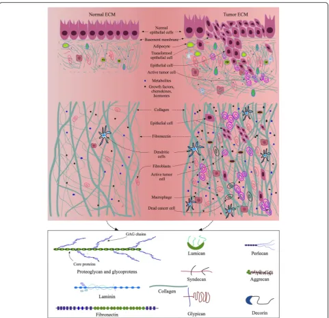
repair [198]. The structure, mechanics, component, organization, orientation, and function of ECM as dis-cussed above not only reflect the peculiar physical char-acteristics of each ECM type, but define specific functional properties required by the cells in their NME or TME (Fig.2). Therefore, to some extent, an ECM mi-croenvironmental niche forges the biological perform-ance of the cells and guides their fate, the differentiation and self-renewal of stem cells for example. Stem cells, tissue specific cells and migratory cells, on the other hand, modify local ECM microenvironment to make it serve the best for cellular functionalities. Bidirectional communications between tissue-specific cells and ECM are thus vital for normal functions of tissues. Currently, most 3D cultures focus on optimizing mechanical and structural properties of scaffolding matrices for cells, with minimal integration of physiological and biochem-ical cues into the culture systems. For instance, collagen type I is a major component of most native tissue ECM, but collagen I alone is an incomplete matrix source for induction of complete cellular functionalities and pheno-types. In addition, the extraction, physiological parame-ters, and reconstitution conditions of collagen are sensitive aspects that may hamper the overall perfor-mances of collagen-derived matrices in 3D tissue cul-tures [199]. Even though hybrid or composite biomaterials have some additional strengths compared to an individual biomaterial in terms of structural and mechanical properties, biodegradability, stability, release of trapped GFs or other factors, they are still far from mimicking NME or TME. Under the current 3D culture status, dECM remains a competitive biomaterial that is able to provide native tissue-like microenvironment and overcome the shortcomings of synthetic polymers, single native ECM proteins, and composite or hybrid hydro-gels. The use of dECM, therefore has been exponentially increased in tissue engineering, regenerative medicine, and cancer studies in recent years [107]. However, the protocols preparing dECM and its derivatives mostly, if not all, involve a variety of detergents, enzymes, acids, or bases that may potentially alter ECM protein structures, configurations, chemical or physical properties, which need to be addressed further. Therefore, challenges re-main to mimic NME or TME at high fidelity using dECM, unless there come revolutionary methods of decellularization and ECM protein extraction from na-tive tissues that can retain all the ECM proteins in their native conformational and functional states.
In vitro microenvironment based on spheroid or organoid model
In addition to the ECM scaffolding methods described above, cell spheroid is a widely used model for 3D tissue culture, drug screening, and personalized medicine
testing [200]. Spheroids are clusters of cells, which ad-here to each other via desmosomes, adad-herens or tight junctions [201–203]. Molecular gradients, cell-cell and cell-ECM interactions are able to be established within spheroids in a way to deliver various signals and mech-anical forces to the cells, influencing the viability, prolif-eration and differentiation of the cells [200,204,205]. A variety of cells including normal pluripotent, mesenchy-mal stem cells, endothelial cells, and cancer cells have been used to form multicellular spheroids (MCSs) for various biomedical studies and tissue engineering appli-cations [206–209]. MCSs may form through loose aggre-gation of cells, direct cell-cell contact, cadherin accumulation at cell membrane and cadherin-cadherin binding [210–212]. During spheroid formation process in tissue culture, cells can secrete ECM proteins as ini-tial scaffolding bed that also serves as part of their living microenvironment [213]. Although questions remain as for whether the cell-cell interactions and signaling mecha-nisms in MCSs are comparable to those in native tissues and whether the cell-generated ECM is sufficient to mimic the heterogeneous native ECM, MCSs are still impressive 3D microenvironment-providing tools to mimic in vivo pathophysiological conditions. Additionally, collagen- and alginate-based multicellular tumor spheroids (MCTS) have been generated and used in evaluating gene expres-sion profiles, signaling pathways, tumor modeling, and drug delivery efficiencies [145,214–216].
component and cell co-culture levels and application of smart scaffolding biomaterials, both cell spheroid and organoid models are rapidly advancing toward more close mimicry of native microenvironments.
Biological factors of native microenvironment
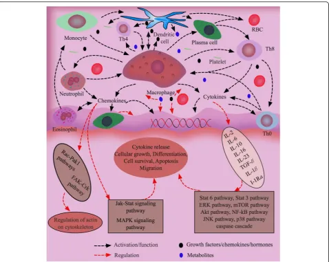
Chemokines, the largest family of cytokines, are highly po-tent factors in immunophysiological regulation of termin-ally differentiated and pluripotent stem cells for their chemotaxis activities (Fig. 3). They are typically divided into endogenous and exogenous soluble small proteins (8–14 kDa), and defined by the presence of four conserved cysteine residues. Generally, G protein-coupled receptors of tissue cells get activated by chemokines, inducing the cells to migrate through concentration gradient in a par-ticular tissue where cells get accumulated for defense mechanisms [227]. There are approximately 50 endogen-ous chemokine ligands in mice and humans that are im-portant to cellular and humoral immune responses and maintenance of tissue homeostasis. For example, CXCL1, CXCL2, CXCL3, CXCL5, CXCL6, CXCL7 and CXCL8 are involved in neutrophil trafficking, CXCL4 in coagulation, and CXCL9, CXCL10 and CXCL11 in the trafficking of natural killer cells, killer cells and helper cells by interact-ing with specific receptors like CCR or CXCR present on the plasma membrane of the cells [228]. Chemokines con-trol not only the residence of immune cells in primary lymphoid organs, but their localization in secondary, ter-tiary lymphoid and periphery organs [229]. Neutrophils, Eosinophils, basophils, mast cells, monocytes, dendritic cells, lymphocytes, regulatory T cells, innate lymphocytes and resident immune cells are all directed and activated by chemokines at different levels for proper responses to antigens depending on defensing mechanisms. Moreover, cytokines are used to differentiate macrophages to den-dritic cells, which express significantly more CD56, CD80, CD86, MHC class I and IL-10 compared to Monocyte-de-rived dendritic cells [230]. CXCL12 produced from bone marrow stromal cells in NME attract lymphocytes, mono-cytes and CD34+hematopoietic precursor cells expressing chemokine receptor CXCR4 [231]. Interestingly, CXCL12 is also expressed by cancer cells in TME and, in coordin-ation with CXCR4, regulates the migrcoordin-ation of the cancer cells for metastasis [232].
T cells grown on 3D scaffold express various chemo-kine receptors such as CXCR1 to CXCR5 and CCR1 to CCR3, CCR5 and CCR6. Ivanoff and colleagues showed that chemokines interacted with 3D collagen I hydrogel substrata, and T cells exhibited migratory response to chemokine stimulation on the gel [233]. However, the chemokines failed to support infiltration of the cells into the collagen gel [233]. These observations were in con-trast with the results that chemokine RANTES (CCL5), a ligand for CCR5, enhanced the generation of T cell
focal adhesions and activated the cells through the FAK, ZAP-70 and paxillin protein complexes, and with that chemokines CCL2, CCL3, and CCL5 stimulated mono-cytes to express MMP-9 on 2D substrata [234, 235]. Since accumulating evidence support the concept that different levels or species of chemokines are expressed in 2D vs. 3D cultures [236], in depth studies on chemo-kine expression by immune cells grown on 3D synthetic or native tissue matrices will provide novel insights in whether different 3D culture models or materials induce similar chemokine production or chemokine receptor expression in the immune cells grown in the cultures.
the stimuli from surrounding environments and ECM perturbations so as to maintain tissue homeostasis [249]. Hormones are important biological factors (BFs) in the body for tissue maintenance and physiological functions through mediating the dynamic balance between cell proliferation and cell death. For example, androgens, prolactin, glucocorticoids, and estrogen stimulate epithe-lial cell proliferations and tissue growth of prostate gland, mammary gland, ovary and uterus, respectively, whereas corticosteroids and glucocorticoids promote apoptosis of thymus gland and bone, respectively [250]. Moreover, effects of hormones on cells depend on the concentrations of hormones since the responses of nor-mal tissues are noticed only when hormones are in
physiological range of concentrations. Dysregulation of concentration or presence of hormones in local tissues or neighboring tissues through autocrine or paracrine mechanisms may lead to abnormal functions and pheno-types of tissue cells, which can be triggered for genetic alternations and transformations into cancerous cells [251,252].
In vitro native-mimicking microenvironment based on biological factors
BFs like chemokines, GFs, and hormones are imperative for the co-ordination of tissue cells within their living microenvironmental networks for their survival, growth, differentiation, and proliferation, (Fig. 3). Almost all
native cells such as MSCs, fibroblast, epithelial cells, endothelial cells, myeloblasts, erythroblasts, megakaryo-blasts, leukocytes and macrophages secret and release BFs into tissue microenvironments as part of the bio-logical functions of the cells and as a mechanism to maintain tissue homeostasis [253]. Secretion of BFs from cells can be detected in in vitro culture systems by means of proliferation, cytotoxicity, chemotaxis, protein induction, and other types of assays [254,255]. The pro-files of BFs in different cultures may substantially vary depending on cell types, culture time, concentration and types of stimulants [256]. Additionally, intercellular communications effect intracellular signaling and release of BFs. Therefore, co-culturing the same or different types of cells in the presence or absence of genetic mod-ifications of the cells for specific biologically phenotypes is sometimes preferred in many studies. Co-culture sys-tems exist in two major types, direct and indirect, where two or more than two different types of cells are allowed to grow under their optimal cultural conditions. BFs re-lease or suppression in direct and indirect co-cultures is different. For example, direct co-culture of ASCs and peripheral blood mononuclear cells (PBMCs) increased the release of IL-6, CXCL9, CXCL10, CCL2 and galectin-1 and decreased the secretion of IFN-γ, TNF-α and galectin-3 from the cells. In contrast, an opposite se-cretion pattern was observed in indirect co-cultures of ASCs and PBMCs [257]. Therefore, cautions need to be taken when planning co-culture experiments (direct vs. indirect) to identify cell-secreted biomolecules in culture environments no matter whether the culture systems are 2D or 3D and made of synthetic or natural materials. Although it is technically challenging to accommodate all the optimal conditions in co-culture systems, sub-stantial progress has been made on mimicking native physiological conditions by introducing different ECM ingredients, cytokines, GFs, and hormones into spatial 3D culture systems that lend promise to establish ad-vanced co-culture systems closely mimicking NME or TME and to study pathological changes within ECM highly resembling human disease conditions.
Physiological conditions of native microenvironment Physiological parameters like temperature, pH, oxygen (O2) and carbon dioxide (CO2) concentration, ions, en-ergy supply, and waste removal play significant roles in tissue homeostasis, growth, and death. Detailed descrip-tion of each of these parameters is beyond the scope of this review, and only their important properties relevant to this topic are discussed. Cells are highly sensitive to temperature, mostly become inactive in temperatures below 4 °C. Depending on the type and the nature of cells, some cells remain active in body temperatures below the regular 37 °C or above 42 °C of very high
fever. Temperature controls cell functions through alter-ations in the types and amounts of intracellular chemi-cals [258]. Satellite cells from different origins have different sensitivities to temperature in terms of prolifer-ation and differentiprolifer-ation. For example, pectoralis major muscle satellite cells were highly proliferative in vitro when temperature changed from 38 °C to 43 °C, whereas biceps femoris muscle cells displayed a different ative manner during the temperature shift and prolifer-ated at a higher rate at 33 °C–39 °C and a lower rate at 43 °C than the pectoralis major muscle satellite cells [259]. Similar to temperature, pH plays a fundamental role in cellular functions by regulating cell cycle and proliferation, and acts as a checkpoint control in various signaling pathways under normal and cancerous condi-tions [260,261]. It has been shown that extracellular pH of cancer cells is slightly acidic (pH 6.2–6.9) than that of normal cells (pH 7.2–7.5). Acidic environment not only promotes cancer cell transcription of tumor-promoting factors such as VEGF, IL-8 and hypoxia-inducible factor (HIF-1) [262–264], but increases the expression of pro-teases like MMPs and cathepsins that facilitate migration of the cancer cells [265].
melanoma has pO2 around 2 mmHg (< 0.3% O2) along with heterogeneous distribution of anoxic or hypoxic tis-sue areas [266]. Thus, optimizing oxygen concentrations during cell cultures are important for optimal perform-ance of the cells and for preventing the cells from oxida-tive stress. Because of overall low oxygen tensions in human tissues, engineering approaches for tissue repair and regeneration have not been very successful as ex-pected [271]. To address this challenge, various func-tional biomaterials, such as oxygen delivery biomaterials, oxygen generating biomaterials, and oxygen releasing biomaterials that provide oxygen and prevent cells from ischemic necrosis, have been developed [104, 272–274]. On the other hand, hypoxia can enhance mechanical properties of engineered tissues as well as increase angiogenesis and deposition of specific ECM compo-nents in cancers.[275,276].
Endogenous CO2within human tissues is generally re-leased during metabolic process as a by-product. Many studies demonstrated that CO2binds with protein compo-nents of tissue cells and regulate various signaling and metabolic processes [277]. CO2 normally travels from its origin in tissues to the lungs via blood circulation either in dissolved form or as carbonic acid after reacting with water or binding with hemoglobin as carboxy-hemoglobin [278]. Carbonic acid and bicarbonate are very important in local tissue environment, contributing to acid-base homeostasis as well as controlling many metabolic and signaling processes. CO2has been found to be required for cell proliferation and growth. However, high concen-trations of CO2may have adverse consequences on cells and tissues by reducing cell proliferation and disturbing O2utilization by the cells, thereby causing less ATP pro-duction without attributing to cell death [279].
In vitro native-mimicking microenvironment based on physiological conditions
The physiological properties of human tissues are com-plex because of dynamic cellular functions and behaviors. Due to signaling changes within tissues or cells, activation and deactivation of cellular functions occur frequently, ac-companied by taking in and secreting molecules from dif-ferent cell populations of the same or difdif-ferent kinds. The full picture of human physiology has yet to be revealed. Indeed, 3D culture systems are able to provide spatial and architectural configurations to the cells. Yet, most of the current 3D culture models are devoid of heterogeneity of native tissue conditions, such as uneven distribution of nutrients and oxygen that creates hypoxic conditions in inner or central portion of scaffolds or hydrogels and zones of uneven cell growth. With the considerations of various physiological conditions such as ions, growth fac-tors, cytokines, hormones, glucose and amino acids that are important for cell survival and growth in tissues, many
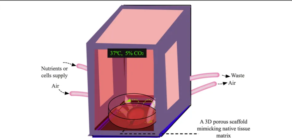
attempts have been implemented to closely mimic tissue microenvironments for in vitro cell cultures. Encour-agingly, different kinds of bioreactors have been developed for dynamic 3D cultures [280, 281]. These systems are equipped with oxygen and nutrient supplies as well as waste removal procedures for optimal cell proliferation, migration and differentiation (Fig.4). Apparently, the per-fusing dynamic 3D culture models are beyond the static culture states and will greatly facilitate biologically rele-vant studies.
Conclusions and future perspectives
Our increasing understanding about tissue microenvi-ronments at structural, mechanical and compositional levels has inspired researchers devising advanced scaf-folds using various native or native-mimicking materials for biomedical and bioengineering spatial tissue cultures. This trend has been growing rapidly in recent years [107] that is benefited from refined methods for native tissue ECM extraction and protein purification, high resolution identifications of ECM components, broad availabilities of biomaterials and synthetic materials, close definitions of basic physical parameters of native ECM, vigorous development of stem cell technologies, advanced instrumental support, robust integration of computational modeling and mathematical algorithms. As a result of the advancement in the ECM modeling field, many biomedical studies have discovered novel molecules, functions, and phenotypes that are otherwise hard to be identified in 2D or non-biologically relevant 3D cultures [4,282]. Meanwhile, bioengineers have com-bined the techniques of scaffold fabrication, bioink pro-duction and 3D printing for advanced tissue repair and regeneration [283, 284]. These encouraging progresses have greatly facilitated the development of novel systems to model pathophysiological conditions and approaches to treat human diseases.[8].
biopolymers have brought them to broader applications as solubilizers, emulsifiers, gelling agents and viscosity en-hancers for more efficient drug development and delivery [289,290]. Recently, drugs are designed as bio-Nano-smart products for personalized medicine and gene therapy using advance biomaterials so as to guide the drugs to tar-get sites, minimizing adverse side effects [291, 292]. Fur-thermore, blending of different types of biopolymers and mixing with treatment agents (e.g. oligonucleotides, gene products, compounds and small molecules) through multi-drug systems formulated and delivered as a single capsule, tablet or nanoparticle will certainly revolutionize therapeutic approaches for patients.
The capability of organoids to form patient-specific tissues makes the model very promising for tissue regen-eration and cost-effective drug screening at personalized medicine levels [293, 294]. A combination of organoid models with optimized biopolymer or disease- and tissue-specific ECM scaffold systems under dynamic cul-turing environments represents a complex yet an ideal platform for future biomedical and bioengineering appli-cations, which will form a new era of personalized medi-cine, precision therapy, effective drug development and delivery, functional artificial tissues and organs for the health of human.
Abbreviations
TNF-α:Tumor necrotic factor; ASC: Adipose derived stem cell; ASXL2: Additional Sex Combs like 2; BFs: Biological factors; BM: Basement membrane; BMP: Bone
morphogenic protein; CAMTA1: Calmodulin-binding transcription activator 1; CCL: Chemokine ligand; CCR: CC chemokine receptor; CD: Cluster of differentiation; CD4: Cluster of differentiation 4, surface protein on helper T cell; CD8: Cluster of differentiation 8, surface protein on cytotoxic memory T cells; CK: Cytokeratins; CLS: Crown-like structure; Colα1: Collagen alpha 1; COX2: Cyclooxygenase-2; CSCs: Cancer stem cells; CSF1: Colony stimulating factor 1; CTLA4: Cytotoxic T-lymphocyte antigen 4; CXCL: Chemokine (C-X-C motif) ligand; DC: Dendritic cell; DCIS: Ductal carcinoma in situ; DDX20: DEAD-box helicase 20; dECM: Decellularized extracellular matrix; DNA: Deoxyribonucleic acid; ECM: Extracellular matrix; EGF: Epidermal growth factor; EMT: Epithelial mesenchymal transition; FGF: Fibroblast growth factors; FOXP3: Forkhead box P3; FXR1: Fragile X-related 2; G-CSF: Granulocyte colony stimulating factor; GFs: Growth factors; GM-CSF: Granulocyte-macrophage colony stimulating factor; HA: Hyaluronic acid; hMSCs: Human mesenchymal stem cells; IFN-α: Interferon alpha; IFN-β: Interferon beta; IFN-y: Interferon gamma; IL: Interleukin; iNOS: Inducible nitric oxide; LATS2: Serine/threonine-protein kinase; LCST: Lower critical solution temperature; MCSs: Multicellular spheroids; MCTSs: Multicellular tumor spheroids; MDSCs: Myeloid-derived suppressor cells; MHC: Major histocompatibility complex; MITF: Mitochondria transcription factor; MMP: Matrix metalloproteinase; MSCs: Mesenchymal stem cells; NHS: N-hydroxysuccinimide; NK: Natural killer cell; NKT: Natural killer T cell; NME: Native microenvironment; PAX7: Paired box protein Pax-7; PCL: Polycaprolactone; PDGF: Platelet-derived growth factor; PDLA: Poly-D-Lactic acid; PDMS: Poly(dimethyl silane); PDO: Polydioxanome; PE: Poly(ethylene); PEEK: Polyether ether ketone; PEG: Poly(ehtylen glycol); PES: Polyether sulfonate; PET: Poly(ethylene terephthalate); pHEMA: Poly(2-hydroxyethyl methacrylate); PLA: Poly(lactic acid); PLGA: Poly(lactic-co-glycolic acid); PLLA: Poly-L-lactic acid; PMMA: Poly(methacrylate); pNIPAm: Poly(N-isopropylacrylamide); PP: Poly(propylene); PRP: Platelet-rich plasma; PVC: Poly(vinyl chloride); PVOH: Polyvinyl alcohol; PVP: Poly(vinlypyrrolidone); RGD: Arg-Gly-Asp; RME: Regenerative microenvironment; SEMA3A: Semaphoring 3A; SIBS: Poly(styrene-b-isobutylene-b-styrene); SMC: Smooth muscle cell; TAF: Tumor associated fibroblast; TAM: Tumor associated macrophage; TGF-β: Transforming growth factor beta; TME: Tumor microenvironment; UHMPE: Ultrahigh molecular weight PE; VEGF: Vascular endothelial
growth factor; VEGFA: Vascular endothelial growth factor A; VEGFC: Vascular endothelial growth factor C; VEGFD: Vascular endothelial growth factor D
Acknowledgements
The authors thank WSU colleagues for discussions and critical comments.
Funding
This work is supported by a WSU Start-up Fund to W.L.
Authors’contributions
G.R. and W.L. wrote and edited the manuscript. Both authors read and approved the final manuscript.
Ethics approval and consent to participate Not applicable.
Consent for publication
The manuscript is approved by the all the authors for publication.
Competing interests
The authors declare that there is no conflict of interests.
Publisher’s Note
Springer Nature remains neutral with regard to jurisdictional claims in published maps and institutional affiliations.
Received: 2 May 2018 Accepted: 30 August 2018
References
1. Hanahan D, Weinberg RA. Hallmarks of cancer: the next generation. Cell. 2011;144(5):646–74.
2. Bissell MJ, Hines WC. Why don’t we get more cancer? A proposed role of the microenvironment in restraining cancer progression. Nat Med. 2011; 17(3):320–9.
3. Yamada KM, Cukierman E. Modeling tissue morphogenesis and cancer in 3D. Cell. 2007;130(4):601–10.
4. Pickup MW, Mouw JK, Weaver VM. The extracellular matrix modulates the hallmarks of cancer. EMBO Rep. 2014;15(12):1243–53.
5. Debnath J, Brugge JS. Modelling glandular epithelial cancers in three-dimensional cultures. Nat Rev Cancer. 2005;5(9):675–88.
6. Sokol ES, Miller DH, Breggia A, Spencer KC, Arendt LM, Gupta PB. Growth of human breast tissues from patient cells in 3D hydrogel scaffolds. Breast Cancer Res. 2016;18(1):19.
7. Rijal GL, W. A versatile 3D tissue matrix scaffold system for tumor modeling and drug screening. Sci Adv. 2017;3(e1700764):16.
8. Egeblad M, Nakasone ES, Werb Z. Tumors as organs: complex tissues that interface with the entire organism. Dev Cell. 2010;18(6):884–901. 9. Gudjonsson T, Adriance MC, Sternlicht MD, Petersen OW, Bissell MJ.
Myoepithelial cells: their origin and function in breast morphogenesis and neoplasia. J Mammary Gland Biol Neoplasia. 2005;10(3):261–72. 10. Eaves CJ. Hematopoietic stem cells: concepts, definitions, and the new
reality. Blood. 2015;125(17):2605.
11. Nolan DJ, Ginsberg M, Israely E, Palikuqi B, Poulos MG, James D, et al. Molecular signatures of tissue-specific microvascular endothelial cell heterogeneity in organ maintenance and regeneration. Dev Cell. 2013;26(2):204–19. 12. Glukhova M, Koteliansky V, Sastre X, Thiery JP. Adhesion systems in normal
breast and in invasive breast carcinoma. Am J Pathol. 1995;146(3):706–16. 13. Dairkee S, Heid HW. Cytokeratin profile of immunomagnetically separated
epithelial subsets of the human mammary gland. In Vitro Cell Dev Biol Anim. 1993;29(5):427–32.
14. Rijal G, Li W. A versatile 3D tissue matrix scaffold system for tumor modeling and drug screening. Sci Adv. 2017;3(9):e1700764. 15. Warburton MJ, Mitchell D, Ormerod EJ, Rudland P. Distribution of
myoepithelial cells and basement membrane proteins in the resting, pregnant, lactating, and involuting rat mammary gland. J Histochem Cytochem. 1982;30(7):667–76.
16. Lazard D, Sastre X, Frid MG, Glukhova MA, Thiery JP, Koteliansky VE. Expression of smooth muscle-specific proteins in myoepithelium and stromal myofibroblasts of normal and malignant human breast tissue. Proc Natl Acad Sci U S A. 1993;90(3):999–1003.
17. Pandey PR, Saidou J, Watabe K. Role of myoepithelial cells in breast tumor progression. Front Biosci. 2010;15:226–36.
18. Kolar Z, Ehrmann J, Turashvili G, Bouchal J, Mokry J. A novel myoepithelial/ progenitor cell marker in the breast? Virchows Arch. 2007;450(5):607–9. 19. Adriance MC, Inman JL, Petersen OW, Bissell MJ. Myoepithelial cells: good
fences make good neighbors. Breast Cancer Res. 2005;7(5):190–7. 20. Man YG. Focal degeneration of aged or injured myoepithelial cells and the
resultant auto-immunoreactions are trigger factors for breast tumor invasion. Med Hypotheses. 2007;69(6):1340–57.
21. Nieto MA. Epithelial Plasticity: A Common Theme in Embryonic and Cancer Cells. Science. 2013;342(6159):1234850.
22. Scadden DT. The stem-cell niche as an entity of action. Nature. 2006;441:1075. 23. Imitola J, Raddassi K, Park KI, Mueller F-J, Nieto M, Teng YD, et al. Directed
migration of neural stem cells to sites of CNS injury by the stromal cell-derived factor 1α/CXC chemokine receptor 4 pathway. Proc Natl Acad Sci. 2004;101(52):18117.
24. Brabletz T, Jung A, Spaderna S, Hlubek F, Kirchner T. Migrating cancer stem cells—an integrated concept of malignant tumour progression. Nat Rev Cancer. 2005;5:744.
25. Schafer M, Werner S. Cancer as an overhealing wound: an old hypothesis revisited. Nat Rev Mol Cell Biol. 2008;9(8):628–38.
26. Hermann PC, Huber SL, Herrler T, Aicher A, Ellwart JW, Guba M, et al. Distinct populations of cancer stem cells determine tumor growth and metastatic activity in human pancreatic cancer. Cell Stem Cell. 2007;1(3):313–23. 27. Magee Jeffrey A, Piskounova E, Morrison SJ. Cancer Stem Cells: Impact,
Heterogeneity, and Uncertainty. Cancer Cell. 2012;21(3):283–96. 28. Hirschi KK, D’Amore PA. Pericytes in the microvasculature. Cardiovasc Res.
1996;32(4):687–98.
29. Carmeliet P, Jain RK. Molecular mechanisms and clinical applications of angiogenesis. Nature. 2011;473:298.
30. Jain RK. Normalization of tumor vasculature: an emerging concept in antiangiogenic therapy. Science. 2005;307(5706):58.
31. Alitalo K. The lymphatic vasculature in disease. Nat Med. 2011;17:1371. 32. Swartz MA, Iida N, Roberts EW, Sangaletti S, Wong MH, Yull FE, et al. Tumor
microenvironment complexity: emerging roles in Cancer therapy. Cancer Res. 2012;72(10):2473.
33. Orimo A, Weinberg RA. Heterogeneity of stromal fibroblasts in tumors. Cancer Biol Ther. 2007;6(4):618–9.
34. Sugimoto H, Mundel TM, Kieran MW, Kalluri R. Identification of fibroblast heterogeneity in the tumor microenvironment. Cancer Biol Ther. 2006;5(12): 1640–6.
35. Wever OD, Demetter P, Mareel M, Bracke M. Stromal myofibroblasts are drivers of invasive cancer growth. Int J Cancer. 2008;123(10):2229–38. 36. Tomasek JJ, Gabbiani G, Hinz B, Chaponnier C, Brown RA. Myofibroblasts
and mechano-regulation of connective tissue remodelling. Nat Rev Mol Cell Biol. 2002;3:349.
37. Cirri P, Chiarugi P. Cancer-associated-fibroblasts and tumour cells: a diabolic liaison driving cancer progression. Cancer Metastasis Rev. 2012; 31(1):195–208.
38. Gascard P, Tlsty TD. Carcinoma-associated fibroblasts: orchestrating the composition of malignancy. Genes Dev. 2016;30(9):1002–19.
39. Li H, Fan X, Houghton J. Tumor microenvironment: the role of the tumor stroma in cancer. J Cell Biochem. 2007;101(4):805–15.
40. Kalluri R. The biology and function of fibroblasts in cancer. Nat Rev Cancer. 2016;16:582.
41. Rajala MW, Scherer PE. Minireview: the adipocyte--at the crossroads of energy homeostasis, inflammation, and atherosclerosis. Endocrinology. 2003; 144(9):3765–73.
42. Wiseman BS, Werb Z. Stromal effects on mammary gland development and breast Cancer. Science. 2002;296(5570):1046.
43. Andarawewa KL, Motrescu ER, Chenard M-P, Gansmuller A, Stoll I, Tomasetto C, et al. Stromelysin-3 is a potent negative regulator of Adipogenesis participating to Cancer cell-adipocyte interaction/crosstalk at the tumor invasive front. Cancer Res. 2005;65(23):10862.
44. Iyengar P, Espina V, Williams TW, Lin Y, Berry D, Jelicks LA, et al. Adipocyte-derived collagen VI affects early mammary tumor progression in vivo, demonstrating a critical interaction in the tumor/stroma microenvironment. J Clin Invest. 2005;115(5):1163–76.