Iranian Biomedical Journal 12 (3): 159-165 (July 2008)
*Corresponding Author; Tel. (+98-21) 6640 5535; E-mail: [email protected]
Comparison of Distribution of Virulence Determinants in Clinical
and Environmental Isolates of Vibrio cholera
Bita Bakhshi
1 & 2, Mohammad Reza Pourshafie
*1, Farahtaj Navabakbar
2, Akbar Tavakoli
2Fereshteh Shahcheraghi
1, Mansoor Salehi
2, Ziba Faradjzadegan
2and Seyed Mohsen Zahraei
31
Dept. of Bacteriology, Pasteur Institute of Iran, Pasteur Ave., Tehran; 2Faculty of Medicine, Isfahan University of Medical Sciences, Hezarjerib Ave., Isfahan; 3Dept. of Enteric Bacteria, 3Center for Disease Control, Tehran, Iran
Received 12 August 2007; revised 10 December 2007; accepted 13 December 2007
ABSTRACT
Background: The virulence of a pathogenic Vibrio cholerae is dependent on a discrete set of genetic
determinants. In this study, we determined the distribution of virulence determinants among the clinical and
environmental isolates of V. cholerae. Methods: The antibiotic resistance profiles of the isolates were
determined using standard disk diffusion assay. PCR assay was performed to analyze the presence of toxin
genes of ctx, zot and ace. The composition of cholera toxin encoding element (CTX) region flanking of the V.
cholerae isolates was also analyzed. Results: All of the clinical isolates (100%) showed a complete set of
virulence genes and also the attachment site of the filamentous bacteriophage CTX
φ
. None of theenvironmental isolates contained the virulence genes and the attachment site of the CTX
φ
. Analysis of theflanking regions including the toxin-linked cryptic element and repeat in toxin genes revealed their integrity in the clinical isolates while in the environmental isolates they were absent or contained incomplete sequences. Comparison of the antibiotic resistance assay of the environmental and clinical isolates showed a significant difference in the resistance profiles of the isolates obtained from the two sites. High rates of resistance to
co-trimoxosol, streptomycin and chloramphenicol were found with clinical isolates. Conclusion: The absence of
all virulence determinants in the environmental strains may suggest that certain ecological features must be
present for V. cholerae to acquire a complete set of virulence determinants and to turn them into pathogenic
strains. Iran. Biomed. J. 12 (3): 159-165, 2008
Keywords: Vibrio cholerae, Virulence determinants, Environment
INTRODUCTION
ibrio cholerae includes both pathogenic and
non-pathogenic strains that vary in their virulence gene contents [1-3]. The
pathogenic and epidemic strains of V. cholerae
possess two essential genetic elements (i) cholera toxin encoding element (CTX) element which is part
of the filamentous bacteriophage (CTX
φ
) genome[4] involved in coding for cholera toxin (CT) as the
most important virulence factor of V. cholerae, and
(ii) the V. cholerae pathogenicity island which
encodes the toxin co-regulated pilus (TCP). The TCP is the adhesin that is coordinately regulated with CT production which functions as an essential colonization factor in the gut as well as the receptor
for CTX
φ
[5]. Upstream of the CTX genetic elementis toxin-linked cryptic (TLC) element and 3' of the
CTX
φ
contains a region which encodes repeat intoxin (RTX) gene cluster composed of the toxin (rtxA), activator (rtxC) and transporters (rtxBD)
[6-8]. It is presumed that CT and TCP elements are
exclusively associated with the clinical strains of V.
cholerae serogroups O1 and O139. Nevertheless, a
few reports on the presence of CT among the
environmental strains of V. cholerae have been
published [9]. TCP has also been shown to be
associated with clinical V. cholerae O1 and O139
and it is rarely found in the environmental isolates.
For example, the presence of tcpA of the TCP gene
cluster has been reported in non-O1 toxigenic [10] and nontoxigenic non-O1 non-O139 strains [11]. It has been shown by some investigators [8, 12] that the conversion of non-toxigenic environmental
V
160 Bakhshi et al. Iran. Biomed. J., July 2008
strains into epidemic strains V. cholerae is possible
if they acquire the appropriate set of virulence genes The aim of this study was to understand the epidemiology and origin of pathogenic strains by determining the prevalence of virulence genes in the
clinical and environmental strains of V. cholerae
isolated in Iran.
MATERIALS AND METHODS
Collecting and processing of environmental samples. Water samples were collected from surface
water located at 6 different locations in Tehran (Iran) including Aminabad, 17-E-Shahrivar, Firoozabad, Beheshti, Yakhchiabad and Afsarieh lakes. The samples were filtered using Whatman no.1 filter paper and subsequently filtered through a 0.45-µm- pore-size membrane by using vacuum
pressure of 15 to 20 lb/in2. The membrane was cut
into eight pieces and vortexed in a 2-ml of 10 mM PBS (pH 7.4) for 3 min. One milliliter of the suspension was added to 10 ml of alkaline peptone water (APW) containing peptone (1%, wt/vol) and NaCl (1%, wt/vol) (pH 8.5) for enrichment with shaking (100 rpm) at 37°C for 16-18 h. Each sample was streaked on thiosulfate-citrate-bile-sucrose agar plates and incubated at 37°C overnight [5]. The resulting yellow colonies were further examined.
The identity of V. cholerae strains was defined by 11
biochemical assays including colony morphology, oxidase, motility, sucrose fermentation, lactose fermentation, growth in 0% NaCl, arginine dehydrolase, ornithine decarboxylase, methyl red, voges-proskauer and indole test [13, 14] and confirmed by PCR amplification of 16s-23s rRNA
intergenic region specific for V. cholerae [15]. The
V. cholerae strains (n = 25) were isolated and
subjected to further analysis.
Clinical strains. V. cholerae strains (n = 25) of
clinical origin, collected from four different provinces in Iran (Tehran, Qom, Golestan and
Zahedan) in 2005, were included in this study. The
specimens were collected on the sterile swabs and were enriched in APW. The suspected samples were then placed in Carry-Blair transport and shipped to the Department of Bacteriology, Pasteur Institute of Iran (Tehran). Biochemical assays were performed
to confirm the identity of V. cholerae strains. The
isolates were identified as described in the above section.
Serology. In this study, serogrouping of all isolates
was performed with polyvalent O1 and monospecific Inaba and Ogawa antisera (Mast Diagnostics Ltd., Bootle, Mersey side, UK).
Anti-microbial susceptibility testing. Antibiotic
susceptibility patterns were tested by the standard disk diffusion technique according to NCCLS (National Committee for Clinical Laboratory Standards, 2001) guidelines with the following antibiotics: gentamicin (10 µg), doxycycline (30 µg),
oxytetracycline (30 µg) and tetracycline (30 µg)
were purchased from Difco laboratories (Detroit,MI,
USA) and ciprofloxacin (5 µg), streptomycin (5 µg),
ampicillin (10 µg), polymixin B (300 U),
chloramphenicol (30 µg) and co-trimaxazole (25 µg)
were purchased from Becton Dickinson and Company (Sparks, MD, USA).
DNA extraction and PCR assay. The
chromosomal DNA from the isolates was obtained by a simplified method whereby one isolated colony was suspended into 200 µl of sterile distilled water and boiled for 5 min and 5 µl of supernatant was used as the template in a PCR reaction [16]. PCR assay was performed using primers specific for the toxin genes inside the CTX genetic element
including ctx, zot and ace [17-19]. The CTX
φ
attachment site was also investigated in the two groups of strains using primers attRS-F or attRS-R specific for 18 nucleotide attachment site with any of the forward or reverse primers inside the core or RS1 regions (data not shown) [20]. The flanking regions including RTX cluster (1448-F, 1448-R and 1451-F, 1451-R) and TLC element (1465-F, 1465-R and 1469-F, 1469-R) were also subjected to analysis due to the important role which may have played in the acquisition of the toxin genes and evolution of the pathogenic strains. The DNA was extracted and
PCR was performed in a 25-µl total reaction volume
containing 20 µl sterile water, 2.5 µl 10× Taq
polymerase buffer, 0.6 µl MgCl2 (25 mM), 0.3 µl
dNTPs (10 mM), 0.5 unit of Taq DNA polymerase,
25 pmol of each primer. The cycling conditions were as follows: preincubation at 94ºC for 5 min, 30 cycles of 1 min at 94ºC for denaturation, 1 min at 58ºC for annealing, 2 min at 72ºC for elongation and incubation at 72ºC for 3 min for final elongation. Primer designation and their sequence used in this
study are depicted in Table 1. V. cholerae ATCC
14035 was used as positive control in each PCR assay [21].
Iran. Biomed. J., July 2008 Distribution of Virulence Determinants 161
http://IBJ.pasteur.ac.ir Table 1. Primers used in this study.
Designation Sequence Reference
prVC-F 5' AGTCACTTAACCATTCAACCCG 3' 15
prVCM-R 5' TTAAGCGTTTTCGCTGAGAATG 3'
ctx-F 5' CGGGCAGATTCTAGACCTCCT 3' 18
ctx-R 5' CGATGATCTTGGAGCATTCCCAC 3'
zot-F 5' TGGCTTCGTCTGCTGCCGGCGATT 3' 19
zot-R 5' CACTTCTACCCACAGCGCTTGCGC 3'
ace-F 5' TAAGGATGTGCTTATGATGGACACCC 3' 17
ace-R 5' CGTGATGAATAAAGATACTCATAG 3'
VC1448-F 5' TGCTTCATCCAAAATCAGCA 3' This study*
VC1448-R 5' TCATCAGCGGTAATCGAGAA 3' This study
VC1451-F 5' TGTTCGGCGATAACATTCAG 3' This study
VC1451-R 5' TTTGTGAACCACGTCTGACCCTT 3' This study
VC1465-F 5' CTTTGGCCGTGTCTATTGGT 3' This study
VC1465-R 5' TAAATACGTGCGGCTCAACA 3' This study
VC1469-F 5' AACTCATGAGCAAGGCGTTT 3' This study
VC1469-R 5' GCTCGGAAGACTTTCGCTTA 3' This study
attRS-F 5' CCTTAGTGCGTATTATGT 3' 16
attRS-R 5' ACATAATACGCACTAAGG 3'
*primers designed according to the V. cholerae strain N16961 whose genome is fully sequenced and is available on BLAST (www.ncbi.nlm.nih.gov).
RESULTS
Identification of isolates and serology. A total of
50 V. cholerae strains including 25 clinical and 25
environmental strains were isolated and analyzed in this study. All of the clinical isolates were O1 Inaba and all of the environmental isolates were non O1-non O139 serotypes (Table 2).
Isolates and antibiotic susceptibility. All 50
isolates including 25 clinical and 25 environmental
strains were identified as V. cholerae O1 serotype
Inaba for clinical and non O1-non O139 serotype for environmental isolates. Antibiotic susceptibility testing of isolates showed 12 and 10 antibiotic resistance patterns among the clinical and environmental isolates, respectively. Pattern one,
SXTr, Sr, CHLr, TEr, PBr, with 24% was the
predominant pattern among the clinical isolates
while the predominant pattern among the environmental isolates was 36% which represents no resistance to any of the antibiotics examined. The
rate of antibiotic resistance among the clinical V.
cholerae strains was significantly higher as
compared with the environmental strains. The most noticeable difference was seen for the SXT, streptomycin and chloramphenicol. Resistance to these three antibiotics was observed in 92%, 92% and 88% of the clinical and 16%, 12% and 8% of environmental isolates, respectively. No resistance was seen to gentamicin and ciprofloxacin in the clinical or environmental isolates.
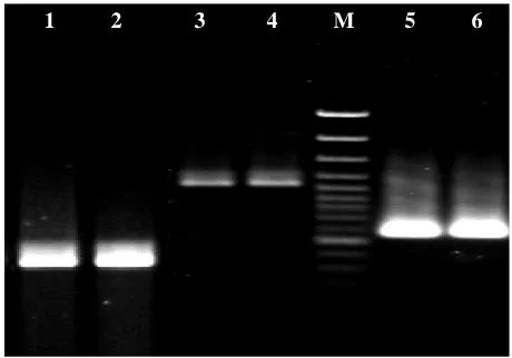
PCR assay. PCR revealed the presence of ctx, zot,
and ace genes in 100% of clinical isolates. None of
the environmental strains contained these toxin genes. Analysis for the presence of attachment sites and the flanking gene clusters revealed that the
162 Ta So Cl En En En En En clin site con chr VC and bot in of iso PC I sel env Te F lan 2
able 2. A summ
ource linical nvironmental nvironmental nvironmental nvironmental nvironmental nical isolate es while n ntained th romosomal R C1451 ORF
d 92% and 9 th of these g the TLC elem
the clinical olates, respec CR products a
In this study, lected virul vironmental ehran, Iran,
Fig. 1. Represe e 5 and 6, VC1
mary of characte
Serology O1 nonO1-no nonO1-no nonO1-no nonO1-no nonO1-no s possessed none of the his nucleo RTX cluster
existed in 10 92% of the e genes. The V
ment were p and 32% a ctively (Tab are shown in
DISCUS
, the occurren lence genes strains of were exami
ntatives of amp 465; lane 7 and
1 2
eristics of V. ch
c onO139 onO139 onO139 onO139 onO139 18 nucleotid e environme otide sequ r genes with 00% and 100 environment VC1465 and V
present in 100 and 0% of e ble 2). Repre n Figures 1 an
SSION
nce and distr s in the
V. cholera
ined. The cl
plified DNA pr d 8, VC1469.
M 3
holerae strains o
ctx ace z
+ + - - - - - - - - - - de attachmen ental isolate uence. Th VC1448 an 0% of clinic tal isolates fo
VC1469 OR 0% and 100% environment
esentatives o nd 2.
ribution of th clinical an
e isolated i
linical strain
roducts of the C
4
Bakhshi et al.
of clinical and e
zot VC1448 + + - + - + - - - + - - nt es he nd al or RF % al of he nd in ns were strains the en where ace, zo Cha 79% o isolate determ Moreo that no non-O Singh presen
ace in
of V.
nonO1 virulen non-O
CTX flanking r
5 environmental o 8 VC1451 + + + + - - V. cholerae
s were non-O nvironmenta eas all clinica
ot and ctx (T
kraborty and of environme es didn’t minants in
over, Brazil
one of the V
O1 serotype and collea nce of virule n the clinical
cholerae, alt
139 strains nce genes in O139 serogro
regions. Lane
6 7
origin. VC1465 + - + + - +
e O1 Inaba
O1, non-O13 l strains car al isolates ha Table 2). d colleagues ental non-O posses any the CTX and colleag
V. cholerae e
contained ct
agues [22] a ence genes i and not the though repo [23] and a ncluding ctx
ups [5] shou
1 and 2, VC14
8 M
Iran. Biomed. J
VC1469 + - - - - - while envi 39 serotype. rried the to arbored all to
[5] have rep 1, non-O139 y of the X genetic gues [11] ha environmenta
tx, zot and a
also have s
including ctx
environment rts on clinic also the pr xAB, in som uld not be ign
448; lane 3 and
3
0
J., July 2008
percent 100 64 24 4 4 4 ironmental None of oxin genes oxin genes: ported that 9 serotype, virulence element. ave shown al isolates,
ace genes.
shown the
tx, zot and
tal isolates cal nonO1-resence of me non-O1,
nored.
d 4, VC1451;
3.0 kb
1.0 kb
0.5 kb
Iran. Biomed. J., July 2008 Distribution of Virulence Determinants 163
http://IBJ.pasteur.ac.ir Fig. 2. Representatives of amplified DNA products of the
genes inside the CTX element. Lane 1 and 2, ace-F/R; lane 3 and 4, zot-F/R,; lanes 5 and 6, ctx-F/R.
The CTX attachment site is a region comprises an 18-bp conserved region adjacent to the CTX element. All clinical strains were shown to carry this region while none of the environmental isolates contained this genetic region. The presence of this
site is necessary for the attachment of CTX
φ
phageinto the genome of V. cholerae [20]. On the
contrary, the absence of this attachment site in the environmental isolates has raised the question of whether the defects in the flanking regions could contribute in hindering the isolates for acquisition of the genes involved in the attachment sites and CTX
element. The DNA from V. cholerae isolates was
subjected to PCR analysis for investigation of flanking regions of the CTX element. The RTX gene cluster which lies at 3' end of the CTX element is a chromosomal cluster and presents in pathogenic and
non-pathogenic strains of V. cholerae [6, 7]. The
presence of VC1448 and VC1451 ORF in the majority of the environmental (92%) and clinical (100%) isolates showed that RTX coding cluster was widely distributed in the genome of both pathogenic
and non-pathogenic V. cholerae isolates (Table 2),
and no significant difference in the presence of this cluster among the two groups can be drown (P>0.05).
The RTX toxins represent a family of important virulence factors that have disseminated widely among Gram-negative bacteria [5], and its presence
in most of the V. cholerae isolates would raise the
question of whether this toxin gene cluster could be
transferred into V. cholerae by other Gram-negative
bacteria.
A development in the evolution of pathogenic V.
cholerae strains in obtaining the CTX genetic
element is the acquisition of upstream TLC element.
It has been suggested that this element could be inserted into the bacterial genome by another
bacteriophage [24]. The V. cholerae strains which
contain CTX element also harbors TLC element simultaneously. On the other hand, the strains which carry TLC element may not harbor the CTX genetic element [24]. This notion is supported by our data that the all toxigenic clinical isolates also carried the two ORF in the TLC element namely VC1465 and VC1469. Moreover, the two ORF were present in 32% and 0% of the environmental isolates, respectively, with significant difference between the two clinical and environmental groups (P< 0.05).
The reason for the less number of environmental isolates to contain the TLC elements could be because of their bacteriophage originality as opposed to the RTX gene cluster which is found in abundance in both environmental and clinical isolates.
Antibiotic resistant pattern was also included in the comparison of the virulence determinants of clinical and environmental isolates. This comparison is summarized in Tables 3 and 4 depicting the significant difference in the resistance patterns of the isolates to antibiotics including SXT, streptomycin
and chloramphenicol (P< 0.05). The resistance to
these antibiotics has been shown to be encoded by the genes in the conjugative SXT transposon. It is, therefore, possible that such transposons were extensively disseminated in the clinical but not environmental isolates.
In conclusion, results of this study shows that
clinical and environmental isolates of V. cholerae
differ in virulence and resistance encoding determinants, in addition to the serogroups and phenotypic characteristics. These differences may show the evolutionary status of non-toxigenic
environmental isolates of V. cholerae to emerge into
pathogenic strains by acquisition of virulence associated genes which in turn, mandates the need to
monitor the occurrence, stability and evolution of V.
cholerae strains in the environment.
ACKNOWLEDGEMENTS
We are very thankful to Tehran Water and Sewage Company (Iran) with special thanks to Mrs. Motallebi and Mrs. Parvar for helping us in collecting surface water samples. This research was funded by Pasteur Institute of Iran (Tehran), grant number 404.
1 2 3 4 M 5 6
164 Bakhshi et al. Iran. Biomed. J., July 2008
Table 3. Percentage of antibiotic resistance patterns among the clinical isolates of V. cholerae.
GM CIP T AP DO PB TE CHL S SXT Percent Pattern S S S R S R R R R R 24 1 S S S R S R R R R R 16 2 S S S S S R S R R R 12 3 S S S S S R R S S S 8 4 S S S S S S R R R R 8 5 S S S S S R R R R R 8 6 S S R R R R R R R R 4 7 S S S R R R R R R R 4 8 S S S S S S R R R R 4 9 S S S R S R S R R R 4 10 S S S S S S S R R R 4 11 S S S S S R R S R R 4 12 0% 0% 4% 52% 8% 84% 80% 88% 92% 92% Percentage of resistance
SXT, co-trimoxazole; S, streptomycin; C, chloramphenicol; TE, tetracycline; PB, polymixin B; DO, doxycycline; AP, ampicillin; T, oxytetracycline; CIP, ciprofloxacin and GM, gentamicin.
REFERENCES
1. Faruque, S.H., Albert, M.J. and Mekalanos, J.J.
(1998) Epidemiology, genetics, and ecology of
toxigenic Vibriocholerae. Microbiol. Mol. Biol. Rev.
62: 1301-1314.
2. Alam, M., Sultana, M., Nair, G.B., Sack, R.B. Sack,
D.A., Siddique, A.K., Ali, A., Huq, A. and Colwell
R.R. (2006) Toxigenic Vibrio cholerae in the aquatic
environment of Mathbaria, Bangladesh. Appl.
Environ. Microbiol. 72: 2849-2855.
3. Begum, K., Ahsan, C.R. Ansaruzzaman, M., Dutta,
D.K., Ahmad, O.S. and Talukder, K.A. (2006) Toxin(s), other than cholera toxin, produced by
environmental non O1 non O139 Vibrio cholerae.
Cell Mol. Immun. 3: 115-121.
4. Waldor, M.K. and Mekalanos, J.J. (1996) Lysogenic
conversion by a filamentous phage encoding cholera
toxin. Science 272: 1910-1914.
5. Chakraborty, S., Mukhopadhyay, A.K., Bhadra, P.K.,
Ghosh, A.N., Mitra, P., Shimada, T., Yamasaki, S., Faruque, S.M., Takeda, Y., Colwell, R.R. and Nair, G.B. (2000) Virulence genes in environmental
isolates of Vibrio cholerae. Appl. Environ.
Microbiol. 66: 4022-4028.
6. Boyd, E.F. and Waldor, M.K. (1999) Alternative
mechanism of cholera toxin acquisition by
Vibrio cholerae: generalized transduction of CTXФ
by bacteriophage CP-T1. Infect. Immun. 67:
5898-5905.
7. Chow, K.H., NG, T.K., Yuen, K.Y. and Yam, W.C.
(2001) Detection of RTX toxin gene in Vibrio
cholerae by PCR. J. Clin. Microbiol. 39: 2594-259.
Table 4. Percentage of antibiotic resistance patterns among the environmental isolates of V. cholerae.
Pattern Percent SXT S CHL TE PB DO AP T CIP GM
1 36 S S S S S S S S S S
2 28 S S S S R S S S S S
3 8 R S S S R S S S S S
4 4 R S S S S S R S S S
5 4 R R R R R R R R S S
6 4 S S R R R R R R S S
7 4 S S S R S R S R S S
8 4 S R S S S S R S S S
9 4 S S S S S S R S S S
10 4 S R S S S S S S S S
Percentage of resistance 16% 12% 8% 12% 44% 12% 16% 12% 0% 0%
SXT, co-trimoxazole; S, streptomycin; C, chloramphenicol; TE, tetracycline; PB, polymixin B; DO, doxycycline; AP, ampicillin; T, oxytetracycline; CIP, ciprofloxacin and GM, gentamicin.
Iran. Biomed. J., July 2008 Distribution of Virulence Determinants 165
http://IBJ.pasteur.ac.ir
8. Boardman, B.K., Meehan, B.M. and Fullner Satchell,
K.J. (2007) Growth phase regulation of Vibrio
cholerae RTX toxin export. J. Bacteriol. 189: 1827-1835.
9. Nair, G.B., Oku, Y., Takeda, Y., Ghosh, A., Ghosh,
S., Chattopadhyay, Pal, S.C., Kaper, J.B. and Takeda,
T. Toxin profiles of Vibrio cholerae non-O1 from
environmental sources in Calcutta, India. Appl.
Environ. Microbiol. 54: 3180-3182.
10. Ghosh, C., Nandi, R.K., Dasgupta, S.K., Nair, G.B.,
Hall, R.H. and Ghose, A.C. (1997) A search for cholera toxin (CT), toxin coregulated pilus (TCP), the regulatory element ToxR, and other virulence factors
in non-O1/non-O139 Vibrio cholerae. Microb.
Pathog. 22:199-208.
11. Brazil, J.M.V., Alves, R.M., Rivera, I.N.G.,
Rodrigues, D.P., Karaolis, D.K.R. and Campos, L.C. (2002) Prevalence of virulence associated genes in
clinical and environmental Vibrio cholerae strains
isolated in Brazil between 1991 and 1999. FEMS
Microbiol. Lett. 215 (1): 15-21.
12. Goel, A.K., Ponmariappan, S., Kamboj, D.V. and
Singh, L. (2007) Single multiplex polymerase chain reaction for environmental surveillance of
toxigenic-pathogenic O1 and non-O1 Vibrio cholerae. Folia.
Microbiol (Praha) 52: 81-85.
13. Choopun, N., Louis, V., Hug, A. and Colwell, R.R.
(2002) Simple procedures for rapid identification of
Vibrio cholerae from the aquatic environment. Appl. Environ. Microbiol. 68: 995-998.
14. Stavric, S. and Bachanan, B. (1995) The isolation and
identification of V. cholerae O1 and non-O1 from
foods. Government of Canada, Health Protection Branch, Ottawa, Polyscience Publication, MFLP-72,
Canada.
15. Chun, J., Huq, A. and Colwell, R.R. (1999) Analysis
of 16S-23S r RNA intergenic spacer region of Vibrio
cholerae and Vibrio mimicus. Appl. Environ. Microbiol. 65: 2202-2208.
16. D.K.R., Karaolis, Lan, R. and Reeves, P.R. (1995)
The sixth and seventh cholera pandemics are due to independent clones separately derived from
environmental, nontoxigenic, non-O1 Vibrio
cholerae. J. Bacteriol. 177: 3191-3198.
17. Truksis, M., Galen, J.E., Michalski, J., Fasano, A.
and Kaper, J.B. (1993) Accessory cholerae
enterotoxin (Ace), the third toxin of a Vibrio cholerae
virulence cassette. Proc. Natl. Acad. Sci. USA 90:
5267-5271.
18. Fields, P.I., Popvic, T., Wachsmut, K. and Olsvic, Q.
(1992) Use of polymerase chain reaction for
detection of Vibrio cholerae O1 strains from Latin
American cholera epidemic. J. Clin. Microbiol. 30:
2118-2121.
19. Tamayo, M., Soblavi, S., Grimont, F., Castaneda, E.
and Grimont, P.A.D. (1997) Molecular epidemiology of Vibrio cholerae O1 isolates from Columbia. J. Med. Microbiol. 46: 611-616.
20. Pearson, G.D., Woods, A., Chiang, S.L. and
Mekalanos, J.J. (1993) CTX genetic element encodes a site specific recombination system and an intestinal
colonization factor. Proc. Natl. Acad. Sci. USA 90:
3750-3754.
21. Lyon, W.J. (2001) TaqMan PCR for detection of
Vibrio cholerae O1, O139, Non-O1, and non-O139 in
pure cultures, raw oysters, and synthetic seawater.
Appl. Environ. Microbiol. 67: 4685-4693.
22. Singh, D.V., Matte, M.H., Matte, G.R., Jiang, S.,
Sabeena, F., Shukla, B.N., Sanyal, S.C. and Hug, A.
(2001) Molecular analysis of Vibrio cholerae O1,
O139, non-O1, and non-O139 strains: clonal relationships between clinical and environmental
isolates. Appl. Environ. Microbiol. 67: 910-921.
23. Bidinost, C., Saka, H.A., Aliendro, O., Sola, C.,
Panzetta-Duttari, G., Carranza, P., Echenique, J., Patrito, E. and Bocco, J.L. (2004) Virulence factors
of non-O1 non-O139 Vibrio cholerae isolated in
Cordoba, Argentina. Revista Argentina de
Microbiologia 36: 158-163.
24. Rubin, E.J., Lin, W., Mekalanos, J.J. and Waldor,
M.K. (1998) Replication and integration of a Vibrio
cholerae cryptic plasmid linked to the CTX
prophage. Mol. Microbiol. 28: 1247-1254.