Available Online at www.ijpret.com 1713
INTERNATIONAL JOURNAL OF PURE AND
APPLIED RESEARCH IN ENGINEERING AND
TECHNOLOGY
A PATH FOR HORIZING YOUR INNOVATIVE WORK
MORPHOLOGY BASED TEXT SEPARATION AND PATHOLOGICAL TISSUE
SEGMENTATION FROM CT IMAGES
MONALI KHACHANE, R. J. RAMTEKE
Yashwantrao Chavan School of Rural Development, Shivaji University, Kolhapur.
Accepted Date: 15/03/2016; Published Date: 01/05/2016
\
0
Abstract: In this paper two different problems patient information separation and
segmentation of CT scan brain images are handled. Medical images carry patient sensitive information. It restricts the public access to such images. Also segmentation of pathological tissues from medical images is also one of the challenging tasks. This paper presented the simple and effective solution to these two problems. Morphological erosion and dilation is used to separate the patient information from the CT scan brain images and the same image is processed further to extract abnormalities from it.
Keywords:Computed Tomography (CT), Morphology
Corresponding Author: MS. MONALI KHACHANE Access Online On:
www.ijpret.com
How to Cite This Article:
Monali Khachane, IJPRET, 2016; Volume 4(9): 1713-1719
Available Online at www.ijpret.com 1714
INTRODUCTION
Image segmentation is one of the important steps in Medical image processing. Segmentation partitioned image into different segments. CT scan imaging is used to visualize the brain abnormalities using x-ray mechanism. Higher density tissues like blood, fresh blood and bone appeared white in CT image. While lower density tissues like air and water appeared dark. Brain tissues are between high and low hence appeared as gray. On this imaging characteristic the Brain image segmented into three main regions White matter (WM), Gray Matter (GM) and Background. Medical Images contain the patient personal information like patient name. The images are sheared online and offline for different purposes like medical education, research. Disclosing such information publically is sensitive issue. So for that the proposed work carried out preprocessing step to separate the patient information from medical images.
I. LITERATURE REVIEW:
Researcher reviewed the literature related to brain tumor segmentation especially using morphological techniques. Many earlier researchers used K-means clustering, watershed segmentation and thresholding for brain image segmentation with morphological erosion and dilation.
Available Online at www.ijpret.com 1715 Morphological closing is used. U. Vanitha et. al [11] proposed technique to extract tumor region from MRI brain images using morphological erosion. They preprocessed images by resizing images to size 120 X 120 and adjusted the contrast of the image. Image is eroded by disk size 20 and applied the Otsu’s method for thresholding. Radha S et. al[12]used K-means clustering for tumor region extraction. They applied strl contrast enhancement filter for image enhancement. Skull stripping is used to create the skull mask and applied K-means clustering on soft tissues.
From above literature by observing the data used and results obtained by these researchers it is found that it is easy to identify the abnormalities lies insides the brain tissues. But no one shows the separation of abnormal tissues and the skull tissues where the abnormalities connected to skull tissues. So for this research such images are chosen and processed for abnormality detection.
II. METHODOLOGY:
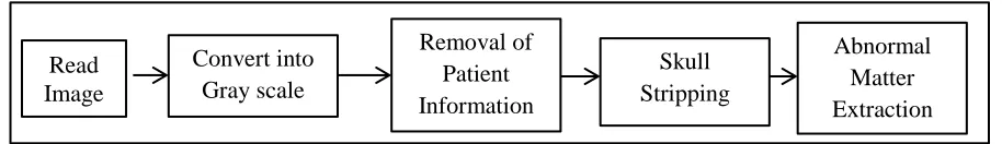
In this work two different tasks are carried together one is separation of patient information from image data and second abnormality detection from CT scan images. The proposed technique is shown in following Fig 1.
Mathematical morphology is a tool for extracting image component and description of region shape. Dilation grows the objects in binary image and Erosion shrinks objects in binary image. Where the thickening and shrinking is controlled by a structuring element of different shapes like disk, line, diamond, square etc.
Mathematical notation of dilation of image I by structuring element s is as follows
𝐼 ⊕ 𝑠 = {𝑧|(𝑠̂)𝑧∩ 𝐼 ≠ ∅} (1)
The mathematical notation of erosion of image I by structuring element s is as follows Read
Image
Convert into Gray scale
Removal of Patient Information
Skull Stripping
Abnormal Matter Extraction
Available Online at www.ijpret.com 1716
𝐼 ⊖ 𝑠 = {𝑧|(𝑠)𝑧∩ 𝐼𝑐 ≠ ∅} (2)
Proposed Algorithm:
1. Read Image:-In this step CT scan image is read.
2. Conversion:-In this step CT scan image is converted into gray scale image.
3. Patient information removal:-Patient information is removed using morphological erosion followed by dilation. Disk type structuring element of size 35 is used for erosion and size 40 is used for dilation.
4. Skull Stripping:-The abnormality lies inside the brain tissues. Hence, In This step Skull tissues are removed from the original image by using the morphological erosion using disk type structuring element of size 35.The resultant image contain the image with brain tissues only.
5. Abnormal matter detection:-In this step abnormal region extracted from brain tissues.
Abnormalities appear brighter than brain normal tissues.
III. RESULTS:
Available Online at www.ijpret.com 1717 (a) (b) (c)
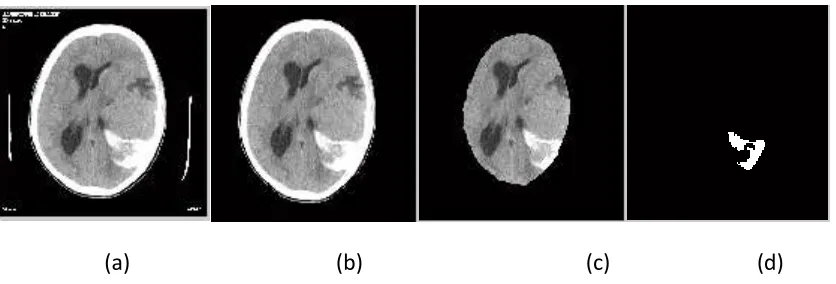
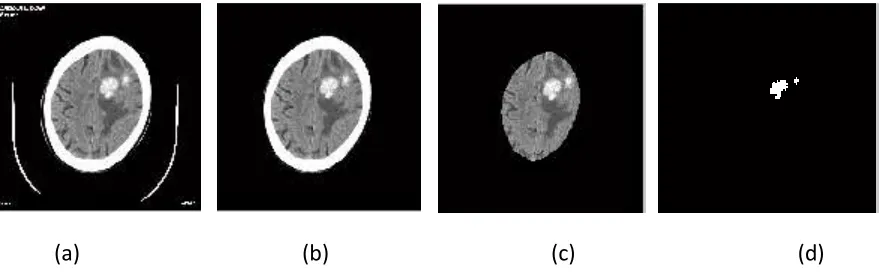
The Image without text is furthure processed for abnormality extraction from CT scan brain images.CT Scan brain image can be segmented into three regions Skull,Brain tissues(Gray Matter GM) and Abnormal tissues(White matter WM).The intensity values of the skull tissues are the brightest tissues in image.Whereas the Brain tissues appear gray and abnormalities appeared as brighter than brain tissues.In this work it is found that abnormal tissues can also of equal intensity of skull.That time if the abnormalities are connected to skull then it is very diffcult to segment abnormal tissues.Hence,morphological erosion is used for skull stripping which separate the skull tissues from the brain matter.After which abnormal tissues are easily segmented from brain tissues as shown in figure Fig.3. Fig 4 shows the results of the proposed technique applied on the CT scan brain image in which the abnormality lies inside the brain tissues.
(a) (b) (c) (d)
Figure 2 Patient information removal (a)original Image (b) Image without text (c) Text separated Image
Available Online at www.ijpret.com 1718
(a) (b) (c) (d)
IV. CONCLUSION:
In this paper morphological erosion and dilation is used for Patient information removal and Abnormality detection. CT scan brain images are segmented to separate the abnormalities from brain. The experimental results a show the proposed technique is effectively separate the brain image and patient information and correctly segment the abnormal tissues from image. Morphological operators convert image into binary and hence processing time is very less.
REFERENCES:
1. Mr. Vishal Shinde, Miss. Priti Kine, Miss. Suchita Gadge, Mr. Shekhar Khatal, “Brain Tumor Identification using MRI Images”, International Journal on Recent and Innovation Trends in Computing and Communication, ISSN: 2321-8169 Volume: 2 Issue: 10 ,PP.3050-3054
2. Abhishek Thakur, Rajesh Kumar, Amandeep Bath, Jitender Sharma, “ Improved Segmentation Technique for Enhancement of Biomedical Images”, International Journal of Electrical & Electronics Engineering, e-ISSN: 1694-2310 | p-ISSN: 1694-2426, PP.04-10
3. Alyaa Hussein Ali, Shahad Imad Abdulsalam, Ihssan Subhi Nema , “Detection and Segmentation of Hemorrhage Stroke using Textural Analysis on Brain CT Images”, International Journal of Soft Computing and Engineering (IJSCE) ,ISSN: 2231-2307, Volume-5 Issue-1, March 2015,PP.11-14
4. Miss. Roopali R. Laddha, Dr. Siddharth A. Ladhake, “Brain Tumor Detection Using Morphological And Watershed Operators”, International Journal of Application or Innovation in
Available Online at www.ijpret.com 1719 Engineering & Management (IJAIEM),Volume 3, Issue 3, March 2014 ISSN 2319 – 4847,PP.383-387
5. Roopali R.Laddha, S.A.Ladhake , “A Review on Brain Tumor Detection Using Segmentation And Threshold Operations”, International Journal of Computer Science and Information Technologies,ISSN:0975-9646 , Vol. 5 (1) , 2014, 607-611
6. Rachana Rana, H.S. Bhadauria, Annapurna Singh ,“Study of Various Methods for Brain Tumour Segmentation from MRI Images” , International Journal of Emerging Technology and Advanced Engineering,ISSN 2250-2459, Volume 3, Issue 6, June 2013,PP.338-348
7. Swe Zin Oo, Aung Soe Khaing , “Brain Tumor Detection And Segmentation Using Watershed Segmentation And Morphological Operation”, IJRET: International Journal of Research in Engineering and Technology eISSN: 2319-1163 | pISSN: 2321-7308,PP.367-374
8. Rohini Paul Joseph, C. Senthil Singh, M.Manikandan , “Brain Tumor Mri Image Segmentation And Detection In Image Processing”, IJRET: International Journal of Research in Engineering and Technology eISSN: 2319-1163 , pISSN: 2321-7308, PP.01-05
9. Alyaa Hussein Ali, Shahad Imad Abdulsalam, Ihssan Subhi Nema , “Detection and Segmentation of Ischemic Stroke Using Textural Analysis on Brain CT Images”, International Journal of Scientific & Engineering Research, Volume 6, Issue 2, February-2015, ISSN 2229-5518,PP.396-400
10. Mohammed Y. Kamil, “Brain Tumor Area Calculation in CT-scan image using Morphological Operations”, IOSR Journal of Computer Engineering, e-ISSN: 2278-0661,p-ISSN: 2278-8727, Volume 17, Issue 2, Ver. V (Mar – Apr. 2015), PP 125-128
11. U.Vanitha, P.Prabhu Deepak, N. Pon Nageswaran, R.Sathappan, “ Tumor Detection In Brain
Using Morphological Image Processing”, Journal of Applied Science and Engineering Methodologies,Volume.1,No.1, 2015, Page.131-136