1556-6811/07/$08.00⫹0 doi:10.1128/CVI.00079-07
Copyright © 2007, American Society for Microbiology. All Rights Reserved.
Aggregating Phenotype in
Lactobacillus crispatus
Determines Intestinal
Colonization and TLR2 and TLR4 Modulation in
Murine Colonic Mucosa
䌤
Sandra Voltan,
1Ignazio Castagliuolo,
1* Marina Elli,
4Stefano Longo,
4Paola Brun,
1Renata D’Inca
`,
2Andrea Porzionato,
3Veronica Macchi,
3Giorgio Palu
`,
1Giacomo C. Sturniolo,
2Lorenzo Morelli,
4and Diego Martines
2Department of Histology, Microbiology, and Medical Biotechnologies,1Department of Gastroenterological Sciences,2
and Department of Human Anatomy and Physiology,3University of Padua, Padua, Italy,
and AAT-Advanced Analytical Technologies S.r.l., Piacenza, Italy4
Received 5 February 2007/Returned for modification 18 April 2007/Accepted 5 July 2007
The colonic microbiota is a major modulator of the mucosal immune system; therefore, its manipulation through supplementation with probiotics may significantly affect the host’s immune responses. Since different probiotics seem to exert various effects in vivo, we tested the relevance of the autoaggregation phenotype on the intestinal persistence of lactobacilli and their ability to modulate the host’s innate immune responses. After 14 days of diet supplementation, the aggregating strainLactobacillus crispatusM247 but not aggregation-deficient isogenic mutant MU5 was recovered from the feces and colonic mucosa of mice. This observation was confirmed by strain-specific PCR amplification and byLactobacillus-specific denaturing gradient gel electrophoresis analysis. Indeed,L. crispa-tusM247 increased Toll-like receptor 2 (TLR2) mRNA levels, while it reduced TLR4 mRNA and protein levels in the colonic mucosa, whereas MU5 was ineffective. In colonic epithelial cells (CMT-93 cells)L. crispatusM247 but not MU5 induced time-dependent extracellular signal-regulated kinase-1 (ERK1) tyrosine phosphorylation and TLR modulation, which were abolished in the presence of PD98059 (an ERK1 inhibitor). To assess the functional relevance of probiotic-induced TLR modulation, we determined the consequences ofL. crispatuspreexposure on TLR4 (lipopolysaccharide [LPS]) and TLR2 [Pam3Cys-Ser-(Lys)4] ligand-mediated effects in intestinal epithelial
cells. Preexposure toL. crispatusM247 blunted LPS-induced interleukin-6 (IL-6) release and inhibition of CMT-93 migration over a wound edge, whereas it enhanced TLR2-mediated IL-10 up-regulation. In summary, the aggre-gation phenotype is required forL. crispatuspersistence in the colon and for modulation of TLR2/TLR4 expression through an ERK-dependent pathway. We speculate that the aggregation phenotype inL. crispatusM247 is required to temper epithelial cell responsiveness to bacterial endotoxins, which thus affects the evolution of intestinal inflammatory processes.
Humans and animals are born germfree, but soon after birth they are colonized with microorganisms. Within a few days, the mucosae and the skin are the homes to a vast and complex community of microorganisms. Indeed, 400 species are estimated to inhabit the gastrointestinal tract and establish life-long inter-actions with the host mucosae to influence a variety of activities of paramount relevance, including the function of the mucosal im-mune system (27). On the other hand, components of the intes-tinal microbiota possess the potential to damage the mucosa either through toxin release or as a cause of detrimental immune responses. Thus, in a variety of animal models, intestinal inflam-mation does not occur when mice are raised under germfree conditions unless their bacterial flora is reconstituted (53). In accordance with the complex effects of the colonic flora on the mucosal immune system, changes in the mucosa-associated mi-crobiota have been related to a variety of diseases (53). Indeed, manipulation of the flora colonizing mucosal surfaces by oral supplementation with live bacteria might influence the host’s
health and has been proposed as a means for the prevention or treatment of a range of diseases, although the mechanism(s) of action of probiotics remains elusive (53). Thus, probiotics can directly suppress the growth of pathogens through the secretion of antimicrobial substances or induce the expression of protective molecules to enhance the mucosal barrier function (56, 59). Fur-thermore, probiotics modulate the mucosal immune system either directly, affecting immune cell activities, or through the manipu-lation of the colonic microbiota. However, different probiotics seem to exert various effects on the host, suggesting the existence of distinctive strain properties (20). Despite the growing number of clinical applications, at the moment no phenotypic markers with which probiotic effects can be predicted in vivo are available. The gut epithelial cells are no longer considered a mechan-ical barrier to the prevention of microbial invasion, as they directly sense the gut environment and activate a variety of intracellular pathways in response to specific bacterium-de-rived products (55). A major breakthrough in the understand-ing of the molecular mechanisms involved in regulatunderstand-ing the bacteria-host interaction was the demonstration that immune and nonimmune cells, including intestinal epithelial cells, rec-ognize several microbial products, referred to as “pathogen-associated molecular patterns,” like the lipopolysaccarides (LPSs) of gram-negative bacteria and the peptidoglycan
frag-* Corresponding author. Mailing address: University of Padua, School of Pharmacy, Department of Histology, Microbiology, and Medical Biotechnologies, Via A. Gabelli 63, Padua 35121, Italy. Phone: 049-827-2360. Fax: 049-827-2355. E-mail: ignazio.castagliuolo @unipd.it.
䌤Published ahead of print on 18 July 2007.
1138
on August 17, 2020 by guest
http://cvi.asm.org/
ments of gram-positive bacteria, through molecules called “pattern recognition receptors” (PRRs) (9). Among the PRRs are the mammalian homologues ofDrosophilaToll receptors, referred to a Toll-like receptors (TLRs), which are transmem-brane proteins characterized by an extracellular domain able to bind different pathogen-associated molecular patterns (5). Thus, TLR4 is the prototype of the gram-negative bacterial LPS receptor, whereas TLR2 is the main receptor for pep-tidoglycan fragments and lipoteichoic acid from gram-positive bacteria. Individual TLRs differentially activate distinct signal-ing events via cofactors and adaptor proteins, leadsignal-ing to the activation and nuclear translocation of transcription factors. These factors modulate the expression of pro- and anti-inflam-matory cytokines and chemokines, which regulate the activities of the innate and the adaptive immune responses (7). These events are involved to control host homeostasis, pathogen sup-pression, and the responses to probiotic ingestion (19, 33, 62). To exert favorable effects on the host, administered pro-biotics are supposed to induce intestinal colonization and to manipulate the colonic microbiota (53). However, to draw a comprehensive picture of the colonic and fecal microbiota of humans and animals, traditional culture-based methods are nowadays considered obsolete for the large number of noncul-tivable microorganisms, whereas several molecular tools allow the identification of strictly anaerobic species, which are usu-ally predominant in the large bowel of mammals (6, 36, 52, 62). Thus, PCR coupled with denaturing gradient gel electrophore-sis (DGGE) was recently applied to the study of complex bacterial communities, with a particular focus on the gut mi-croflora (24, 64) and its fluctuations in diseases or following probiotic administration (17, 29, 31).
Since in a previous study we observed that aggregation-deficientLactobacillus crispatusMU5 was devoid of therapeu-tic effects in a colitis model, as opposed to wild-type strain M247, which has an aggregation phenotype, we hypothesized that the aggregation phenotype might give the probiotic strains advantages that are relevant to their in vivo effects (13). Thus, in the study described here, we assessed the impact of oral supplementation with two isogenic strains ofL. crispatus, spon-taneously aggregating strain M247 and aggregation-deficient strain MU5, on the colonic microbiota and the associated ef-fects on the mucosal level of PRRs, with the view that the levels of these receptors contribute to the establishment of the responsiveness of the mucosa-associated immune system to bacterium-derived products and regulate the amplitudes of the inflammatory responses.
MATERIALS AND METHODS
Isolation, characterization, and culture ofLactobacillus crispatus.Lactobacillus crispatusstrain M247 was isolated and characterized as described previously (14). Cells grown in De Man-Rogosa-Sharpe (MRS; Difco) medium appeared to the naked eye as discernible clumps which sediment at the bottom of the tube, leaving the upper part of the medium clear. A spontaneous nonclumping mutant of M247, named MU5, was isolated from the lower aqueous phase during a hydrophobic assessment test based on the water-hexadecane partition assay. Strains were grown in MRS broth or agar at 37°C under microaerophilic condi-tions (10% CO2in air [21% O2]).
Preparation of orally administered cultures.L. crispatusM247 and MU5 were grown in MRS medium at 37°C for 18 h. The cells were harvested by centrifu-gation at 8,000 rpm for 5 min, washed twice with sterile distilled saline, and finally suspended in GG solution (20% glucose plus 10% glycerol) to obtain a final concentration of 108CFU per 100l of bacterial suspension.
Administration ofL. crispatusM247 and MU5 to mice.BALB/c mice (age, 8 to 10 weeks) purchased from Charles River (Charles River Laboratories, Lecco, Italy) were used in all experiments. The animal studies were approved by the Institutional Animal Care and Use Committee of the University of Padua. The animals, housed in groups of four mice per cage, were randomly divided into three experimental groups of 8 to 12 elements each. The mice received 108CFU ofL. crispatusM247 or MU5, whereas control animals received only GG solution (vehicle). The microorganisms were administered daily intragastrically via a polyethylene cannula in a total volume of 100l. During the treatment period the animals had free access to food and water. After 14 days the animals were killed by use of an overdose of anesthesia (xylazine-ketamine; 100 mg/kg of body weight), the abdomen was immediately opened, and the proximal colon was removed. For each mouse a colon sample was frozen in liquid nitrogen for RNA extraction. Full-thickness specimens of the colon were fixed in 4% paraform-aldehyde and embedded in paraffin, and 10-m-thick sections were stained with hematoxylin-eosin for routine histological examination. An adjacent colon seg-ment was placed in a cryoembedding matrix (OCT) and frozen in isopentane at
⫺110°C. In addition, tissue samples were placed in ice-cold RPMI 1640 medium (Gibco, Milan, Italy) and were immediately processed for epithelial cell isolation. Persistence study.Fecal samples were collected immediately before the first administration (time zero) and after 7 days and 14 days of M247 and MU5 supple-mentation. A sample of the colonic mucosa was also collected after 14 days of probiotic supplementation. Fecal pellets and colonic tissue specimens were imme-diately placed in Amies medium (3) and stored at 4°C until they were processed (within 24 h). Fecal samples were then serially diluted with sterile saline solution, plated onto MRS medium (Difco), and incubated in an anaerobic atmosphere (85% N2, 10% H2, 5% CO2) at 37°C for 48 h. To recover bacteria adherent to the mucosa, colonic tissue samples were washed twice in sterile saline and subjected to hypotonic lysis, and then the debris was collected by centrifugation and seeded onto MRS plates and incubated under anaerobic conditions. White colonies were then replica plated and incubated anaerobically at 37°C for 48 h. The isolated strains were then identified by strain-specific PCR with primers specific forL. crispatusM247 and MU5, as described by Cesena et al. (14).
DNA extraction, PCR amplification, and DGGE analysis. The DNA was directly extracted from fecal samples by means of a PSPSpin stool kit (Invitrogen, Germany). The eluted DNA was amplified by nested PCR with two different primer pairs. The PCR products obtained by using primers S-D-Bact-0011-a-A-17 and S-G-Lab-0677-a-S-D-Bact-0011-a-A-17 (24) were then used as templates in nested PCR with primers S-G-Lab-0159-A-S-20 and S-Univ-0515-a-A-24-GC (31). The cy-cling conditions were those previously indicated by Heilig et al. (24) and Kon-stantinov et al. (31). The amplicons were then analyzed by gel electrophoresis and were visualized by ethidium bromide staining.
DGGE analysis was performed with an Ingeny2⫻2 system (IngenyPhor, Den-mark), which was run at 60°C and a voltage of 120 V for 16 h. An 8% poly-acrylamide gel with a 30% to 60% urea gradient was loaded with 40l of amplified sample and run with 1⫻TAE (Tris-acetate-EDTA) buffer. The gel was stained with SYBR green dye (Bio-Rad) and was viewed under UV transillumi-nation. The amplified fragments were then excised from the gel, and the DNA was eluted in sterile distilled water after 18 h incubation at 4°C. A small amount of the eluted DNA was submitted to PCR amplification with primers S-G-Lab-0159-A-S-20 and S-Univ-0515-a-A-24-GC, which were described previously (31). The amplicons were purified with a Microclean kit (Microzone, United King-dom), and the eluted DNA was sequenced with primer S-G-Lab-0159-A-S-20 at CRIBI, University of Padua. The sequences were then analyzed as described by Knarreborg et al. (29).
Isolation of epithelial cells from mouse colon.Pieces of colon tissue from the control and theL. crispatus-supplemented mice were surgically removed and washed with RPMI 1640 medium to remove mucus and fecal matter and were placed into Hank’s balanced salt solution (HBSS; GIBCO BRL) containing Ca2⫹, Mg2⫹, and 1 mM dithiothreitol for 20 min at room temperature with
occasional agitation. To remove and recover epithelial cells, the specimens of colonic tissues obtained from the three different experimental groups were then cut into small pieces (2 by 2 mm) and placed in HBSS supplemented with 1 mM EDTA but without Ca2⫹and Mg2⫹. The specimens were incubated for 30 min
at 37°C with agitation (170 rpm), and then the medium was collected and the epithelial cells were pelleted by centrifugation (2,000⫻gfor 10 min). Contam-inating intraepithelial lymphocytes were removed by centrifugation (2,000⫻g for 20 min) through a Percoll gradient (Amersham-Pharmacia, Milan, Italy). The epithelial cells were collected, washed twice in ice-cold phosphate-buffered saline (PBS), and stored at⫺80°C for subsequent RNA extraction.
Cell culture.The CMT-93 mouse colonic epithelial cell line was obtained from the European Collection of Cell Cultures (ECACC no. 89111413). The CMT-93 cells were maintained in Dulbecco’s modified Eagle medium (GIBCO)
on August 17, 2020 by guest
http://cvi.asm.org/
mented with 10% heat-inactivated fetal calf serum (GIBCO), 100 U/ml penicil-lin, and 100g/ml streptomycin (complete medium). Subconfluent monolayers were trypsinized, resuspended at 106cells/ml, and seeded in 6- or 12-well tissue culture plates (Costar). At 7 days postconfluence, the medium was removed; and the cells were washed twice in antibiotic-free medium (AFM) and refed fresh AFM alone or AFM containing 108
CFU/mlL. crispatusM247,L. crispatusMU5, or enteropathogenicEscherichia coliATCC 49106. When it was so indicated, the monolayers were treated with the specific extracellular signal-regulated kinase-1 (ERK1) inhibitor (PD98059; Calbiochem) 30 min before exposure to the bacte-ria. After 1 h of coincubation withL. crispatusat 37°C, the monolayers were washed three times with AFM and refed with complete medium, and when it was so indicated, the monolayers were treated with TLR2 [Pam3Cys-Ser-(Lys)4 (Pam3CSK4)] or TLR4 (LPS) ligand (10g/ml). After an additional 0 to 24 h of incubation at 37°C, the cells were removed with a cell scraper, washed with ice-cold PBS, centrifuged (1,500⫻gfor 10 min), and used for RNA or protein extraction.
RNA extraction and quantitative real-time RT-PCR analysis.Samples of the colonic mucosa or purified epithelial cells were placed in 175l of SV RNA lysis buffer from the SV total RNA isolation system kit obtained from Promega Corporation (Madison, WI) and homogenized with a Retsch MM300 apparatus (QIAGEN, Milan Italy). The total RNA was then purified according to the manufacturer’s protocol, and the contaminating DNA was removed by DNase I digestion. RNA purity was confirmed by assessing the optical density at 260 nm (OD260) and the OD280. Samples (3g of total RNA) with OD260/OD280ratios of between 1.8 and 2 were used to generate randomly primed cDNAs with Moloney murine leukemia virus reverse transcriptase (Applied Biosystems, Fos-ter City, CA).
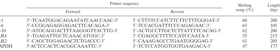
Real-time quantitative reverse transcription-PCR (RT-PCR) analysis for in-terleukin-6 (IL-6), IL-1, IL-10, TLR4, and TLR2 mRNAs was performed on an ABI Prism 7700 sequence detector (Applied Biosystems, Milan, Italy). The oligonucleotide primer sequences and PCR conditions used are reported in Table 1. Quantitative RT-PCR analysis was performed with a SYBR green PCR core reagents kit (Applied Biosystems, Milan, Italy), according to the manufac-turer’s protocol. Standard curves were obtained by amplification of the corre-sponding cDNAs subcloned into the pGEM-T vector (Promega, Italy). The expression of the target genes was normalized to that of the endogenous control gene, glyceraldehyde-3-phosphate dehydrogenase (GAPDH).
Immunoprecipitation and Western blotting.To extract total proteins from CMT-93 cells following the treatments, the cells were washed twice with ice-cold PBS and then lysed (45 min on ice) with nondenaturing RIPA buffer (150 mM NaCl, 50 mM Tris-HCl, 0.25% sodium deoxycholate, 0.1% Nonidet P-40, 100
M NaVO4, 1 mM NaF, 1 mM phenylmethylsulfonyl fluoride, 10g/ml apro-tinin, and 10g/ml leupeptin). Particulate material was removed by centrifuga-tion (15,000⫻gfor 5 min at 4°C), the supernatants were collected, and the protein concentrations were determined by the Bradford method (Pierce, Cram-blington, United Kingdom). To assess ERK1 phosphorylation following L. crispatusexposure, cell lysates (2 mg/ml) were incubated with a rabbit anti-ERK1 polyclonal antibody (10g/mg cell lysate; Santa Cruz Biotechnology, Santa Cruz, CA) for 2 h at 4°C. Then, protein A-agarose (Santa Cruz Biotechnology) was added and the mixture was incubated for 1 h at 4°C. Beads were washed twice by centrifugation (20 s, 12,000⫻g) with ice-cold RIPA buffer, followed by one wash with ice-cold PBS, and were then boiled in 25l of sample loading buffer (62.5 mM Tris, pH 6.8, 10% glycerol, 2% sodium dodecyl sulfate [SDS], 5% -mer-captoethanol, and 0.1% bromophenol blue). The immunoprecipitated proteins were then fractionated on an SDS-polyacrylamide gel and transferred to and immobilized on a nitrocellulose membrane. To determine the TLR2 expression level, 40g of total proteins was boiled in loading buffer and then fractionated on an SDS-polyacrylamide gel and transferred to and immobilized on a nitro-cellulose membrane. The membranes were blocked overnight at 4°C in 5% skim
milk in PBS containing 0.05% Tween 20 and were then incubated for 2 h with the proper antibody (anti-TLR2, anti-phosphotyrosine pY99; Santa Cruz Biotech-nology). Bound antibody was then detected by incubating the nitrocellulose membrane with horseradish peroxidase-conjugated donkey goat (for anti-TLR2) immunoglobulin G (IgG) antibody (Santa Cruz Biotechnology), and the immunocomplexes were visualized by using enhanced chemiluminescence West-ern blot analysis detection reagents (Santa Cruz Biotechnology). The membranes were photographed with a VersaDoc imaging system (Bio-Rad), and the images were digitally stored with Quantity One (Bio-Rad) software.
Immunohistochemistry.Immunohistochemistry was performed with colonic tissue sections (10m thick) obtained from control andL. crispatus -supple-mented mice. The frozen sections were fixed in methanol (5 min at⫺20°C) and then washed twice (5 min each) in Tris-buffered saline (TBS), and nonspecific binding was blocked by incubation with 2% donkey serum in TBS for 20 min. The sections were then incubated with properly diluted primary antibody (rabbit polyclonal anti-TLR4 or goat anti-TLR2 antibody; Santa Cruz Biotechnology) for 2 h at 22°C. Nonbound antibody was removed by extensive washes with TBS. Immunocomplexes were detected with goat Alexa Fluo 488-labeled anti-rabbit IgG (Invitrogen Corporation, Italy) or a rabbit Alexa Fluo 488-labeled anti-goat IgG (Invitrogen Corporation, Italy). The sections were then washed, mounted, analyzed, and photographed with a Leica TCSNT/SP2 confocal microscope (⫻63 objective). The images were digitally stored by using Leica software and were then elaborated by using a graphics program (Adobe).
IL-6 release.Following 1 h incubation with AFM alone or AFM supplemented with 108CFU/mlL. crispatusM247 or MU5, the CMT-93 cell monolayers were exposed to LPS (10g/ml) for 6 h. Then, the culture medium was collected, centrifuged (2,000 rpm for 10 min) to remove the deattached cells, and stored at⫺20°C. IL-6 release was measured by a commercially available enzyme-linked immunosorbent assay (Biosource, Camarillo, CA), following the manufacturer’s protocol. The results were expressed as ng/ml.
Migration (restitution) assays.Assays for wound healing were carried out as described previously (34). The CMT-93 cells dissociated with trypsin-EDTA were seeded onto microscope coverslips and grown in complete medium. At 7 days postconfluence, the monolayers were washed and then incubated with AFM alone or AFM supplemented with 108CFU/ml ofL. crispatusM247 or MU5. One hour later, the medium was removed and the cells were washed with sterile PBS and incubated with complete medium. The monolayers were incubated for an additional 2 h at 37°C in 5% CO2before linear wounds were made with a sterile razor blade. Then, the cells were incubated in AFM alone or AFM supplemented with LPS (10g/ml) for an additional 24 h. Finally, the mono-layers were washed in cold PBS, fixed in buffered 2% paraformaldehyde for 5 min, and then mounted on microscope slides. Migration was assessed in a blinded fashion by determining the number of CMT-93 cells across the wound border in a defined wound area by taking photomicrographs at a fivefold mag-nification with a Leica DM-LB inverted microscope connected to a digital Leica DC-100 camera. The experiments were performed in triplicate, and at least 10 wound areas were used to quantify the migration.
Statistical analysis.The data are expressed as means⫾standard errors (SEs). Statistical analysis was performed by using attest for unpaired samples. Statis-tical significance was considered for aPvalue of⬍0.05.
RESULTS
Persistence ofL. crispatusM247 in feces and adherence to colonic mucosa of mice.Fecal samples from each animal were collected before the treatment withL. crispatuswas started and after 7 and 14 days of daily supplementation with 108CFU of
TABLE 1. PCR primers and conditions used in the study
Primer sequence Melting
temp (°C)
Length (bp)
Forward Reverse
IL-1 5⬘-TCAATGGACAGAATATCAACCAAC-3⬘ 5⬘-CTTTTCCATCTTCTTCTTTGGGAT-3⬘ 60 200
IL-6 5⬘-CCGGAGAGGAGACTTCACAGA-3⬘ 5⬘-TCCACGATTTCCCAGAGAAC-3⬘ 63 105
IL-10 5⬘-ATGCAGGACTTTAAGGGTTACTTG-3⬘ 5⬘-ACTGCCTTGCTCTTATTTTCACAG-3⬘ 62 206
TLR4 5⬘-TGAGATTGCTCAAACATGGC-3⬘ 5⬘-CGAGGCTTTTCCATCCAATA-3⬘ 55 213
TLR2 5⬘-CAGCTGGAGAACTCTGACCC-3⬘ 5⬘-CAAAGAGCCTGAATGGGGAG-3⬘ 61 205
GAPDH 5⬘ACTCCACTCACGGCAAATTC-3⬘ 5⬘-TCTCCATGGTGGTGAAGACA-3⬘ 47 177
on August 17, 2020 by guest
http://cvi.asm.org/
eitherL. crispatusM247 or MU5.L. crispatusM247 or MU5 was not identified in the feces of any animal at the beginning of the experiment. Viable M247 cells were retrieved in the feces of 3 of 17 mice after 7 days of supplementation and in the fecal samples of 12 of 17 mice at the end of the treatment period (Table 2). However, viableL. crispatusMU5 cells were iden-tified only at day 14 in 2 of 14 fecal samples from the treated animals. The adherence of the probiotic strains to the colonic
mucosa was evaluated at day 14, when the mice were killed; and as shown in Table 2,L. crispatusM247 was identified in 9 of 17 colonic tissue specimens, whereas we were not able to retrieveL. crispatusMU5 in any colonic tissue specimen.
Recovery ofL. crispatusDNA from fecal samples.Following supplementation of the diet withL. crispatusM247 and MU5, the colonic microbiota of the BALB/c mice was analyzed by PCR-DGGE. DNAs extracted from fecal samples before and after probiotic supplementation were amplified and analyzed by PCR with universal primers S-D-Bact-0968-A-S-GC and S-D-Bact-1401-a-A-17 (32) in order to amplify the V6 to V8 region of the 16S RNA gene. However, the probiotic strains administered were not retrieved in the DGGE profiles ob-tained with these primers (data not shown).
Mice receiving M247 and MU5 were therefore studied by using the DGGE primers designed by Konstantinov et al. (31, 32) and Knarreborg et al. (29) to monitor theLactobacillus -specific bacterial community in the gastrointestinal tract. The profiles obtained for the fecal samples collected at day 0 and those collected at day 14 were compared. Several DNA frag-ments were excised from the gel, and their sequences were found to correspond to those of theLactobacillus genus, as shown in Fig. 1. Indeed,L. murinus(100% identity; GenBank accession no. AF157049), as well asL. johnsonii(100% iden-tity; GenBank accession no. AE017198), was commonly de-tected in the microbiota of both nontreated and probiotic-treated mice. Moreover, M247-probiotic-treated mice revealed DGGE bands identified as L. intestinalis/L. crispatus (98% identity; GenBank accession no. AM117143) because of the high degree of similarity in the 16S rRNA gene sequences of these two species. However, these fragments were not retrieved in the profiles of MU5-treated mice.
TABLE 2. Recovery of viableL. crispatusM247 and MU5 from mouse feces and tissues
Sample type and no. of detections per mouse
L. crispatusM247 L. crispatusMU5
No. of mice whose feces contained
viable probiotic
strain
Mean log10 CFU/g (wet wt) of
sample
No. of mice whose feces contained
viable probiotic
strain
Mean log10 CFU/g (wet wt) of
sample
Feces
Twicea 3 7.30⫾0.44 0 NDb
Oncec 9 8.22⫾0.25 2 8.67⫾0.05
Never 5 ND 12 ND
Colonic tissued
Oncec 9 7.09⫾0.21 5 7.24⫾0.64
Never 8 ND 9 ND
Total no. of mice
17 14
aStrains M247 and MU5 were detected at days 7 and 14. bND, not determined.
cStrains M247 and MU5 were retrieved only at day 14.
dOnly one colonic tissue sample, obtained when the mice were killed at day
14, was analyzed.
FIG. 1. Oral supplementation with aggregating strainL. crispatusM247 influences colonic microbiota. Mice received orally 108CFU ofL. crispatusM247, aggregation-deficient mutant MU5, or GG solution (vehicle) in a total volume of 100l for 14 days. Then, total DNA was extracted from fecal samples and amplified by nested PCR with species-specific primer pairs to identifyLactobacillusspp. The amplicons were then analyzed by PCR-DGGE. The fragments were then excised from the gel, and the eluted DNA was sequenced.
on August 17, 2020 by guest
http://cvi.asm.org/
L. crispatussupplementation modulates the mRNA level of Th1 and Th2 cytokines in the colonic mucosa.Since we re-cently reported that spontaneously aggregating strainL. crispa-tus M247 but not isogenic nonaggregating mutant MU5 re-duced the severity of dextran sodium sulfate colitis in mice (13), we evaluated the effect ofL. crispatusM247 and MU5 supplementation on the proinflammatory and the anti-inflam-matory cytokine levels in the colonic mucosa. Thus, total RNA was extracted from the mucosa of mice supplemented for 2 weeks withL. crispatusM247 or MU5, and the amounts of the mRNAs coding for IL-6 and IL-10 were estimated by real-time quantitative RT-PCR. As shown in Fig. 2, the level of IL-6 mRNA in the colonic mucosa was significantly reduced after 2 weeks ofL. crispatusM247 supplementation, whereas the level of IL-10 mRNA was significantly increased compared to that in the controls. Indeed, as shown in Fig. 2, supplementation of the diet withL. crispatusMU5 did not significantly modify the mucosal level of steady-state IL-6 and IL-10 mRNAs.
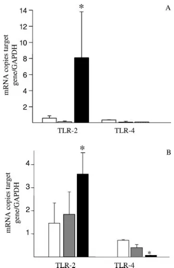
L. crispatus M247 but not aggregation-deficient mutant MU5 modulates TLR2 and TLR4 levels in the colonic mucosa.
Since conserved bacterial structures modulate the activity of mucosal immune system through pattern recognition receptors such as TLRs, we decided to assess the effect ofL. crispatus
supplementation on the TLR2 and TLR4 levels in the colonic mucosa and in epithelial cells. As shown in Fig. 3, supplemen-tation of the diet withL. crispatusM247 significantly increased the TLR2 mRNA levels both in the colonic mucosa and in epithelial cells, whereas the TLR4 mRNA levels were reduced in epithelial cells. These effects were evident in the colonic mucosa (Fig. 3A), as well in purified colonic epithelial cells (Fig. 3B). However, oral supplementation with the aggrega-tion-deficient strainL. crispatusMU5 had no effect on either TLR2 and TLR4 mRNA levels (Fig. 3).
We next determined whether L. crispatussupplementation modified TLR4 and TLR2 expression and/or their distribution in the colonic mucosa. As expected, the levels of TLR4- and TLR2-specific immunostaining in the colonic mucosa of the mice were low and were localized mainly in the epithelium (Fig. 4). Indeed, following 2 weeks of oral supplementation withL. crispatusM247, a striking increase in TLR2 immuno-reactivity was evident, but this was not the case withL. crispa-tusMU5. TLR2 staining was mainly localized in the epithe-lium, whereas we did not observe a significant change in mononuclear cell staining within the lamina propria. However, the intensity and distribution of TLR4-specific immunostaining were substantially unaffected in the mucosa.
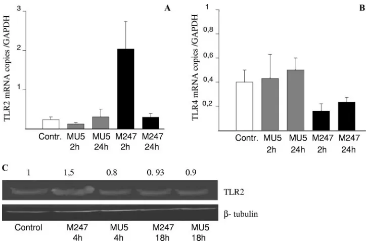
L. crispatus M247 directly modulates TLR expression in CMT-93 cells.Since oral supplementation withL. crispatusnot only modified the colonic microbiota but also was able to modulate cytokines and TLR expression in the colonic mucosa, we next determined whetherL. crispatus directly influenced TLR levels in intestinal epithelial cells. As shown in Fig. 5, following 1 h of coculture withL. crispatusM247, CMT-93 cells showed time-dependent increases in TLR2 mRNA levels in association with a decrease in TLR4 mRNA levels, whereas aggregation-deficient mutant MU5 had no significant effects
FIG. 2. Oral supplementation withL. crispatusmodifies cytokine mRNA levels in the colonic mucosa. Mice received orally 108CFUL. crispatusM247 (black bars), aggregation-deficient mutant MU5 (gray bars), MU5 in a 30% sucrose solution that was able to reestablish the aggregation phenotype (bars with vertical stripes), or GG solution (vehicle; open bars) in a total volume of 100l for 14 days. Total RNA was extracted from the mucosa, and steady-state IL-6 and IL-10 mRNA levels were determined by quantitative RT-PCR and are ex-pressed as the numbers of copies of the target gene/number of copies of the GAPDH gene (used as an internal standard) in 3g total RNA. For each condition, from six to eight determinations were performed, and the values are expressed as means⫾SEs.ⴱ,P⬍0.01 versus the results for the respective control.
FIG. 3. Supplementation of the diet withL. crispatusmodifies TLR mRNA levels in the colonic mucosa. Mice received 108CFUL. crispa-tusM247 (black bars), aggregation-deficient mutant MU5 (gray bars), or vehicle only (open bars) for 2 weeks. cDNA was prepared from total RNA extracted from the colonic mucosa (A) or from colonic epithelial cells (B), and the steady-state TLR2 and TLR4 mRNA levels were determined by quantitative RT-PCR. Values were expressed as the number of copies of TLR2 and TLR4 mRNA and were normalized to the number of copies of GAPDH mRNA (the endogenous RNA control) in 3 g total RNA. For each condition, from six to eight determinations were performed, and values are expressed as means⫾ SEs.ⴱ,P⬍0.01 versus the results for the control animals.
on August 17, 2020 by guest
http://cvi.asm.org/
on TLR2 and TLR4 mRNA levels. As shown in Fig. 5C, West-ern blot analysis demonstrated a similar time-dependent in-crease in the TLR-2 level in CMT-93 cells exposed toL. crispa-tusM247 but not in cells exposed to MU5. Indeed, incubation of CMT-93 cells with 108CFU/ml enteropathogenicE. colidid
not cause any significant change in the levels of TLR2 and TLR4 expression (data not shown).
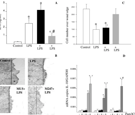
Functional relevance of L. crispatus M247-induced TLR modulation in CMT-93 cells.Recent studies reported the abil-ity of bacterium-derived products to modulate several activities in intestinal epithelial cells, including epithelial cell migration over the wound edge and cytokine release (15). Since we ob-served a striking change in TLR2 and TLR4 expression in the colonic mucosa of mice as well as in CMT-93 cells following exposure toL. crispatusM247, we assessed the functional rel-evance of this effect.
First, we determined the consequence ofL. crispatus expo-sure on LPS-induced proinflammatory cytokine release from CMT-93 cell monolayers (Fig. 6A). As expected, LPS induced a significant release of IL-6 from CMT-93 cell monolayers. However, monolayers preincubated withL. crispatusM247, but not with aggregation-deficient mutant MU5, showed a blunted LPS-induced IL-6 release.
Second, we assessed the healing properties of L. crispatus
treatment of CMT-93 cell monolayers in the presence of high
concentrations of LPS. As expected, CMT-93 cell migration over a wound edge was significantly inhibited in the presence of LPS (Fig. 6B and C). However, in CMT-93 cell monolayers preexposed toL. crispatus M247 but not to aggregation-defi-cient mutant MU5, the LPS-mediated effects were abolished and significant cell migration over the wound edge was ob-served.
Finally, we determined the effect of Pam3CSK4, a
TLR2-specific ligand, on the IL-10 mRNA level in CMT-93 cells preexposed toL. crispatusM247 or MU5. As shown in Fig. 6D, preincubation of the CMT-93 cells withL. crispatusM247, but not MU5, caused a significant increase in the IL-10 mRNA levels in the epithelial cells for up to 12 h. However, following exposure of the CMT-93 cells toL. crispatusM247, incubation with the TLR2-specific ligand resulted in a further increase in the IL-10 mRNA level that was still evident after 24 h.
TheL. crispatusM247-induced TLR modulation in CMT-93 cells involves ERK activity.Since recent studies suggested that nonpathogenic bacteria can activate specific intracellular signal cascade pathways (49, 50), we investigated the role of ERK1 in
L. crispatus-induced TLR modulation. As shown in Fig. 7, incubation of CMT-93 cell monolayers withL. crispatusM247 induced a strong and time-dependent ERK1 tyrosine phosphor-ylation. Interestingly, aggregation-deficient mutantL. crispatus
MU5, which was unable to induce significant effects on TLR
FIG. 4. Oral supplementation withL. crispatusmodulates TLR4 and TLR2 protein levels in the colonic mucosa. Immunofluorescence analysis was performed with frozen sections (thickness, 10m) of large intestine tissue specimens collected from control mice and animals supplemented with eitherL. crispatusM247 orL. crispatusMU5 for 14 days. The sections were fixed in cold acetone and were incubated with an anti-TLR4 monoclonal antibody or an anti-TLR2 monoclonal antibody. Specific immunocomplexes were detected by using fluorescein isothiocyanate-labeled secondary antibodies and were visualized on a Leica TCS-NT/SP2 confocal microscope with a⫻63 objective. The images are representative of four separate experiments. The arrows indicate specific immunostaining.
on August 17, 2020 by guest
http://cvi.asm.org/
mRNA and protein levels (Fig. 3 and 4), did not induce sig-nificant changes in the ERK1 tyrosine phosphorylation level. To investigate the functional relevance of ERK1 phosphoryla-tion in M247-induced TLR modulaphosphoryla-tion, we treated the CMT-93 cell monolayers with the specific ERK1 inhibitor PD98059. As depicted in Fig. 7, the inhibition of ERK1 activity significantly inhibited the effects ofL. crispatusM247 on the TLR2 and TLR4 mRNAs levels in CMT-93 cell monolayers.
DISCUSSION
An imbalance in the endogenous intestinal microflora is now considered a critical component in the chain of events contrib-uting to the development of dysfunctional immune responses by the host’s mucosa-associated immune system, leading to the onset of many clinically relevant diseases (60). Indeed, several clinical trials that used live bacteria (i.e., probiotics) to manip-ulate the intestinal flora for the treatment of acute and chronic diseases have obtained encouraging results (8, 54). However, the choice of biotherapeutic agents is still based on empirical approaches, and comprehensive knowledge of the bacteria’s relevant phenotypic traits necessary to induce beneficial effects on the host’s microbiota and mucosal immune system is lack-ing. In this study we identified a phenotypic characteristic as-sociated with a probiotic strain required to guarantee its per-sistence in the gastrointestinal tract, to shape the intestinal microflora, and to modulate in intestinal epithelial cells the expression of TLRs, a class of receptors able to deeply affect
the activities of the innate and adaptive mucosal immune re-sponses following microbe recognition.
Probiotic candidates for therapeutic applications are generally screened by using heterogeneous models on the basis of their ability to survive in the presence of gastric acid, tolerate bile salts, and adhere to gut mucus and epithelial cell monolayers in vitro but by paying no attention to the identification of the bacterial characteristics required to exert the beneficial effects in vivo (35, 53). Therefore, it is not surprising that the probiotic bacteria chosen by this strategy often fail to exert the desired biological effect (21). Here we report that the aggregation phenotype, often observed withinLactobacillusspp., is associated with a stronger ability to colonize the intestine and to produce immunomodula-tory effects in vitro and in vivo. Indeed, in a previous study we reported that the aggregation phenotype was associated with pro-tective effects in a colitis model in vivo (13). The ability to aggre-gate or coaggreaggre-gate has been demonstrated in several bacterial species colonizing harsh environments, such as the oral cavity and the intestinal mucosa (38, 48). Indeed, severalLactobacillusspp. show a strong aggregating phenotype and the property of coag-gregation withE. colistrains and enterococci (14, 30). This phe-notypic property may provide greater chances for survival, persis-tence, and colonization of the host’s mucosal surfaces and, therefore, to come into strict contact with the mucosal immune system.
The indigenous commensal microflora that initiates innate immune responses plays an active role in host mechanisms that
FIG. 5.L. crispatusM247 but notL. crispatusMU5 modulates TLR4 and TLR2 levels in CMT-93 cells in vitro. CMT-93 cell monolayers were exposed toL. crispatusM247 (black bars) or MU5 (gray bars) or to medium only as a control (open bars). After 1 h of culture the medium was removed and the cells were washed and incubated for additional 1 to 24 h in fresh complete medium. (A and B) The cells were collected, the total RNA was extracted, steady-state TLR2 and TLR4 mRNA levels were determined by quantitative RT-PCR, and the values were normalized to those for GAPDH mRNA (the endogenous RNA control) in 3g total RNA. For each condition, nine determinations (three experiments with triplicate determinations) were performed, and the values are expressed as means ⫾ SEs.ⴱ, P ⬍ 0.01 versus the results for control and MU5-treated cells. (C) The cells were removed, proteins soluble in RIPA buffer were extracted, and the TLR2 level was determined by Western blotting analysis. A representative blot of three different experiments with similar results is shown. The relative densitometric units of the band, with the density of the control band arbitrarily set at 1.0, are reported. The results presented here are for one of three experiments, with similar results obtained in each experiment. Beta-tubulin was used as an internal standard to confirm equal loading.
on August 17, 2020 by guest
http://cvi.asm.org/
maintain tissue homeostasis (16, 26, 39). Since commensal bacteria differ in their ability to promote the development and activity of gut-associated lymphoid tissue, changes in the co-lonic ecosystem may have profound effects on the host’s health (61). In this scenario, membrane and cytoplasmic receptors in intestinal immune and nonimmune cells play a key role recog-nizing repetitive patterns of nonpathogenic gram-positive and gram-negative microbes since PRR-derived signals influence a
variety of physiological activities. In fact, TLR-induced signal-ing regulates the synthesis of cytoprotective factors (47) essen-tial for intestinal barrier function and repair (47) and stimulates the release of antimicrobial peptides and immuno-modulatory cytokines (40, 44, 59). However, excessive TLR stimulation can have deleterious effects on the host (i.e., it can trigger persistent inflammation); therefore, TLR signaling is carefully regulated in the healthy gut by several mechanisms,
FIG. 6.L. crispatusM247 but notL. crispatusMU5 modulates LPS-mediated IL-6 release and cell migration across the wound edge. CMT-93 cell monolayers were exposed toL. crispatusM247 (gray bars) or MU5 (black bars) or to medium only (open bars) as a control. After 1 h the culture medium was removed and the cells were washed and incubated in fresh complete medium. (A) Cells were incubated for an additional 6 h in medium alone or in medium containing LPS (10g/ml). Then, the medium was collected and the IL-6 concentration (expressed as ng/ml) was measured by enzyme-linked immunosorbent assay. For each condition, nine determinations (three experiments with triplicate determinations) were performed, and the values are expressed as means⫾SEs.ⴱ,P⬍0.01 versus the results for the control cells; °,P⬍0.05 versus the results for the control cells; #,P⬍0.01 versus the results for the cells exposed only to LPS. (B and C) Linear wounds were made with a sterile razor blade, and the cells were then incubated at 37°C in the presence of medium alone or medium supplemented with LPS (10g/ml). After 24 h the cells were fixed, and the nuclei were stained with hematoxylin and visualized on a Leica DM-LB inverted microscope by using a⫻5 objective connected to a Leica DC-100 camera. (B) Images from a typical experiment of CMT-93 cell migration are shown, in which the line indicates the position of the cells at the wound edge. (C) Quantification of the migration rate, calculated as the mean number of epithelial cells over the wound edge, expressed as means⫾SEs of three separate experiments, with 10 wound areas used to quantitate cell migration across the wound edge. (D) Cells were then incubated in medium alone or medium containing Pam3CSK4(20g/ml). After 8 to 24 h, the cells were collected, total RNA was extracted, and the IL-10 mRNA level was determined by quantitative RT-PCR and expressed as the number of copies of the target gene/ the number of copies of the GAPDH gene (which was used as an internal standard) in 3g total RNA. For each condition, from six to eight determinations were performed, and the values are expressed as the means⫾SEs.ⴱ,P⬍0.01 versus the results for the respective control.
on August 17, 2020 by guest
http://cvi.asm.org/
including the anatomical distribution of receptors, as well as the level distribution of accessory and regulatory molecules (41). Thus, the down-regulation of TLR cell surface expression and the inhibition of intracellular signaling might contribute to the tolerance to normal bacterial products observed in intes-tinal epithelial cells (42). Conversely, abnormal TLR expres-sion/signaling has been associated with inflammatory bowel diseases (1, 12) and increased sensitivity to the development of colitis in mice (46, 55). As a matter of fact, in this study we report that following oral supplementation with L. crispatus
M247, the TLR mRNA and protein levels in the intestinal mucosa and epithelial cells were profoundly modified, since the level of TLR4 was drastically reduced, whereas TLR2 was up-regulated. Indeed, this may represent an additional mech-anism involved in the regulation of the intestinal microbiota of the host’s mucosal immune system, since regulation of the level of expression of a receptor determines the sensitivity of a system to a biological stimulus (22). Therefore, probiotics like
L. crispatus M247 and L. casei, which increase the level of mucosal TLR2 expression over that of TLR4, might establish a higher level of mucosal sensitivity to commensal nonpatho-genic gram-positive bacteria, such as lactic acid bacteria, bifidi,
andEnterococcusspp., generally considered to exert favorable effects on the mucosal immune system function (27).
Although TLR signaling is required to promote tissue ho-meostasis, the role of different receptors may be quite different in this regard. Thus, TLR2-derived signaling mainly promotes Th2-type cytokine release, whereas TLR4 activation by LPS primarily stimulates Th1-type responses (2). In addition, TLR2 stimulation induces dendritic cell maturation (45) and protec-tion from pathogens through the secreprotec-tion of antimicrobial peptides (37) and enhances the mucosal barrier function by up-regulating the expression of ZO1 (10). On the other end, the down-regulation of TLR4 expression protects intestinal epithelial cells from the deleterious responses triggered by gram-negative commensal bacteria by inducing the release of an excess of proinflammatory cytokines and inhibits epithelial cell migration over a wound edge (1, 46). Therefore, it is not surprising that in a healthy intestine the level of TLR4 expres-sion in intestinal epithelial cells is low and that the increased level of expression that occurs in patients with inflammatory bowel disease is associated with the loss of tolerance toward commensal bacteria (12). In addition, the modulation of TLR expression might result in the observed immunomodulatory
FIG. 7.L. crispatusmodulates TLR levels through a MAPK-dependent pathway. (A) Confluent CMT-93 cell monolayers were exposed toL. crispatusM247 or MU5 (108CFU/ml) or to medium alone as a control for 15 to 60 min, and then the medium was removed and the cells were washed and lysed by addition of RIPA buffer. The cell lysates (2 mg/ml) were then incubated with a rabbit anti-ERK1 polyclonal antibody (2 h at 4°C), and the immunoprecipitated proteins were then fractionated on an SDS-polyacrylamide gel and transferred to a nitrocellulose membrane. Phosphorylated ERK1 was detected by using anti-phosphotyrosine pY99. A representative blot of three different experiments with similar results obtained in each experiment is shown. (B and C) Confluent CMT-93 cell monolayers were incubated for 30 min in the presence of the ERK1 inhibitor PD98059 (10M) (gray bars) or medium alone (open bars). Then, the monolayers were exposed toL. crispatusM247 or MU5 or to medium only as a control. After 1 h of culture the medium was removed and the cells were washed and incubated for additional 1 h in fresh complete medium. The cells were then collected, the total RNA was extracted, the steady-state TLR2 and TLR4 mRNA levels were determined by quantitative RT-PCR, and the values were normalized to the mRNA level of GAPDH, which acted as an endogenous RNA control. For each condition, nine determinations (three experiments with triplicate determinations) were performed, and the values are expressed as the means⫾ SEs.ⴱ,P⬍0.01 versus the results for the control and the MU5-treated cells.
on August 17, 2020 by guest
http://cvi.asm.org/
effects on mucosal cytokine levels associated with probiotic administration that lead to increased IL-10 levels as opposed to reduced IL-1 levels (18). In fact, we observed that the undesired effects of TLR4 stimulation, such as IL-6 release and inhibition of epithelial cell migration over the wound edge, and the beneficial consequences of TLR2 stimulation, like IL-10 expression, in intestinal epithelial cells are profoundly affected byL. crispatusM247 exposure as a consequence of TLR mod-ulation, thus suggesting that at least part of the beneficial effects of lactobacilli are mediated through the fine-tuning of the TLR expression profile in intestinal epithelial cells. Indeed, a paper by Cario et al. published while this paper was under revision demonstrated that the administration of a TLR2 syn-thetic ligand is effective at reducing the inflammatory damage in mice, further supporting the relevance of TLR2 modulation following probiotic administration (11).
Intestinal epithelial cells release a number of factors affect-ing the mucosa-associated microbiota: mucus, defensins, and enzymes which contribute to shape the colonic flora (23). How-ever, bacteria profoundly influence intestinal epithelial cell and mucosa-associated lymphoid tissue function by directly trans-ferring to the epithelial cells or releasing a variety of molecules in the extracellular environment (23, 51). Thus, bacterium-derived products (i.e., LPS, muramyl dipeptide, lipoteichoic acid, and esotoxins) bind to specific receptors and trigger dif-ferent intracellular signal transduction cascades, leading to phenotypic modifications (62) or to the release of an array of soluble mediators, such as cytokines and prostaglandins. Thus, the typology of the soluble mediators released is strikingly different, depending on the bacteria, either pathogens or com-mensal anti-inflammatory bacteria, from which they arise (25). The physiological significance of the distinct modes of action of pathogenic and anti-inflammatory gut bacteria is not fully ap-preciated, since they seem to share signal transduction com-ponents, such as the NF-B signaling pathway, which is also involved in a variety of functions, such as cellular integrity, survival, and repair (4, 20, 28). However, recent studies suggest that nonpathogenic bacteria may activate specific intracellular signal cascades. Thus, probiotic bacteria such asLactobacillus rhamnosusGG andBacteroides lactisinduce NF-B activation and p38 mitogen-activated protein kinase (MAPK) signaling cascades, whereas Bacteroides vulgatus, a commensal able to trigger colitis in genetically predisposed hosts, triggers NF-B activation but not p38 MAPK phosphorylation (50). Here, we show that aggregation-deficient strainL. crispatusMU5, which is devoid of protective and immunomodulatory efficacy in vivo and in vitro, as opposed to aggregating strain M247, was un-able to activate the ERK1 signaling pathway, supporting a key role for MAPK signaling in the epithelium in response to
Lactobacillusstrains showing probiotic activity. Furthermore, a recent study by Resta-Lenert and Barrett has reported that probiotic-mediated protection of the epithelial cell damage produced by inflammatory cytokines also requires MAPK sig-naling, underscoring the key role of this signal pathway in the effects of probiotic bacteria on intestinal epithelial cells (49).
In conclusion, we identified a phenotypic trait for a probiotic strain,Lactobacillus crispatus, that is associated with the ability to persist and colonize the host’s colon as well as to signifi-cantly modify the colonic microbiota. Aggregation-competent strainL. crispatusM247, which is able to exert favorable effects
on a model of intestinal inflammation in vivo (13), significantly modifies the expression in colonic epithelial cells of TLR4 and TLR2, two key receptors in the innate immune system, follow-ing in vivo or in vitro exposure. Therefore, probiotic strains able to affect the mucosal expression of key receptors for bacterial conserved structures can establish the responsiveness of the mucosa-associated immune system to the commensal flora and therefore modulate the feedback mechanisms which regulate mucosal immune responses to the constant challenge by luminal bacteria (62).
ACKNOWLEDGMENT
This study was supported in part by grant 2004063577_004 from MIUR to G.C.S.
REFERENCES
1.Abreu, M. T., P. Vora, E. Faure, L. S. Thomas, E. T. Arnold, and M. Arditi. 2001. Decreased expression of Toll-like receptor-4 and MD-2 correlates with intestinal epithelial cell protection against dysregulated proinflammatory gene expression in response to bacterial lipopolysaccharide. J. Immunol. 167:1609–1616.
2.Agrawal, S., A. Agrawal, B. Doughty, A. Gerwitz, J. Blenis, T. Van Dyke, and B. Pulendran.2003. Cutting edge: different Toll-like receptors agonist in-struct dendritic cells to induce distinct Th responses via different modulation of extracellular signal-regulated kinase-mitogen-activated protein kinase and c-Fos. J. Immunol.171:4984–4989.
3.Amies, C. S.1967. A modified formula for the preparation of Stuart’s trans-port medium. Can. J. Public Health58:296–300.
4.Bambou, J. C., A. Giraud, S. Menard, B. Begue, S. Rakotobe, M. Heyman, F. Taddei, N. Cerf-Bensussan, and V. Gaboriau-Routhiau.2004. In vitro and ex vivo activation of the TLR5 signaling pathway in intestinal epithelial cells by a commensal Escherichia coli strain. J. Biol. Chem.279:42984–42992. 5.Beutler, B.2004. Inferences, questions and possibilities in Toll-like receptor
signaling. Nature430:257–263.
6.Bibiloni, R., M. A. Simon, C. Albright, B. Sartor, and G. W. Tannock.2005. Analysis of the large bowel microbiota of colitic mice using PCR/DGGE. Lett. Appl. Microbiol.41:45–51.
7.Blum, S., and E. J. Schiffrin.2003. Intestinal microflora and homeostasis of the mucosal immune response: implications for probiotic bacteria? Curr. Issues Intest. Microbiol.4:53–60.
8.Broekaert, I. J., and W. A. Walker.2006. Probiotics and chronic disease. J. Clin. Gastroenterol.40:270–274.
9.Cario, E.2005. Bacterial interactions with cells of the intestinal mucosa: Toll-like receptors and NOD2. Gut54:1182–1193.
10.Cario, E., G. Gerken, and D. Podolsky.2004. Toll-like receptor 2 enhances ZO-1-associated intestinal epithelial barrier integrity via protein kinase C. Gastroenterology127:224–238.
11.Cario, E., G. Gerken, and D. K. Podolsky.2007. Toll-like receptor 2 controls mucosal inflammation by regulating epithelial barrier function. Gastroenter-ology132:1359–1374.
12.Cario, E., and D. K. Podolsky. 2000. Differential alteration in intestinal epithelial cell expression of Toll-like receptor 3 (TLR3) and TLR4 in in-flammatory bowel disease. Infect. Immun.68:7010–7017.
13.Castagliuolo, I., F. Galeazzi, S. Ferrari, M. Elli, P. Brun, A. Cavaggioni, D. Tormen, G. C. Sturniolo, L. Morelli, and G. Palu`.2005. Beneficial effect of auto-aggregatingLactobacillus crispatuson experimentally induced colitis in mice. FEMS Immunol. Med. Microbiol.43:197–204.
14.Cesena, C., L. Morelli, M. Alander, T. Siljander, E. Tuomola, S. Salminen, T. Mattila-Sandholm, and A. von Wright.2001.Lactobacillus crispatusand its non aggregating mutant in human colonization trials. J. Dairy Sci.84: 1001–1010.
15.Cetin, S., H. R. Ford, L. R. Sysko, C. A. Wang, M. D. Neal, C. Baty, G. Apodaca, and D. J. Hackam.2004. Endotoxin inhibits intestinal epithelial restitution through activation of rho-GTPase and increased focal adhesions. J. Biol. Chem.279:24592–24600.
16.Coyne, M. J., B. Reinap, M. M. Lee, and L. E. Comstock.2005. Human symbionts use a host-like pathway for surface fucosylation. Science307: 1778–1781.
17.Deplancke, B., K. Finster, W. V. Graham, C. T. Collier, J. E. Thurmond, and H. R. Gaskins.2003. Gastrointestinal and microbial responses to sulfate-supplemented drinking water in mice. Exp. Biol. Med.228:424–433. 18.Dieleman, L. A., M. S. Goerres, A. Arends, D. Sprengers, C. Torrice, F.
Hoentjen, W. B. Grenther, and R. B. Sartor.2003. Lactobacillus GG pre-vents recurrence of colitis in HLA-B27 transgenic rats after antibiotic treat-ment. Gut52:370–376.
19.Galdeano, C. M., and G. Perdigon.2006. The probiotic bacterium
on August 17, 2020 by guest
http://cvi.asm.org/
cillus caseiinduces activation of the gut immune system through innate immunity. Clin. Vaccine Immunol.13:219–226.
20.Galdiero, M., M. Vitiello, E. Sanzari, M. D’Isanto, A. Tortora, A. Longa-nella, and S. Galdiero.2002. Porins fromSalmonella entericaserovar Typhi-murium activate the transcription factors activating protein 1 and NF-B through the Raf-1-mitogen-activated protein kinase cascade. Infect. Immun. 70:558–568.
21.Gaudier, E., C. Michel, C. Cherbut, and C. Hoebler.2005. The VSL#3 probiotic mixture modifies microflora but does not heal chronic dextran-sodium sulfate-induced colitis or reinforce the mucus barrier in mice. J. Nutr. 135:2753–2761.
22.Harper, P. A., D. S. Riddick, and A. B. Okey.2006. Regulating the regulator: factors that control levels and activity of the aryl hydrocarbon receptor. Biochem. Pharmacol.72:267–279.
23.Hect, G.1999. Innate mechanisms of epithelial host defense: spotlight on intestine. Am. J. Physiol.277(3 Pt 1):C351–C358.
24.Heilig, H., E. G. Zoetendal, E. E. Vaughan, P. Marteau, A. D. L. Akkermans, and W. M. de Vos.2002. Molecular diversity ofLactobacillusspp. and other lactic acid bacteria in human intestine as determined by specific amplifica-tion of 16S ribosomal DNA. Appl. Environ. Microbiol.68:114–123. 25.Kagnoff, M. F., and L. Eckmann.2001. Analysis of host responses to
micro-bial infection using gene expression profiling. Curr. Opin. Microbiol.4:246– 250.
26.Kelly, D., J. I. Campbell, T. P. King, G. Grant, E. A. Jansson, A. G. Coutts, S. Pettersson, and S. Conway.2004. Commensal anaerobic gut bacteria attenuate inflammation by regulating nuclear-cytoplasmic shuttling of PPAR-gamma and RelA. Nat. Immunol.5:104–112.
27.Kelly, D., S. Conway, and R. Aminov.2005. Commensal gut bacteria: mech-anisms of immune modulation. Trends Immunol.26:326–333.
28.Kim, Y. G., T. Ohta, T. Takahashi, A. Kushiro, K. Nomoto, T. Yokokura, N. Okada, and H. Danbara.2006. ProbioticLactobacillus caseiactivates innate immunity via NF-B and p38 MAP kinase signaling pathways. Microbes Infect.8:994–1005.
29.Knarreborg, A., M. A. Simon, R. C. Engberg, B. B. Jensen, and G. W. Tannock.2002. Effect of dietary fat source and subtherapeutic levels of antibiotic on the bacterial community in the ileum of broiler chickens of various ages. Appl. Environ. Microbiol.68:5918–5924.
30.Kolebrander, P. E.1991. Co-aggregation: adherence in the human oral microbial ecosystem, p. 303–309.InM. Dworkin (ed.), Microbial cell-cell interactions. American Society for Microbiology, Washington, DC. 31.Konstantinov, S. R., A. Awati, H. Smidt, B. A. Williams, A. D. L. Akkermans,
and W. M. de Vos.2004. Specific response of a novel and abundant Lacto-bacillus amylovorus-like phylotype to dietary prebiotics in the guts of weaning piglets. Appl. Environ. Microbiol.70:3821–3830.
32.Konstantinov, S. R., W.-Y. Zhu, B. A. Williams, S. Tamminga, and W. M. de Vos.2003. Effect of fermentable carbohydrates on faecal bacterial commu-nities as revealed by DGGE analysis of 16S rDNA. FEMS Microbiol. Ecol. 43:225–235.
33.Lan, J. C., S. M. Cruickshank, J. C. Singh, M. Farrar, J. P. Lodge, P. J. Felsburg, and S. R. Carding.2005. Different cytokine response of primary colonic epithelial cells to commensal bacteria. World J. Gastroenterol.11: 3375–3384.
34.Lotz, M. M., I. Rabinovitz, and A. M. Mercurio.2000. Intestinal restitution: progression of actin cytoskeleton rearrangements and integrin function in a model of epithelial wound healing. Am. J. Pathol.156:985–996.
35.Mao, Y., S. Nobaek, B. Kasravi, D. Adawi, U. Stenram, G. Molin, and G. Jeppson.1996. The effect of Lactobacillus strains and oat fibre on metho-trexate-induced enterocolitis in rats. Gastroenterology111:334–344. 36.McCraken, V. J., J. M. Simpson, R. I. Macie, and H. R. Gaskins.2001.
Molecular ecological analysis of dietary and antibiotic-induced alterations of the mouse intestinal microbiota. J. Nutr.131:1862–1870.
37.Melmed, G., L. S. Thomas, N. Lee, S. Y. Tesfay, K. Kukasek, K. S. Michel-sen, Y. Zhou, B. Hu, M. Arditi, and M. T. Abreu.2003. Human intestinal epithelial cells are broadly unresponsive to Toll-like receptor 2-dependent bacterial ligands: implication for host-microbial interaction in the gut. J. Im-munol.170:1406–1415.
38.Millsap, K. W., H. C. van der Mei, R. Bos, and H. J. Busscher.1998. Adhesive interactions between medically important yeasts and bacteria. FEMS Microbiol. Rev.21:321–336.
39.Neish, A. S., A. T. Gewirtz, H. Zeng, A. N. Young, M. E. Hobert, V. Karmali, A. S. Rao, and J. L. Madara.2000. Prokaryotic regulation of epithelial responses by inhibition of IkappaB-alpha ubiquitination. Science289:1560– 1563.
40.Netea, M. G., J. W. Van der Meer, and B. J. Kullberg. 2004. Toll-like receptors as an escape mechanism from the host defense. Trends Microbiol. 12:484–488.
41.Ortega-Cava, C. F., S. Ishihara, M. A. K. Rumi, K. Kawashima, N. Ishimura, H. Kazumori, J. Udagawa, Y. Kadowaki, and Y. Kinishita.2003. Strategic compartmentalization of Toll-like receptor 4 in the mouse gut. J. Immunol. 170:3977–3985.
42.Otte, J. M., E. Cario, and K. Podolsky.2004. Mechanism of cross hypo-responsiveness to toll-like receptor bacterial ligands in intestinal epithelial cells. Gastroenterology126:1054–1070.
43. Reference deleted.
44.Pasare, C., and R. Medzhitov.2003. Toll pathway-dependent blockade of CD4⫹CD25⫹T cell-mediated suppression by dendritic cells. Science299: 1033–1036.
45.Pulendran, B.2005. Modulating vaccine response with dendritic cells and Toll-like receptors. J. Immunol.1:2457–2465.
46.Qureshi, F. G., C. Leaphart, S. Cetin, J. Li., A. Grishin, S. Watkins, H. R. Ford, and D. J. Hackam.2005. Increased expression and function of inte-grins in enterocytes by endotoxin impairs epithelial restitution. Gastroenter-ology128:1012–1022.
47.Rakoff-Nahoum, S., J. Paglino, F. Eslami-Varzaneh, S. Edberg, and R. Medzihitov.2004. Recognition of commensal microflora by Toll-like recep-tors is required for intestinal homeostasis. Cell118:229–241.
48.Reniero, R., P. Cocconcelli, V. Bottazzi, and L. Morelli.1992. High frequency of conjugation in Lactobacillus mediated by an aggregation-promoting fac-tor. J. Gen. Microbiol.138:763–768.
49.Resta-Lenert, S., and K. E. Barrett.2006. Probiotics and commensals reverse TNF-alpha- and IFN-gamma-induced dysfunction in human intestinal epi-thelial cells. Gastroenterology130:731–746.
50.Ruiz, P. A., M. Hoffmann, S. Szcesny, M. Blaut, and D. Haller.2005. Innate mechanisms for Bifidobacterium lactis to activate transient pro-inflamma-tory host responses in intestinal epithelial cells after the colonization of germ-free rats. Immunology115:441–450.
51.Ruppoli, R., and M. G. Pizza. 2000. Bacterial toxins, p. 193–280.InP. Cossard, P. Boquet, S. Normark, and G. Ruppoli (ed.), Cellular microbiol-ogy. American Society for Pharmacology and Biology, Washington, DC. 52.Salzman, N. H., H. de Jong, Y. Paterson, H. J. Harmsen, G. W. Welling, and
N. A. Bos.2002. Analysis of 16S libraries of mouse gastrointestinal microflora reveals a large new group of mouse intestinal bacteria. Microbiology148: 3651–3660.
53.Sartor, R. B.1997. Enteric microflora in IBD: pathogens or commensal? Inflamm. Bowel Dis.3:230–235.
54.Sazawal, S., G. Hiremath, U. Dhingra, P. Malik, S. Deb, and R. Black. Efficacy of probiotics in prevention of acute diarrhoea: a meta-analysis of masked, randomised, placebo-controlled trials. Lancet Infect. Dis.6:374– 382.
55.Sebastiani, G., G. Laveque, L. Lariviere, L. Laroche, E. Skamene, and P. Gros.2000. Cloning and characterization of the murine Toll-like receptor (Tlr5) gene: sequence and mRNA expression studies in salmonella suscep-tible MOLF/Ei mice. Genomics64:230–240.
56.Shen, L., and J. R. Turner.2006. Role of epithelial cells in initiation and propagation of intestinal inflammation. Eliminating the stating tight junction dynamics exposed. Am. J. Physiol. Gastrointest. Liver Physiol.290:G577– G582.
57. Reference deleted. 58. Reference deleted.
59.Thoma-Uszynski, S., S. Stenger, O. Takeuchi, M. Engele, P. A. Sieling, P. F. Barnes, M. Rollinghoff, P. L. Bolcskei, M. Wagner, S. Akira, M. V. Norgard, J. T. Belisle, P. J. Godwoski, B. R. Bloom, and R. L. Modlin.2001. Induction of direct antimicrobial activity through mammalian Toll-like receptors. Sci-ence291:1544–1547.
60.Tlaskalova-Hogenova, H., R. Stepankova, T. Hudcovic, L. Tuckova, B. Cuk-rowska, R. Lodinova-Zadnikova, H. Kozakova, P. Rossmann, J. Bartova, D. Sokol, D. P. Funda, D. Borovska, Z. Rehakova, J. Sinkora, J. Hofman, P. Drastich, and A. Kokesova.2004. Commensal bacteria (normal microflora), mucosal immunity and chronic inflammatory and autoimmune diseases. Im-munol. Lett.93:97–108.
61.Umesaki, Y., H. Setoyama, S. Matsumoto, A. Imoaka, and K. Itoh.1999. Differential roles of segmented filamentous bacteria and clostridia in devel-opment of the intestinal immune system. Infect. Immun.67:3504–3511. 62.Uzzau, S., and A. Fasano.2000. Cross-talk between enteric pathogens and
the intestine. Cell. Microbiol.2:83–89.
63.Vaathvuo, J., P. Toivanen, and E. Eerola. 2001. Study of murine faecal microflora by cellular fatty acid analysis; effect of age and mouse strain. Antonie Leeuwenhoek80:35–42.
64.Zoetendal, E. G., A. D. L. Akkermans, and W. M. de Vos.1998. Temperature gradient gel electrophoretic analysis of 16S rRNA from fecal samples reveals stable and host-specific communities of active bacteria. Appl. Environ. Mi-crobiol.64:3854–3859.