T
T
h
h
e
e
r
r
a
a
n
n
o
o
s
s
t
t
i
i
c
c
s
s
2011; 1:154-188 ReviewIntegrin Targeted Therapeutics
Melissa Millard, Srinivas Odde and Nouri Neamati
Department of Pharmacology and Pharmaceutical Sciences, University of Southern California, School of Pharmacy, PSC304 1985 Zonal Avenue, Los Angeles, CA 90089
Corresponding author: Dr. Nouri Neamati, Department of Pharmacology and Pharmaceutical Sciences, University of Southern California, School of Pharmacy, PSC304 1985 Zonal Avenue, Los Angeles, CA 90089; E-mail: [email protected]
© Ivyspring International Publisher. This is an open-access article distributed under the terms of the Creative Commons License (http://creativecommons.org/ licenses/by-nc-nd/3.0/). Reproduction is permitted for personal, noncommercial use, provided that the article is in whole, unmodified, and properly cited.
Published: 2011.02.17
Abstract
Integrins are heterodimeric, transmembrane receptors that function as mechanosensors, adhesion molecules and signal transduction platforms in a multitude of biological processes. As such, integrins are central to the etiology and pathology of many disease states. Therefore, pharmacological inhibition of integrins is of great interest for the treatment and prevention of disease. In the last two decades several integrin-targeted drugs have made their way into clinical use, many others are in clinical trials and still more are showing promise as they ad-vance through preclinical development. Herein, this review examines and evaluates the var-ious drugs and compounds targeting integrins and the disease states in which they are im-plicated.
Key words: Integrin-targeted therapeutics, abciximab, tirofiban, eptifibatide, natalizumab, cilen-gitide, abegrin, acute coronary syndromes, Multiple Sclerosis, Crohn’s Disease, cancer, angiogen-esis, osteoporosis, age related macular degeneration, integrin-targeted small molecules, integ-rin-targeted peptides, integinteg-rin-targeted therapeutic antibodies
1. Introduction
Integrins are heterodimeric cell surface receptors found in nearly all metazoan cell types, composed of non-covalently linked and subunits. In mammals, eighteen -subunits and eight -subunits have been identified to date [1]. From this pool, 24 distinct het-erodimer combinations have been observed in vivo that confer cell-to-cell and cell-to-ligand specificity relevant to the host cell and the environment in which it functions [2]. Integrin-mediated interactions with the extracellular matrix (ECM) are required for the attachment, cytoskeletal organization, mechanosens-ing, migration, proliferation, differentiation and sur-vival of cells in the context of a multitude of biological processes including fertilization, implantation and embryonic development, immune response, bone resorption and platelet aggregation. Integrins also
function in pathological processes such as inflamma-tion, wound healing, angiogenesis, and tumor metas-tasis. In addition, integrin binding has been identified as a means of viral entry into cells [3].
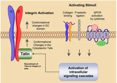
of integrin tails are also capable of activating the high affinity state. Intracellular signals that result in integ-rin activation are referred to as ‘inside-out’ signaling events.
Integrin ligation via cell-to-cell and cell-to-matrix contacts transduces intracellular signaling events in a process called ‘outside-in’ signaling (Fig. 2). The in-tegrin tails have no intrinsic kinase activity but rather serve as a site for the docking of various kinases and related adaptor proteins that comprise focal adhe-sions. The -tail is the primary docking site in the formation of focal adhesions, but the alpha tail in some cases may also serve as a signaling scaffold for calcium dependent signaling events. Signals emanat-ing from focal adhesions have been shown to promote survival, differentiation and proliferation [5]. In the absence of integrin ligation, these processes are abro-gated therefore pharmacological inhibition of integrin ligation is of great interest for the therapy of numer-ous diseases resulting from abberant integrin medi-ated signaling.
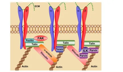
Through cell-cell and cell-ECM contacts, integ-rins behave as mechanosensors, bi-directionally transducing information both into and out of the cell,
directing the strength, stability and growth of focal adhesions to promote cell adhesion, spreading and motility (Fig. 3). Talin binding to activated integrin
-tails promotes cytoskeletal organization via the crosslinking of both vinculin and actin. Association of talin with vinculin promotes focal adhesion growth, while crosslinking of viculin and actin through talin provides stable adhesion [5]. Disruption of focal ad-hesions prevents integrin mediated cell adhesion and impaired cell motility and migration. Prolonged in-tegrin inhibition in adhesion dependent cells results in anoikis, apoptotic cell death due to ECM depriva-tion [6].
By integrating and transducing information both into and out of the cell, integrins mediate cell locali-zation, shape, spreading and motility and thus are critical determinants of both health and disease. In-tegrins have been implicated in the pathogenesis of inflammatory disease, platelet aggregation, tumor progression as well as osteoporosis and macular de-generation. The role of integrins in pathological con-ditions coupled with their ‘druggability’ by means of cell surface accessibility makes them attractive phar-macological targets.
Figure 2. Integrin “Outside-in” signaling governs cellular processes. Ligand binding serves as the initiation point for transduction of intracellular signaling cascades that regulate a multitude of biological processes. Figure adapted from ref-erence 5 with permission.
2. Integrin structure, ligand binding and
ac-tivation
The structural classification of integrins is based on the presence or absence of an extracellular A-type domain in the - subunit. A-type domain integrins contain an ~180 amino acid metal ion coordinating insert that is required for ligand binding [7]. Within this domain a five amino acid motif termed the metal ion dependent adhesion site (MIDAS) acts to coordi-nate a divalent metal ion. Ligand binding places a glutamate residue within MIDAS to participate in metal coordination [8]. Integrins lacking the A-type domain contain a similar domain within the
-subunit, termed the A domain. Ligand binding to
A integrins occurs through a divalent cation de-pendent site that requires glutamate or a similar acidic residue within the ligand to stabilize the metal coor-dination [9]. In contrast to the A-type domain, in the absence of ligand a proximal glutamate can serve as an alternate coordinating residue.
Information regarding integrin structure has been obtained by NMR, electron microscopy and ro-tary shadowing techniques. High resolution crystal structures are the method of choice for structural de-termination for the development of therapeutics. High-resolution crystal structures of membrane spanning proteins are a challenge to obtain due to the fluid nature of most biological membranes. For this reason there are no entire integrin crystal structures. The most complete information regarding integrin atomic structure to date has been derived from the crystal structures of the ectodomains of v3 and
IIb3 in both the presence and absence of ligand [10-12]. Due to the homology observed between
subunits and to a lesser degree, subunits, and sup-porting NMR and crystal structure data it can be con-cluded that most integrins adopt a similar structural conformation to those seen in the v3, IIb3 crystal structures.
The and integrin subunits form a non-covalently associated dimer, each consisting of an NH2-terminal extracellular domain, a transmembrane domain (TM) and a cytoplasmic tail. The extracellular domain protrudes from the cell surface to interact with and bind, in a divalent cation-dependent man-ner, ligands of the extracellular matrix. The domains that comprise the extracellular portion of v3 adopt a conformation that is often likened to a head and two tails. The transmembrane domain traverses the cell membrane linking the extracellular domain and cyto-plasmic tail. The cytocyto-plasmic tails act as adaptor pro-teins for intracellular signaling molecules as well as a
scaffold for the formation of focal adhesions and in-teractions with the cytoskeleton.
Each individual integrin pair recognizes its own specific set of ECM proteins. There is to some extent overlap between integrins in their ligand recognition, as they generally recognize a single sequence motif common among matrix components. Based on the
v3 crystal stucture, the head-piece is the site of ligand binding and is formed by the -propeller of the
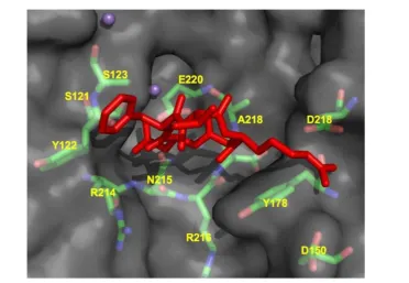
v subunit and the hybrid and A-domains of 3. The ECM ligand binding site appears to be formed more or less equally by both subunits (Fig. 4). Ligand recognition in the v3 integrin is through the argi-nine-glycine-aspartate (RGD) sequence of ECM mol-ecules such as vitronectin and fibronectin (Fig. 5). The arginine residue forms salt bridges with aspartate residues within the -propeller while the carboxylate group within the aspartate residue of the RGD se-quence forms a series of polar interactions in the A domain, and the glycine residue appears to form hy-drophobic contacts solely with the v subunit.
The seven-bladed, -propeller is formed by the arrangement of 4-stranded anti-parallel -sheets. The radial arrangement of the -sheets forms a channel or cup-like structure composed mainly of aromatic res-idues in a consensus sequence termed the cage motif. Within the -propeller are 4 Ca2+ coordination sites that may impart structural rigidity in the contacts between domains. The A-domain is a six stranded beta sheet flanked by eight alpha helices. In these and other crystal structures, the integrin appears in a bent or folded over conformation that is believed to repre-sent the low affinity state. In addition to the 4 Ca2+ binding sites, another metal ion dependent adhesion site exists is the A domain which mediates a magne-sium ion necessary for ligand binding.
re-tains its bent conformation but allows movement of the transmembrane domain in a manner that opens
the ligand binding site to heighten receptor avidity.
Figure 4. CycloRGDFV bound to v3 (RCSB code: 1L5G). Metal ions Mn2+ are depicted as spheres.
3. Approaches to integrin targeting
The functionality of integrins is dependent on activation, ligand binding, focal adhesion formation and cytoskeletal contacts. Inhibition of any of these events abrogates integrin regulated functions and thus each comprise a valid pharmacological target. Based on a survey of current anti-integrin therapeu-tics and preclinical literature the most common ap-proach to integrin antagonism involves targeting at or near the receptor binding site but knowledge of in-tegrin activation and downstream signaling suggests that alternate approaches are possible.
Targeting the ligand binding site has been ac-complished through the use of antibodies, cyclic pep-tides, disintegrins, peptidomimetics and small mole-cules antagonists. These compounds are meant to bind the targeted integrin in a manner similar to the endogenous ligand thereby displacing it and pre-venting integrin ligation. This method of antagonism has been widely utilized for several reasons. The paucity of informative structural data regarding the relevant integrins allows the rational design of an-tagonists based on known ligands. In addition, extra-cellular regions are preferable protein targets as they circumvent the problems associated with intracellular drug delivery such as lysosomal degradation and activation of drug efflux pumps. In addition the ex-tracellular domains are the only regions accessible using antibody-based therapeutics, which constitute a large portion of the antagonists in clinical use and under development. Finally, the majority of high throughput screening methods utilize solid phase receptor binding assays that screen for compounds that are able to inhibit ligand binding and thus by design favor the identification of compounds showing affinity to the ligand binding site. A similar method of antagonism involves the binding of molecules or an-tibodies in close proximity to the natural ligand binding site. This strategy provides steric hindrance of the ligand binding site such that endogenous binding site is obscured and ligand binding is prevented.
Proposed crystallographic and NMR-based models maintain integrin activation is dependent on global structural rearrangements. Targeting sites of allosteric control may represent an alternate means by which to antagonize integrin function. Our current understanding of integrin activation postulates that both extra- and intra-cellular cues can propagate sig-naling cascades that act on intracellular portions of integrins to activate inside-out signaling. The pres-ence of metal ion dependent adhesion sites (MIDAS) and calcium coordination sites further supports the assertion that function is dependent on specific
con-formational arrangements. Therefore dependence of activation on conformational arrangements suggests that sites of allosteric control exist either intra- or ex-tracellularly on integrins. This is significant for the design of anti-platelet integrin antagonists for which conformational changes induced by ligand mimetics have been proposed to lead to the adverse effects as-sociated with paradoxical platelet activation.
Targeting the intracellular ‘tails’ of integrins represents another underutilized therapeutic ap-proach to antagonism. Focal adhesion formation oc-curs at integrin tails and is the earliest intracellular event following ECM ligation. The formation of the signaling platform at this level impinges on cellular fate. Post-translational modification of integrin tails determines the composition of the signaling platform which ultimately regulates the nature of the signals transduced downstream. Pharmacological modifica-tion of integrin tails therefore has the potential of regulation the nature and extent of integrin signaling to mediate the potential outcome of ligand engage-ment. Integrin tails are also extensively crosslinked with the actin cytoskeleton via interactions with talin and vinculin. Pharmaceutical inhibition of cytoskele-tal organization can inhibit cell proliferation and mi-gration for the treatment of inflammatory disorders or inhibition of tumor growth.
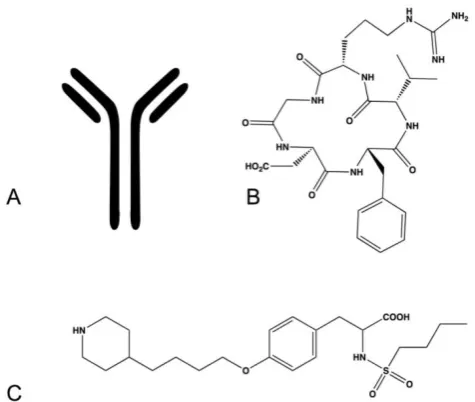
Integrin targeting therapeutics come in many forms, each type having its own inherent advantages and limitations. In addition the approach to integrin specific drug design is dependent on multiple factors including the expected clinical application and the availability of reliable structural information. In this review we will primarily discuss three types of integ-rin therapeutics, targeted antibodies, peptide-based drugs and small molecule, peptidomimetics.
therapy, that depending on the intended application may or may not be advantageous. The disadvantages associated with antibody therapeutics are the high cost of production, the need for intravenous admin-istration in a clinical setting, and the propensity for host immunogenicity and infusion reactions.
Figure 6. Examples of integrin antagonists (A) therapeutic antibody (B) cyclic peptide (C) small molecule antagonist.
Peptide-based drugs (Fig. 6B) represent another class of drugs designed to target integrins. These drugs incorporate peptide sequences similar to ligand recognition sequences in endogenous integrin ligands and thus compete for the ligand binding site within integrins. This type of drug is advantageous for drug design as most of the endogenous ligands and their recognition sequences have been determined for each integrin pair. Peptide-based integrin targeting drugs have moderate to high target affinity but may lack specificity, as the same ligand recognition sequences are shared amongst many integrins. Varying the identity of specific peptides flanking the recognition sequence has been demonstrated to impart greater specificity between integrins by exploiting the subtle variations in topology amongst the dimer interfaces. Steric constraints imparted by peptide cyclization can also improve specificity and protect against proteo-lytic cleavage in vivo. The addition of D-amino acids is another means of prolonging circulation half-life and protecting against hydrolysis. Peptide-based drugs have additional disadvantages such as requir-ing injection or intravenous administration, high production cost, and limited stability.
Small molecule compounds (Fig. 6C) represent another class of integrin targeting drugs. Small
mol-ecule drugs are synthetic compounds with low mo-lecular weight having favorable biopharmaceutical properties. They are readily synthesized, are less costly to manufacture than antibody and protein based drugs, have better stability profiles, and in many cases can be administered orally. Despite these advantages the development of small molecule an-tagonists that target integrins remains a challenge. Several methods have been applied for the develop-ment of small molecule integrin antagonists, includ-ing pharmacophore based modelinclud-ing, bioisosteric substitution and high-throughput library screening. Most compounds in the clinic and under development retain structural features that preserve the molecular interactions observed between the integrin and its native ligand. These molecules tend to be zwitterionic in nature and generally encounter limitations in bio-availability, serum protein binding and integrin se-lectivity.
4.
IIb
3 integrin
The IIb3 integrin is comprised of a 136 kDa
IIb subunit non-covalently associated with the 92kDa 3 subunit. The IIb3 integrin exists in vari-ous states of activation. Activated, ligand occupied and resting states have been demonstrated. Activating signals are required for ligand binding and lead to a conformational change in IIb3 integrin that increase ligand affinity, receptor clustering leading to in-creased ligand avidity. IIb3 integrin recognizes two separate consensus sequences, the argi-nine-glycine-aspartic acid (RGD) sequence [15] in fi-brinogen, fibronectin, and Von Willebrand Factor and the lysine-glutamine-alanine-glycine-aspartate-valine sequence found in the gamma chain of fibrinogen [16-17]. As with v3 integrin, coordination of Ca2+ ions within MIDAS and ADMIDAS are required for ligand binding.
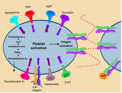
IIb3 integrin (GPIIa/IIIb) is highly expressed on the surface of platelets and to a lesser degree, on megakaryocytes [18]. IIb3 integrin comprises ap-proximately 80% of the total surface proteins found on platelets. Engagement of this integrin is induced by platelet activation and represents the final common pathway in blood coagulation (Fig. 7). Under normal conditions integrin IIb3 is maintained in the inac-tivated state. Soluble factors in the blood such as thrombin, ADP and thromboxane A2 bind their re-spective platelet receptors to activate inside-out sig-naling pathways that cause conformational changes in
re-ceptor affinity and avidity promote platelet aggrega-tion and clot formaaggrega-tion through increased cell-to-cell contacts and cell-matrix contacts mediated by integrin
IIb3. Aberrant platelet aggregation or thrombosis is central to the pathophysiology of multiple Acute
Coronary Syndromes (ACS), unstable angina, is-chemic stroke and sickle cell anemia. Inhibition of
IIb3 prevents platelet aggregation and therefore has shown efficacy in the prevention of thrombosis for the treatment of ACS.
Figure 7. Mechanisms of platelet activation. Binding of secreted factors to cognate ligands promotes platelet activation leading to αIIbβ3 activation. Activation of αIIbβ3 promotes integrin clustering and binding of fibrinogen and von Willebrand Factor leading to platelet aggregation and thrombus formation.
IIb3 integrin antagonists in Percutaneous Cardiac Intervention, Acute Coronary Syndromes and Myocardial Infarction. Currently, there are three FDA approved therapeutics targeting IIb3 integrin, Abciximab, eptifibatide and tirofiban. These therapeutics are di-rected against IIb3 integrin and act in the preven-tion of platelet aggregapreven-tion. They are utilized during stent placement and other percutaneous coronary intervention (PCI) procedures for the treatment of both ST-elevated myocardial infarction (STEMI) and non ST-elevated myocardial infarction (nSTEMI). All three antagonists have been extensively studied in large randomized, placebo controlled clinical trials and demonstrated advantages over previous an-ti-platelet treatment modalities such as aspirin and the thienopyridines in preventing thrombosis and
mor-tality. Although all three compounds act at the level of integrin IIb3 to prevent thrombus formation, they represent separate classes of drug and thus differ in their pharmacokinetic and pharmacodynamic prop-erties. The variations between each of these antago-nists determine the extent of their utility in the treat-ment of various ACS. The clinical success of these agents in the prevention of platelet adhesion have prompted evaluation for the treatment of disorders and disease states in which abberant platelet aggre-gation is central to the pathology, such as ischemic stroke and sickle cell disease.
an-tagonist to be approved for clinical use. Abciximab is the Fab fragment of a monoclonal, chimeric mouse-human antibody with specificity towards the
IIb3 integrin as well as the v3 [19] and M2 (MAC-1) integrins [20]. The platelet integrin (IIb3) epitope for Abciximab has been identified as a region within the -chain proximal to the RGD-sequence binding site and the secondary fibrinogen binding site [20]. Hence Abciximab inhibits platelet aggregation via steric hindrance of ligand binding.
For use in PCI, the treatment of MI and ACS, Abciximab is administered parenterally and the un-bound fraction undergoes rapid proteolytic cleavage in the bloodstream. Due to short plasma half-life it is administered as a single bolus followed by continuous infusion over a period of 12-96 hours. The plasma concentrations drop rapidly upon discontinuation, the first phase half-life is close to 10 minutes and the second phase half-life is about 30 minutes due to rapid receptor occupancy. A slow decline in drug concentration results in an overall half-life of ap-proximately 7h and continued low-grade receptor blockade has been noted for periods up to 10 days post infusion [21]. Abciximab is recommended for the treatment of both nSTEMI and STEMI [22].
Eptifibatide. Eptifibatide (Fig. 8) is a synthetic cy-clic heptapeptide derived from barbourin, a snake venom disintigrin. Modifying the KGD sequence of barbourin, to homo-ArgGD and cyclization by intro-duction of a disulfide bridge produced a competitive inhibitor that shows high selectivity for IIb3 at lower concentrations than the linear peptide [23]. Eptifibatide has low binding affinity and rapidly dis-sociates from IIb3 in circulation, such that by 4 hours post administration, platelet binding ability is restored. Dosing is usually administered parenterally as a single or double bolus in combination with con-tinuous infusion. The plasma half life is approxi-mately 2.5 h and the drug is cleared mainly by the kidneys as epitifabitide, deamidated eptifabitde or other polar metabolites. Due to the extensive renal clearance, dosing modifications must be considered for renal impaired patients [24].
Tirofiban. Tirofiban (Fig. 8) is a non-peptide, small molecule RGD-mimetic antagonist specific for
IIb3 integrin. Tirofiban was developed through sequential bioisosteric substitutions of a candidate molecule pharmacophore chosen to approximate the bond distances observed between the carboxy and amino portions of the RGD-scaffold. This produced a small molecule, tyrosine-derivative with increased potency and selectivity for IIb3. Tirofiban prevents fibrinogen binding and platelet aggregation in vitro by 50% at concentrations of 5 and 11 nM, respectively
[25]. Tirofiban is administered intravenously. As with eptifibatide, this small molecule competes with fi-brinogen and von Wilebrand Factor for the RGD-sequence binding pocket of platelet integrin
IIb3. It binds with higher affinity than eptifibatide but has a comparable volume of distribution and plasma half-life. Tirofiban is not highly bound to plasma in the bloodstream and the volume of distri-bution at steady state suggests that a large portion distributes within the extracellular space. Tirofiban has a plasma half-life of 1.5-2h and is excreted mostly as unchanged compound in the urine and feces. Renal insufficient patients require dose reductions and se-vere renal failure prohibits use. Hepatic failure does not require dose adjustment [26].
Figure 8. Structures of IIb3 antagonists Eptifibatide and Tirofiban.
de-termined that optimal receptor occupancy was achieved with single bolus dosing followed with con-tinuous infusion. Weight-adjusted heparin dosing reduced the propensity for bleeding events. Subse-quent large-scale randomized trials examined the impact of abciximab on endpoints such as mortality, need for revascularization and occurrence of myocar-dial infarction [27]. Meta-analysis of the eleven major Phase III trials of abciximab showed significant over-all decreases in death at 30 day endpoint, decreased need for revascularization and reduced occurance of myocardial infarction in patients receiving abciximab during percutaneous coronary intervention, as com-pared to fibrinolytic agent in myocardial infarction and during stent placement for the treatment of un-stable angina [28]. Due to possible immunogencity related to the chimeric nature of abciximab, the safety of administration was examined in the ReoPro re-administration trial. Abciximab was found to be safe for repeat administration [29]. Trials of eptifibatide were designed in a similar manner as the abciximab trials. Phase I studies examined various dosing levels alone and in combination with weight-adjusted hepa-rin. Initially dosing was under estimated as the use of citrate anticoagulant chelated calcium ions necessary for receptor ligand binding and activation and pro-duced falsely decreased readings of platelet aggrega-tion that led to lower than anticipated performance in meeting protocol endpoints for survival, restenosis and myocardial infarction [30-31]. Subsequent trials utilized anti-coagulants that did not perturb meas-urements of platelet aggregation and dosing was in-creased from single bolus 135 mg/ kg to double bolus 180 mg/kg with 2 mg/kg/min infusion for up to 24 h [32]. This dosing resulted in significant reduction in mortality, restenosis and myocardial infarction when used in the ESPRIT trial [33]. The Randomized Effi-cacy Study of Tirofiban for Outcomes and Restenosis (RESTORE) trial evaluated tirofban versus placebo in patients at risk for arterial obstruction due to multiple acute coronary syndromes and those undergoing an-gioplasty for myocardial infarction. Significant reduc-tion in primary endpoints were noted at day 2 but decreased by day 30 [34]. Overall meta-analysis of 12 trials of IIb3 antagonists in over 20,000 patients demonstrated a significant reduction in mortality and myocardial infarction at 30 days [35].
Factors impacting the therapeutic efficacy of IIb3 antagonists. The efficacy of IIb3 antagonists is modulated by the total number of receptors available for drug binding. It is generally accepted that roughly 80% of the total platelet IIb3 integrins should be bound to obtain therapeutic dosing [36]. Platelet count is the primary determinant of receptor availability as
IIb3 integrins are expressed almost solely on the surface of platelets, at levels of approximately 50-100,000 receptors per platelet. Although the in vivo significance of platelet count on drug efficacy has not been clearly established ex vivo studies suggest an inverse relationship between platelet count and in-tegrin inhibition [37]. Additional factors that contrib-ute, albeit less so, to receptor occupancy are the ratio of internalized receptors versus receptors at the platelet surface, the degree of receptor activation, and blood plasma fibrinogen levels.
Resting platelets typically maintain an internal receptor pool that upon activating stimuli such as ADP and Thromboxin A2 binding can be externalized to increase surface expression by 40-50%. Of this in-ternalized pool, a portion may emerge at the platelet surface bound to soluble fibrinogen released from
-particles within the platelet. Abciximab is unable to completely inhibit this internal pool and the mimetic antagonists as competitive inhibitors may also lose efficacy, but likely to a lesser degree in light of their rapid dissociation kinetics [38].
greater than 90% receptor occupancy are associated with higher risk of adverse bleeding events [40].
Drug-induced thrombocytopenia following IIb3 antagonist treatment. A small percentage of patients receiving IIb3 antagonists experience thrombocy-topenia, a severe drop in platelet number (platelet count < 50,000) that is associated with increased inci-dence of ischemic events, bleeding related complica-tions and requirement of plasma transfusions [41]. The primary cause of thrombocytopenia in patients treated with IIb3 antagonists appears to be immune mediated [42], although non-immune mediated mechanisms have been reported for the RGD-mimetics, eptifibatide and tirofiban [43]. Bleed-ing events can range from minor occurrences such as leakage at the site of IV insertion, to systemic, life-threatening episodes requiring rapid intervention. As a humanized chimeric antibody, abciximab has demonstrated a propensity for the induction of hu-man antichimeric antibody response upon first ad-ministration in approximately 1% of patients and in 4% upon repeat administration [29]. Thrombocytope-nia caused by immune-mediated platelet destruction following Abciximab treatment may be of rapid or delayed onset [44]. Acute thrombocytopenia occurs within hours of infusion and may be accompanied by fever, drop in blood pressure, and anaphylaxis. Rap-id, immune-mediated platelet destruction occurs through the binding of IgG and IgM antibodies that recognize murine sequences of the Fab’ fragment or platelet conformational changes induced by abcixi-mab binding. Delayed-type thrombocytopenia has also been noted to occur up to 8 days following cessa-tion of treatment and may be caused by newly formed antibodies or through the stimulation of existent an-tibodies [45]. The rate of drug-induced thrombocyto-penia is of lower frequency in patients receiving the ligand mimetics, Tirofiban and Eptifabitide compared to Abciximab treated patients [46]. The onset is usu-ally acute, within hours of treatment and can be ac-companied by symptoms similar to those accompa-nying abciximab immune thrombocytopenia. In con-trast to Abciximab, the clearance rate of the RGD mimetics is much faster, thus limiting to the time pe-riod in which patients are at risk of bleeding events due to thrombocytopenia. The severity of complica-tions varies from asymptomatic to minor bleeding to major hemorrhage. Immune mediated thrombocyto-penia is believed to result from the clonal expansion of haptens, antibodies that recognize drug-bound receptors.[44] Monitoring platelet counts prior to and
following administration of all three platelet inhibi-tors is warranted considering these issues. Depending on the severity of related bleeding events, platelet transfusion may be necessary to restore haemostasis.
Oral IIb3 antagonists as preventive treatment for coronary syndromes. Prolonged inhibition of platelet aggregation using aspirin and thienopyredine has been demonstrated to significantly reduce the risk of ischemic events [47]. It was hypothesized that, as more potent inhibitors of platelet aggregation, the
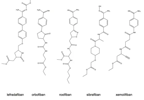
IIb3 antagonists would show increased benefit in long-term ischemia risk reduction. Based on the suc-cess and clinical efficacy of the parenterally adminis-tered IIb3 antagonists, an effort to develop orally available small molecule antagonists for the long-term prevention of cardiovascular disease was undertaken and led to the clinical evaluation of several antago-nists having similar structural characteristics and pharmacological properties (Fig. 9). Most of the drugs from this class are prodrugs, requiring hepatic con-version to the active state and having limited bioa-vailability in the range of 7-38% [48]. The plasma elimination half-life for these compounds is generally in the range of 4-20 h with the exception of roxifiban that has an elimination half-life of up to 5 days. Renal excretion accounts for the primary means of drug elimination within this class. Pharmacokinetic studies established that efficacious dosing was in the range of 5-30 mg. Higher doses given once daily were associ-ated with increased bleeding events, lower more fre-quent dosing provided similar effect and reduced the bleeding risks. Renal insufficient patients required lower dosages to prevent increased risk of bleeding presumably to decreased clearance rates [48]. Despite the dose adjustment efforts taken in risk reduction, the outcomes of dose ranging studies revealed overall increase in the risk of bleeding amongst all antago-nists tested [48].
Figure 9. Structures of orally active, small molecule IIb3 antagonists.
The mechanism leading to increased mortality is confounding based on evaluation of the meta-analysis of concurrently measured endpoints such as myocar-dial infarction, need for urgent revascularization and major bleeding events. The incidence of myocardial infarction was not significantly different between pa-tients receiving study drug and those receiving aspi-rin alone. The incidence of urgent need for revascu-larization was significantly reduced in 2 of 4 trials analyzed, and not significantly different in the re-maining two trials [49]. These results suggest throm-bosis may not be the primary factor leading to in-creased mortality. A statistically significant three percent increase in major bleeding events due to platelet inhibition contradicts lack of antagonist effi-cacy as a contributing factor to increase in mortality.
Alternate mechanisms have been proposed to account for the increased mortality observed with oral antagonists including drug related toxicity, partial agonist activity, non-platelet mediated mechanisms and patient inter-variability. No one mechanism has been explicitly proven to account for the increased
mortality and it is likely that the cause is mul-ti-factorial.
IIb3 antagonists for the treatment of ischemic stroke. Similar to acute coronary syndromes, inhibi-tion of platelet aggregainhibi-tion and thrombus formainhibi-tion using aspirin has shown benefit in ischemic stroke by reducing recurrence and mortatlity, albeit with mod-est effects. As potent antagonists of platelet aggrega-tion and thrombus formaaggrega-tion, is was expected that
IIb3 inhibitors would demonstrate increased bene-fits over aspirin for the reduction of stroke related complications. Contrary to expected, meta-analysis of several large randomized clinical trials failed to demonstrate conclusive evidence supporting the use of IIb3 antagonists for the treatment of ischemic stroke [50]. Furthermore, a few recent trials have ter-minated enrollment due to increased incidence of intracranial hemmorhage.
leading to decreased pliability, perturbations in ion balance and increased adhesion and viscosity in blood vessels. Sickle cell anemia is characterized by episodes of ischemia-repurfusion [51], increased levels of in-flammatory mediators [52] and abnormal activation of granulocytes and monocytes [53]. Activated platelets are thought to contribute to the pathology of Sickle cell anemia through multiple processes. Activation of platelets increases endothelial cell activation [54], P-selectin exposure, the exposure and release of CD40L [55] and it promotes vasoconstriction. Platelet released factors increase adhesion of RBC to vascular endothelium, promote coagulation [56], induce vaso-constriction [57]. Exposure and release of CD40L triggers immune and inflammatory responses, trig-gers B-cell proliferation, increases the expression of endothelial cell adhesion molecules and upregulates tissue factor. Platelet adhesivity and adhesion is me-diated through IIb3 integrin as well as granule se-cretion and procoagulant activity [56, 58]. Blockade of integrin IIb3 with eptifibatide, abciximab or tirofi-ban prevents thrombus formation, prothrombin acti-vation and the release of CD40L from activated platelets, events which are central to the pathology of sickle cell anemia and therefore, may offer benefit in the treatment of sickle cell anemia. Phase I trials evaluating the safety and pharmacokinetics of eptifi-batide in sickle cell anemia patients in the non-crisis state have shown promising results. Eptifibatide ad-ministered as two 180 mg/kg boluses separated by 10 minute interval combined with 2 µg/kg/min infusion reduced levels of inflammatory cytokines and myo-globin, a marker of muscle injury. The vasodialotors MMP-2 and MMP-9 were increased with eptifibatide treatment, as was the adipokine leptin. Eptifibatide treatment had no effect on platelet reactivity or ag-gregate formation. The observed effects of eptifibatide were optimal by 6 h and fully reversible as early as 24h following infusion, suggesting that IIb3 an-tagonist therapy may be beneficial for the treatment of the acute painful crises characteristic of sickle cell anemia [59].
5.
4 integrins
4 integrin chains associate with 1 or 7 integ-rin family members, via non-covalent linkages, to form dimers. The 41integrin, also known as VLA-4, Very Late Antigen-4, and CD49d/CD29, is constitu-tively expressed on the surface of lymphocytes and most leukocytes [3]. The primary ligands of 41 are endothelial VCAM1 and the extracellular matrix (ECM) glycoprotein, fibronectin. 41 interacts with VCAM1 through the QID(40)SPL site in domain 1, in
which aspartate 40 appears to be essential for ligand binding. Interaction with the ECM occurs through the LDV sequence of the connecting segment-1 (CS-1) region of fibronectin. Other 41ligands have been identified, including the acidic glycoprotein osteo-pontin, juntional adhesional molecule B and molecule B and mucosal addressin cellular adhesion molecule (Mad-CAM) [60].
Similarly, integrin 47 (LPAM, Lymphocyte Peyer’s Patch Adhesion Molecule) is also expressed on the surface of lymphocytes and leukocytes. Inter-actions of this integrin with VCAM-1 and fibronectin occur through the same epitope as 41integrin, alt-hough the 47 avidity for VCAM-1 is considerably lower than that of 41. In addition, 47binds the mucosal addressin cellular adhesion molecule -1 (MAdCAM-1) expressed on high endothelial venules of gut mucosal lymphoid tissue such as Peyer’s Patches and mesenteric lymph nodes, as well as lam-ina propria venules [61].
The 4integrin family members, 41and 47, function in leukocyte recruitment from the peripheral circulation to sites of inflammation within tissue [62-63] (Fig. 10A). The local release of chemokines at sites of inflammation causes upregulation of 41and
Figure 10. Mechanism of immune-cell mediated inflammation and its inhibition by 4 antagonists. Left Panel- 4 integrins are up-regulated on the surface of activated immune cells. Through contacts with endothelial cell adhesion molecules such as selectins and glycoproteins, these cells are captured from the circulation. The intial contacts are strengthened when 4 integrins make contact with MAdCAM and VCAM on endothelial cells. Extravasation and migration to sites of inflammation are mediated in part through 4 integrins. Right Panel- In the presence of 4 antagonists such as natalizumab, firm adhesion and subsequent migration are inhibited preventing immune cell infiltration within tissue.
The infiltration of leukocytes and lymphocytes is central to the pathology of many inflammatory dis-eases. The 4 integrin family members, 41 and 47 act both in the formation of initial contacts and firm adhesion and therefore, are key constituents in the process of cellular influx to inflamed tissue. Suppres-sion of 4integrin binding inhibits cellular migration, thereby attenuating the inflammatory response (Fig.
10B). The therapeutic potential of 4 antagonists in the attenuation of inflammatory response has been investigated for the treatment of several chronic dis-eases.
Multiple Sclerosis (MS) is an autoimmune dis-ease affecting the central nervous system (CNS). The pathology of MS is caused by increased entry of acti-vated immune cells through the blood-brain barrier (BBB) into the CNS. Activated immune cells initiate processes of inflammation, edema and demyelination leading to the formation of acute CNS lesions [64]. The progressive accumulation of lesions in the CNS is responsible for the debilitating symptoms of MS, such as fatigue, imbalance, loss of mobility, sensory symptoms, bladder and bowel dysfunction, memory loss and concentration deficits, spasticity, visual problems, pain and sexual dysfunction [65].
Contacts between circulating immune cells and the BBB is an early and critical event in the patho-genesis of MS. Antibody directed inhibition of 41 mediated contacts between immunocompetent cells in the circulation and VCAM-1 on the vascular endothe-lium of the BBB has been demonstrated to prevent the
development and even reverse disease progression by preventing the infiltration of inflammatory cells through the BBB. Targeting 4 integrin mediated cell binding to attenuate immune cell migration therefore represents a significant means by which to prevent disease progression.
Crohn’s disease and ulcerative colitis are idio-pathic inflammatory diseases of the intestinal tract characterized by focal or continuous regions of mu-cosal inflammation, respectively, which produce symptoms of abdominal pain, diarrhea and rectal bleeding and over time physical manifestations such as strictures and perforations of the intestinal wall. Dysregulated immune responses contribute to the pathogenesis of both types of IBD causing increased infiltration of immune cells into muscosal tissue of the gut. Interactions between immune cell 47 and VCAM and MAdCAM-1 on the gut mucosa facilitate transmigration and are believed to contribute to the pathogenesis of IBD [65].
Asthma is a chronic inflammatory disease of the lung characterized by increased infiltration of acti-vated T-cells and eosinophils resulting in broncho-constriction, airway hyperresponsiveness and airway remodeling. Inhibition of T-cell migration via block-ing of 4 mediated cell interactions may reduce the severity of asthma symptoms.
arthri-tis. The rationale for targeting 4 integrin to modulate aberrant immune cell migration and activation is supported by in vitro as well as cell- and ani-mal-based studies. Antibodies that block the function of 4 and 1 integrin chains decrease T cell and monocyte binding to VCAM-1 in frozen sections of inflamed blood vessels [66]. Inhibition of activated T-cell migration across the BBB using 41 blocking antibodies prevented and in some cases, reversed inflammation-induced neurological symptoms in mouse, rat and guinea pig models of experimental autoimmune encephalopathy. Similar results were obtained by blocking the 4 ligand VCAM-1 [67]. In-hibition of 47 reduces the development of sponta-neous ulcerative colitis in tamaran monkeys [68] and alleviates inflammation in mouse models of colitis [69]. Inhibition of the 47 ligand, VCAM-1, produces similar results in mouse models of induced colitis [70]. Studies using monoclonal antibodies directed toward
4 integrins or VCAM in animal models of anti-gen-induced asthma suggest a critical role for this interaction in disease pathophysiology [71]. Various small molecule 4 antagonists have shown efficacy in allergen-induced asthma models.
4 antagonists for the treatment of inflammatory diseases. To date, there is only one 4 antagonist approved by the U.S. Food and Drug Administration (FDA). Natalizumab is approved for treatment of re-lapsing, remitting multiple sclerosis and Crohn’s dis-ease. Natalizumab is a humanized mouse monoclonal antibody directed against the 4 integrin. Natali-zumab binds 41 and 47 integrin on the surface of circulating T lymphocytes preventing interaction with cellular adhesion molecules that facilitate extravasa-tion and migraextravasa-tion from the circulaextravasa-tion to tissue.
Following the administration of 300 mg I.V. bo-lus Cmax was 110 52 µg/mL. Mean average steady state concentrations were between 23 and 29 µg/mL and the time to steady state was 24 weeks after 4 weeks dosing. The mean t1/2 is approximately 11 days, with a volume of distribution of 5.7 and clearance of 16 ml/hour. Clearance is influenced by bodyweight and the presence of neutralizing antibodies, increases in both results in a faster rate of clearance. Age and sex do not affect the rate of clearance [72-73].
Natalizumab underwent a series of clinical trials for the treatment of relapsing-remitting multiple sclerosis, and Crohn’s disease. Natalizumab showed remarkable efficacy for the treatment of MS in Ran-domized clinical trials. 4-integrin blockade resulted in a decrease in the number of CD4+ and CD8+ T-cells, CD19+ B-lymphocytes and CD138+ plasma cells in CSF for up to 6 months following treatment
cessation.[74] Based on measurements of 2-year out-comes, natalizumab decreased clinical measures of disease such as relapse rates and the risk of sustained disability progression by 68 and 54% respectively and reduced MRI based disease measures such as the ap-pearance of new T2 lesions, new gadolinium positive lesions and lowered overall disease burden by 83, 92 and 18 % respectively [75]. The efficacy of natali-zumab treatment appeared to improve with the dura-tion of treatment. The difference in clinical and MRI measures of disease and progression between treated and placebo groups were significantly greater in the second year compared to the first. By the second year of treatment greater than 35% of natalizumab treated patients were free of all disease activity and more than 40% showed only one measure of disease activity [76]. In both highly active disease and non-highly active disease subgroups, the proportion of patients free of disease activity was significantly higher than mem-bers of the same group that received placebo only. Health related quality of life measures relating to pa-tient perceptions regarding disease status were also improved in natalizumab treated patients [77]. Clcal measures of efficacy in Crohn’s disease were ini-tially less significant than those observed in the treatment of MS. Although treatment with natali-zumab demonstrated efficacy in disease response and remission among a high percentage of treated patients in the ENACT 1 and ENACT 2 randomized phase III trials, both failed to meet their primary endpoints of statistically significant reduction in disease activity across the total population. However, the follow-up trial ENCORE was able to demonstrate disease sponse and remission in active Crohn’s patients re-ceiving 300 mg intravenous infusion every two weeks over a period of 12 weeks. In this randomized, pla-cebo controlled, phase III trial, 51% of natalizumab treated patients showed response by week 4 after one infusion. The percent of patients in response at weeks 8 through 12 was similar to the initial results, 50% of natalizumab treated patients sustained response ver-sus only 37% of patients receiving placebo [78].
to be caused by impaired immune surveillance in the CNS as a direct result of 4 inhibition on circulating immune cells [80]. The risk of developing PML is be-lieved to increase with duration of Natalizumab treatment and may be hastened in patients receiving prior or concurrent immunosuppressive therapies. The incidence of PML is calculated to be 1 case per 1000 patients treated with natalizumab for 17.9 months. To date, 11 cases of PML including 3 fatalities have been reported in patients receiving Natalizumab either as monotherapy or in combination [79, 81-83]. Additional opportunistic infections have been ob-served in patients receiving natalizumab, although the role of natalizumab in causation is as yet unde-termined. Infections reported include viral meningitis and encephalitis (2), acute cytomegalovirus (2), pul-monary aspergillosis (2), cryptosporidial gastroenter-itis (1), Pneumocystis carinii pneumonia (1), varicella pneumonia (1), mycobacterium avium intracellulare complex pneumonia (1), and Burkholderia cepacia pneumonia (1). Values in parentheses represent the number of reported cases. In an effort to decrease the inherent risks accompanying the obvious benefits to MS patients, FDA recommendations suggest the use of natalizumab in limited populations including those refractory to other therapies or those with particularly aggressive disease under strict clinical supervision.
Because the increased incidence of PML is be-lieved to be caused as a direct result of 4 antagonism, improvement of the biopharmaceutical properties of compounds targeting 4 integrin would therefore have no bearing on the likelihood of developing PML. Though, presumably, a small molecule or pep-tidomimetic antagonist could present lower risk; the shorter plasma half-life of these molecules compared to antibodies would prevent the need for costly and invasive plasma exchange procedures currently uti-lized for the removal of natalizumab from the blood in the event PML symptoms are manifest [84]. Ulti-mately, due to the serious risks that increase with duration of administration, 41 integrin antagonist based therapies should be limited for the treatment of conditions that are life threatening or do not respond to first-line treatments.
Antagonism of VLA-4 may also have implica-tions for fetal and embryonic development. Cell-to-cell interactions mediated by 41 integrin are critical to reproductive events such as fertilization, implantation, placental formation and cardiac devel-opment. 41 knockout mice are embryonic lethal and show disruption in placentation, defects in al-lantois-chorion fusion, cardiac abnormalities, mal-formation and hemorrhage. VCAM-1 knockout mice
show similar defects further supporting the role of 4 integrins in embryonic development.
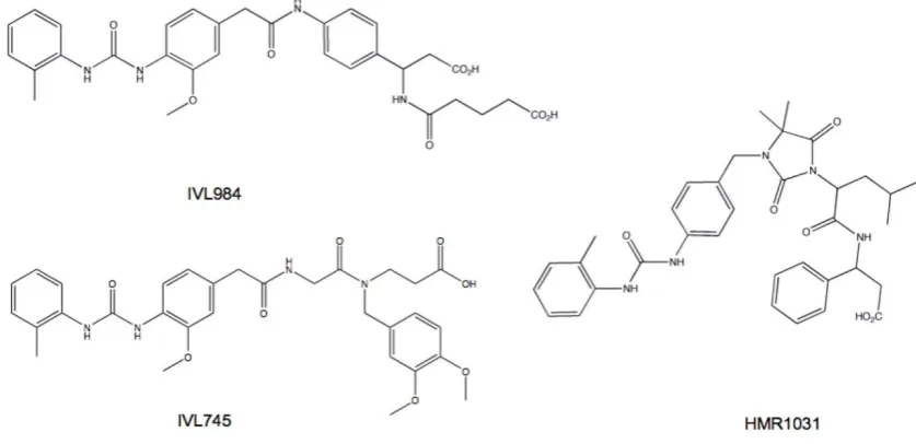
Based on animal studies of embryonic and post-natal development it has been deemed that pro-longed, high dose natalizumab treatment caused no developmental defects [85-87]. These results cannot be extrapolated to other 4 antagnoists. In fact, basic teratogenicity assays of several small molecule 41 antagonists in preclinical development showed vari-ability in propensity to cause defects (Fig. 11). The teratogenic potential of 41 antagonists appears to be related to the affinity state of the integrin to which the compound binds. Integrins exist in multiple acti-vation states wherein ligand binding stability is pro-portional to the affinity or activation status of the re-ceptor. Compounds that bind to VLA-4 at both low and high affinity states appear carry a greater risk of toxicity and embryonic defects compared to com-pounds selective for the activated integrin [88]. Therefore integrin activation state affinity is a major consideration for the development of successful clin-ically useful inhibitors.
4 antagonists under clinical evaluation. MLN-00002 MLN-0002 is a human antibody specific for the 47 integrin dimer. In phase II trials, MLN-00002 showed dose dependent efficacy as in-duction therapy for the treatment of active Crohn’s disease and ulcerative colitis. Treatment consisted of two infusions, at day 1 and 29. Primary endpoint was a greater than 70 point decrease in the CDAI (Crohn’s Disease Activity Index) score achieved in both stud-ies. In the phase II trials patients were screened for the presence of latent JC virus prior to the administration of MLN-0002 and were monitored for signs of PML through out the course of treatment [89].
Firategrast. Firategrast is a small molecule 4 antagonist developed by Glaxo Smith Kline that has recently completed Phase I clinical trials for the treatment of Relapsing, Remitting Multiple Sclerosis. This trial evaluated pharmacokinetic parameters fol-lowing oral administration of 900 mg once daily in men and women diagnosed with multiple sclerosis. Further Phase II studies have been initiated to evalu-ate the effect of firevalu-ategrast on white blood cell counts in the cerebrospinal fluid.
biliary route [91]. These limitations in biopharmaceu-tical properties typify the problems inherent with most small molecule 4 antagonists. Bioavailability is improved when administered by inhalation. Safety studies in both healthy and asthmatic volunteers demonstrated tolerability by inhalation at dosing up
to 20 mg BID. Subsequent trials in human asthma patients failed to demonstrate efficacy greater than that observed with placebo [92-93] suggesting that the role of 4 integrins in asthma and allergen induced air inflammation may be more complicated or less in-fluential than previously predicted [94].
Figure 11. Small molecule 4 antagonists vary in their propensity for teratogenic effects. IVL984 (potent teratogen), HMR1031 (mild teratogen) and IVL745 (non-teratogenic) are structurally similar yet have different teratogenicity profiles. The difference is attributed to their affinity for non-activated 4 integrin. Compounds that bind 4 in both activated and resting states appear to be more teratogenic.
6.
v integrins
Integrins v3 and v5 are highly expressed on the surface of osteoclasts, angiogenic endothelial cells, and some solid tumors. Their antagonism has been explored for the purpose of preventing/reversing osteoporosis, inhibition of angiogenesis and induction of tumor regression. Currently there are no FDA-approved antagonists of v3 and/or v5 in-tegrins on the market, but several compounds have shown efficacy in preclinical evaluation and are cur-rently under evaluation in clinical trials for the treat-ment of malignancies, rheumatoid-arthritis and os-teoporosis.
v integrin antagonists as anti-angiogenic agents. Angiogenesis is the process of neovascularization in which new blood vessels sprout from existing ones. The ‘angiogenic switch’ is an early event required for tumor progression. Initially tumor growth is sup-ported by nutrients and oxygen derived from proxi-mal vasculature. As tumor mass increases, local blood supply becomes inadequate, resulting in a hypoxic, nutrient-deprived tumor environment. The inducible
mar-row-derived endothelial cell progentior to terminal, committed endothelial cell, v3 integrin expression increases. Integrin v3 and v5 recognize the RGD sequence in ECM components such as fibronectin, fibrinogen, and vitronectin. Through interactions between v3 and v5 integrins and the ECM, mi-grating endothelial cells are captured from the circu-lation and participate in the formation of vasculature to feed the growing tumor. The association of integrin
v3 with matrix proteases including matrixmetallo-protease -2 (MMP-2), matrixmetallomatrixmetallo-protease-9 (MMP-9) and urokinase plasminogen activator (uPA) facilitates ECM degredation allowing tissue extrava-sation. Integrins on the leading edge of lamelapodia use ECM components to generate the traction neces-sary for cell migration. Adhesion and spreading are mediated through integrin binding to ECM. Cell-to-cell contacts are mediated through integrins during tube formation and vessel maturation. During angiogenesis integrins stimulate the processes of en-dothelial cell migration, proliferation, differentiation through FAK/Src/p130Cas and Raf/MEK/Erk sig-naling pathways [95].
Endothelial survival in both nascent and estab-lished tumor vasculature is supported by integrin mediated signaling events that are dependent on li-gation with the ECM. Integrin lili-gation promotes cell survival primarily through activation of the PI3K/Akt pathway. Cross talk between integrins and growth factor receptors, possibly through trimeric interaction between growth factors, growth factor receptors and integrins [96], inhibits the activation of intrinsic and extrinsic pathways of apoptosis. In addition, integrin ligation promotes the increased expression of the pro-survival molecules Bcl-2 and FLIP, increases NFkB signaling and decreases p53 activation. Hy-per-stimulation of pro-survival signaling mechanisms aids endothelial cells as they form vasculature in the hypoxic tumor microenvironment. Conversely, un-ligated integrins can promote apoptosis through the interruption of pro-survival signaling and the initia-tion of pro-apoptotic signaling cascades [6, 97].
The significance of v3 and v5 integrins in the processes of angiogenesis is supported by a wealth of experimental data obtained using in vitro, cell culture and in vivo methods. Angiogenic factors and tumor-secreted cytokines increase the expression of v3 integrin in animal and human models of an-giogenesis. Inhibition of v3 and v5 integrins prevents endothelial cell migration, adhesion, differ-entiation, tube formation and survival in cell culture models and has been demonstrated to inhibit angio-genesis and vascular tube formation in animal mod-els. Mice null of v integrin expression show a high
rate of embryonic lethality due to bleeding and pla-cental defects, as do 3 knockout mice. Surviving
v-knockout mice die prematurely due to complica-tions of vessel malformation in the brain and gut. In contrast, surviving 3 knockout mice exhibit normal vasculature and increased angiogenic potential, pre-sumably through up-regulation of VEGFR2 mediated compensatory mechanisms. The reasons for the pro-angiogenic effect of 3 knockdown and the im-plications for the role of v3 integrins in angiogene-sis is a source of continued debate. It is important to keep in mind that the observed effects of
3-knockdown during development may differ from those seen post-natally [97-98].
The primary goal of anti-angiogenic therapy is to prevent tumor growth by inhibiting the formation of new vasculature and destroying established tumor blood vessels that feed oxygen and nutrients to the growing neoplasm. Additional evidence suggests that anti-angiogenic therapies may act to transiently stabi-lize tumor vasculature increasing oxygenation and perfusion. The resulting increase in tumor blood flow may explain the seemingly paradoxical potentiation in efficacy observed when anti-angiogenic agents are combined with standard chemotherapies [99]. An-ti-angiogenic agents are generally well tolerated and can be administered at high doses with few dose-limiting toxicities. With low levels of toxicity, anti-angiogenic agents can easily be combined with standard drug regimens with little or no additional adverse effects. The anti-angiogenic effects of radia-tion therapy can synergize with angiogenic inhibitors to increase cell kill [100]. Additionally, as non-transformed cells, the genetic stability of endo-thelial cells precludes the development of drug re-sistance, making them an attractive target.
between integrins v3 on tumor cells and IIb3 on activated platelets facilitates circulating tumor cell capture from the blood, a critical initial step in the metastatic cascade. Extravasation, migration, adhe-sion and proliferation are supported by integrin liga-tion with the extracellular matrix. Associaliga-tion of v3 integrin with Src promotes anchorage independent cell growth. v3 mediated signaling through focal adhesion kinase (FAK) supports the survival of cancer stem cells. Inhibtion of v3 is associated with de-creased tumor cell growth in breast and prostate cancers, melanomas and gliomas. Supression of tumor
v3 integrin expression in animal models decreases the ability of tumor to migrate and metastasize [6].
v integrin antagonists for the treatment of osteopo-rosis. Osteoporosis is a disease characterized by the progressive loss of bone density due to imbalances in the rate of bone resportion by osteoclasts and bone formation by osteoblasts. The disease is common in post-menopausal women, as age-related declines in estrogen levels increase osteoclast activity. Osteopo-rosis imparts increased risk of fractures leading to augmented rates of morbidity and mortality among osteoporotic women [65]. The development of anti-resorptive agents that prevent osteoclast function to successfully reverse bone loss and increase bone mineral density (BMD) are of great interest for the treatment of osteoporosis. v3 integrins are highly expressed on the bone surface and have been impli-cated in cell spreading, migration and signaling
pro-cesses central to osteoclast function. Inhibition of
v3 has been demonstrated to prevent bone resorp-tion both in vitro and in vivo. The RGD-disintegrin, echistatin prevents bone loss in hypercalcemic mice on a low calcium diet [101]. It also inhibits serum cal-cium level increases following parathryroid hormone treatment in parathyroid/thyroidectomized mice and prevents bone resporption in ovarectomized mice [102-103]. Similarly, 3 function blocking antibody, mAb F11 blocks the effect of parathyroid hormone on blood calcium levels [104]. At the genetic level, dis-ruption of 3 induces late onset osteoporosis in knockout mice. Osteoclasts derived from these mice are unable to spread, lack cytoskeletal organization and show decreased capacity to resorp bone [105]. The exact mechanisms by which v3 integrin inhibi-tion prevent bone resorpinhibi-tion are not clear as the role of v3 in osteoclast function are not completely de-lineated. In vitro studies suggest that v3 antago-nism prevents bone resorption through inhibition of osteoclast migration [106-107]. A few orally available, small molecule v3 antagonists have been devel-oped for the treatment of osteoporosis and are in the early stages of clinical development.
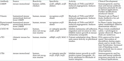
Therapeutic antibodies. A series of therapeutic an-tibodies targeting v integrins are currently under development and in clinical trials as antiangiogenic and anti-tumor agents. These antibodies are summa-rized in Table 1.
Table 1. v3 targeted therapeutic antibodies.
Antibody Source Reactivity Specificity Activity Clinical Development LM609 mouse monoclonal human, rabbit,
chicken αvβ3, αIIbβ3, αvβ5 dimers Blockade of TNFα and bFGF induced angiogenesis. Induces apoptosis.
Immunogenicity and hu-man anti-mouse antibody responses preclude its use in clinical applications. Further modifcations produced vitaxin. Vitaxin humanized mouse
monoclonal derived from LM609
human, mouse recognizes αvβ3
dimer Blockade of TNFα and bFGF induced angiogenesis. Induces apoptosis
Deemed safe in Phase I trials; Ineffective for ad-vanced cancers. Etaracizumab
(Abegrin) humanized mouse monoclonal derived from LM609
human, mouse recognizes αvβ3
dimer Blockade of TNFα and bFGF induced angiogenesis. Induces apoptosis
In Phase I trials, long term safety established; failed to demonstrate efficacy. CNTO 95 humanized IgG1 human,
does not recog-nize murine
αv integrin specific
αvβ3, αvβ5 Inhibits melanoma cell adhesion, migration and invasion in vitro. Inhibits tumor growth in mouse melanoma xenografts
Phase I trials established safety, 1 prolonged re-sponse observed. Phase II trials are underway. c7E3 chimeric mouse
hu-man human, murine αIIbβ3, αvβ3, MAC-1 Potenet antiplatelet drug. Shows anti-cancer activity in preclinical studies.
FDA approved for treat-ment/ prevention of thrombosis during angio-plasty and in Acute Coro-nary Syndromes. Increased risk of bleeding events may preclude use in cancer drug regimens.
17E6 mouse monoclonal human, does not recog-nize murine
αv integrin specific
αvβ3, αvβ5, αvβ1 Inhibits tumor growth in αvβ3 positive tumor xenografts. Ac-tivity attributed to blockade of tumor integrins.
LM609 LM609 was developed in the laboratory of David Cheresh and was subsequently used in the discovery and characterization of v3 integrin as adhesion molecules in endothelial and melanoma cell lines. As such it is a mouse monoclonal antibody that recognizes human v3 integrin dimers. It displays cross reactivity with cognate rabbit and chicken but not murine v3 integrins [108]. LM609 binds v3 integrin and prevents the adhesion and spreading of endothelial cells to ECM proteins fibrinogen, vitron-ectin, von Wilebrand Factor mulitmers, and RGD-peptide coated surfaces but not collagen or laminin as these interactions are mediated through alternate adhesion receptors [98]. LM609 is unable to lift attached endothelial cells from the former men-tioned ECM components presumably due to epitope masking as a result of intergrin interactions with ma-trix proteins. In cell based models of bone resorption LM609 supresses osteopontin-induced cytosolic cal-cium reduction and subsequent osteoclast function through the inhibition of v3 signaling [109]. In quail embryo models of embryonic neovasculogenesis, LM609 effectively disrupts the process of vessel for-mation [110]. LM609 also inhibits angiongenesis in mouse-human skin graft tumor models [111]. The anti-angiogenic and anti-tumor properties observed with LM609 treatment in experimental models occurs primarily via the inhibition of v3 mediated TNF
and bFGF signaling pathways. The murine origin of LM-609 limits its clinical utility, raising the concern of antigenic responses when administered to humans. Humanization of LM-609 using phage display strat-egy produced the second generation v3 antibody, Vitaxin.
Vitaxin (MEDI-523, Applied Molecular Evolution, San Diego, CA) Vitaxin is the humanized version of LM609 containing the mouse CDR region grafted to human IgG1 Kappa. Humanization using phage dis-play libraries produced greater affinity while retain-ing epitope specificity and reduced antigenicity [112]. In pre-clinical evaluation Vitaxin inhibited growth-factor mediated endothelial cell proliferation and prevented angiogenesis. Vitaxin has been evalu-ated in two Phase I clinical trials for the treatment of advanced, treatment refractory solid tumors. The ini-tial Phase I trial evaluated dosing in range of 0.1 to 4 mg/kg administered weekly as a 90 minute intrave-nous infusion over a period of six weeks among 17 patients. Adverse effects were observed in 88% of the patients but were mild and of low grade in the initial studies. The most common adverse event was infu-sion reaction including fever, nausea and chills but preadministration of aspirin and diphenhydramine