4598 FORMULATION AND EVALUATION OF PRONIOSOME BASED DRUG
DELIVERY SYSTEM OF VALCYCLOVIR FOR IN-VITRO AND EX-VIVO
PERFORMANCE ANALYSIS
Ashok Mateti1*, Mohammed Habibuddin2, Subhash Jadhav3, Sravanthi Gandu1 and Srinivas Reddy Devireddy4
1Department of Pharmaceutics, Trinity College of Pharmaceutical Sciences, Telangana. 2Department of Pharmaceutics, Shadan College of Pharmacy, Hyderabad, Telangana 3Department of Pharmaceutics, Sri Kakatiya Institute of Pharmaceutical Sciences, Warangal
4Department of Pharmaceutics, Vageswari College of Pharmacy, Karimnagar, Telangana
*Corresponding author E-mail:[email protected]
ARTICLE INFO ABSTRACT
Key Words
Proniosomes, Valcyclovir, Cholesterol, Transdermal, Permeation
A proniosome based drug delivery system of Valcyclovir as planned for
development and characterisation for in-vitro performance. Proniosomes
containing valcyclovir were prepared by co-acervation method using (span 20, span40, span60, span 80), cholesterol and lecithin at different concentrations. All the proniosomes formulation evaluated for
entrapment efficiency, drug content, shape and size distribution, in-vitro
and ex-vivo studies. The method of proniosomal encapsulation resulted
as 96%-82%. Morphology of F1 Proniosomal formulation was
characterised by using scanning electron microscopy (SEM), in-vitro
studies showed the prolonged release of entrapped Valcyclovir and
ex-vivo studies revealed the Proniosomes prepared with span 80 showed a
significantly lower enhancement effect then those prepared with span 20.
INTRODUCTION:
The Transdermal drug delivery system (TDDS) can defined as a delivery device, which upon application on a suitable skin surface will be able to deliver the drug in to systemic circulation at
sufficient concentration to ensure
therapeutic efficacy, an additional
limitation to oral drug delivery, can be avoided with transdermal administration. Provide suitability for self-administration [1]. The transdermal route has many advantages for the administration of drugs for local and systemic therapy. The outermost layer of skin, the stratum corneum (SC), forms a strong barrier to
most exogenous substances including drugs. The barrier function of the SC is attributed to its multilayered wall-like
structure, in which terminally
differentiated keratin- rich epidermal cells
(corneocytes) are embedded in an
intercellular lipid-rich matrix. Various
approaches were put forward for
overcoming it. Of these, colloidal carrier is an efficient one as it acts as drug containing reservoirs and can loosen the stratum corneum, thereby modifying the barrier, and can adjust the release rate at the target site. Among the various colloidal carriers, liposome and niosome were the
An Elsevier Indexed Journal ISSN-2230-7346
4599
popular ones as they can efficiently
encapsulate both hydrophilic and
hydrophobic drugs [2]. Although the
vesicular carriers are promising in
providing the alternative routes of drug delivery and also provide a sustained action due to prolonged release yet on other hand these carriers also suffer from some shortcomings at industrial and
clinical levels [3].
Proniosomes are dry formulation of the water soluble carrier particles that are coated with surfactant and can be measured out as needed and dehydrated to form niosomal dispersion immediately before use on brief agitation in hot aqueous media within minutes. The resulting niosomes are very similar to conventional niosomes and uniform size[4] The proniosomes approach minimize problems associated with the liposome’s and niosomes by using dry, free flowing product, which is more stable during
sterilization and storage. Ease of
transportation, distribution, measuring,
dosing and storage make proniosomes a versatile delivery system with potential for were with a wide range of active
compounds proniosomes not only do they
offer a promising means of drug delivery, but also but it could enhance the recovery
rate of skin barrier [3].
This study is aimed to incorporate valcyclovir in proniosomal gel system for topical administration to enhance the permeation through skin as well as the bioavailability of the drug. The prepared
system was optimized and evaluated
ex-vivo. Valcyclovir is a nucleoside antiviral
drug in pharmacologic studies, valcyclovir has shown antiviral activity. Valcyclovir is used in the treatment of Herpes Simplex Virus. In the present study proniosome concept was developed to encapsulate valcyclovir in surfactant vesicles and
evaluate for their ex-vivo characteristics
and an attempt to improve the oral bioavailability of the drug with less frequency of dose.
MATERIALS AND METHODS
Valcyclovir was obtained as a gift
from (Dr. Reddy,s Lab, Hyderabad), Span -20, 40, 60 and 80 was procured from
(Central Drug House Ltd., Delhi).
Cholesterol (S.D. Fine-Chem. Ltd.,
Mumbai), lecithin (lipo. Germany),
Ethanol, Methanol (Merck Ltd., Mumbai), Potassium dihydrogen phosphate purchased from (Himedia Laboratories Pvt. Ltd), Mumbai. Sodium hydroxide (Merck Ltd., Mumbai) and other chemicals are analytical reagent grade.
Preparation of proniosomal gel
Proniosomes were prepared using a
modified literature method[5], precisely
weighed amounts of different non-ionic surfactant, lipid and drug (valcyclovir-30mg) are taken in a clean and dry wide mouth glass vial of 5.0 ml capacity and alcohol (0.5 ml) is added to it. After warming, all the ingredients are mixed well with a glass rod the open end of the glass bottle is covered with a lid to prevent the loss of solvent from it and warmed over water bath at 60-70°C for about 5 min until
the surfactant mixture is dissolved
completely. Then the aqueous phase (7.4pH phosphate buffer) is added and warmed on a water bath till a clear solution was formed which is then converted into proniosomal gel on cooling [2].
Evaluation of Proniosomal Gel Particle and size distribution study:
Niosomes are prepared from
proniosomes upon hydration. 100 mg of proniosomal gel was hydrated in a small glass test tube using 10 ml of 7.4 pH phosphate buffer solution. The dispersion was observed under optical microscope at
40x magnification. Size and size
distribution of 200–300 niosomes were noted using calibrated stage and ocular
micrometers (Elico Instruments,
Hyderabad). Similarly, size was noted for niosomes formed spontaneously from
proniosomes after hydration without
4600 Entrapment efficiency study
0.4 g of proniosomal gel weighed in a glass tube, 2 ml phosphate buffer pH 7.4 was added. The aqueous suspension was then sonicated. Niosomes containing drug were separated from unentrapped drug by centrifugation at 9000 rpm for 45 min at 4° C. The supernatant was
recovered and assayed
spectrophotometrically using UV-visible spectrophotometer (UV-1800 Shimadzu,
Japan), at 254nm. The encapsulation
percentage of drug (EP) was calculated by the following equation [7]
EP = [(Ct – Cr)/ Ct] * 100
Where
Ct is concentration of total drug and Cr is
concentration of free drug.
Vesicle physical analysis
The shape, surface characteristics, and size of the niosomes were observed by scanning electron micros copy. 0.2 g of the proniosomal gel in a glass tube was diluted with 10 ml of pH 7.4 phosphate buffer. The niosomes were mounted on an
aluminium stub using double-sided
adhesive carbon tape. Then the vesicles were sputter-coated with gold palladium (Au/Pd) using a vacuum evaporator (Edwards) and examined using a scanning electron microscope (Hitachi 3700N, Germany) equipped with a digital camera, at 10 kV accelerating voltage[8].
Scanning Electron microscopy
The surface morphology and size distribution of proniosomes were studied by scanning electron microscopy (SEM). Proniosomes were sprinkled on to the double-sided tape that was affixed on aluminium stubs [6]. The aluminium stub was placed in vacuum chamber of a scanning electron microscope (Shimadzu, Germany).
Drug -Excipient Interaction
Fourier Transform infrared
spectroscopy (FTIR) of pure drug
Valcyclovir and mixture of drug with
excipients (Span 20, lecithin and
cholesterol) was taken using Perkin Elmer
FTIR spectrophotometer (RXIFT- IR system, USA). Sample was prepared with potassium bromide and data were collected
over a spectral range of 450-4000 cm−1[9].
In Vitro Release
In vitro release studies on
proniosomal gel were performed using vertical unjacketed Franz-diffusion cell
with diffusional surface area of 5.722 cm2.
The dialysis cellophane membrane was soaked in 7.4 pH phosphate buffer to get equilibrium. The receptor compartment contains 20ml of 7.4 pH phosphate buffer. Place entire unit on magnetic stirrer (temperature at 37±2°C) with 300 rpm. A weighed amount 0.2 g of formulation reference formulation equivalent to 15 mg of drug of proniosomal gel was placed on superior side of the dialysis membrane. Aliquots of 5 ml were withdrawn periodically at different time intervals (0.5, 1, 2, 3, 4, 6, 8, 10, 12, and 24 hr respectively) and replaced with equal volume to maintain constant receptor phase volume. At the end of the study, the samples were suitably diluted and the
amount of drug was determined
spectrophotometrically at 254 nm.
Ex vivo Permeation Study using albino Rat Abdominal Skin
Male albino rats (150-200 g) were
used for the experiment [10]. Ex vivo
permeation studies were carried out using unjacketed vertical Franz diffusion cells with a diffusional surface area of 5.722
cm2 and 20 ml of receptor cell volume.
4601
compartment was constantly stirred at 300rpm. The donor chamber and the sampling port were covered with lid to prevent evaporation during the study. Aliquots of 5 ml were withdrawn periodically at different time intervals (0.5, 1, 2, 3, 4, 6, 8, 10, 12, and 24 hr) and replaced with equal volume to maintain constant receptor phase volume. At the end of the study, the samples were suitably diluted and the amount of drug was determined spectrophotometrically at 254
nm. The cumulative amount of drug
permeated through a unit area of skin was plotted as a function of time. The Steady state Flux was calculated by using the
slope of the graph Where, Jss =
(dQ/dt)*(1/A) Jss is Flux (µg/cm2/hr), A is
Surface area and dQ/dt is Cumulative amount permeated per unit area per unit time. Permeability co-efficient which represents the correlation between the flux and initial drug load was calculated using
the following equation.[16] Kp = Jss/C
Where, Kp is Permeability co efficient
(cm/hr), Jss is transdermal flux and C is
Initial concentration of Valcyclovir. The
penetration enhancing effect of various formulations containing proniosomal gel was calculated in terms of Enhancement Ratio (ER) by using the following equation [11].
ER = Jss of formulation / Jss of reference
Skin irritancy test
The skin irritancy potential of the proniosome formulations was evaluated in albino rats. The hair was removed on the back of the animal and the formulations were applied, and the animals were examined for any signs of skin irritation and erythema for a period of 1 week [5].
Stability Studies
Proniosome gel was subjected to stability testing as per ICH Q1AR and Q6A guidelines, were placed in a glass vials with aluminium foil and kept in a programmable environmental test chamber
(Remi instrument, CHM-16S, India)
maintained at 40 ± 2ºC and 75 ± 5% RH for 3 months.
The formulations stored in glass vials covered with aluminium foil were kept at room temperature and in refrigerator (4◦ C) for a period of 30 days. At definite time intervals (10, 20, and 30 days), samples were withdrawn and hydrated with phosphate-buffered saline (pH 7.4) and observed for any sign of drug crystallization under optical microscope [4]. Furthermore, the samples were also evaluated for particle size and percent retention of valcyclovir.
Statistical Analysis
Significance of difference among formulations was calculated by one way analysis of variance (ANOVA) using Newman Keuls (compare all pairs) with Instant Graph Pad Prism software. The
difference was considered to be
statistically significant at p<0.05[10].
RESULTS AND DISCUSSION
Drug content
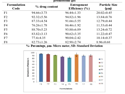
Valcyclovir content estimation in the proniosomes gel of different non-ionic surfactant, with or without lecithin, cholesterol is carried out the drug content was found to be in between 94.66±3.73
and 98.76±4.59 indicates that the
percentage of the drug in formulation
within acceptable limits. Entrapment efficiency
The ability of the vesicles to entrap
Valcyclovir was investigated. The
proniosomes containing Valcyclovir using non- ionic surfactant (Span 20, 40, 60 and 80) along with cholesterol and with or without lecithin content. Results indicated proniosomal gel formulations of Valcyclovir with lecithin and cholesterol showed higher entrapment efficiency as compared to formulation prepared without lecithin and with cholesterol. Therefore, it was concluded that incorporation of lecithin imparted superior effect on entrapment efficiency of the proniosomes. Results indicated entrapped the drug in the
range of 96.44±1.33, 94.82±1.96,
4602
and 82.98±2.74 represented in Table- 2 and Fig-1. Span 20 has higher HLB value of 8.6 and vesicles of largest size but faster drug release was obtained perhaps due to its low transition temperature [12, 13].
Particle size analysis
The particle size of Proniosomes was determined by optical microscopy. The prepared f ormulations were studied under 40x magnifications to observe the formation of vesicles. About 300 particles were measured and the results are shows in Table -2. The Proniosomes were observed to be spherical vesicles with smooth surface. The vesicles were discrete and separate with no aggregation. Size range of the all formulations was found to be is 20.02±0.45 to 8.96±0.68. F1 formulation showed greater particle size due to higher HLB value of surfactant has great particle size.
Scanning Electron Microscopy
Surface morphology confirms the coating of surfactant in carrier. Results that
niosomes from F1 proniosomes
formulation shows the vesicles are well identical spherical and discreet with sharp boundaries are represented in Figure 2.
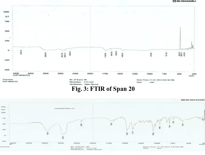
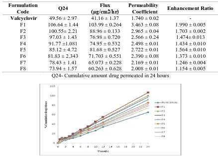
Drug and excipient interaction study
Fourier transforms infrared
spectroscopy (FTIR) of pure drug
valcyclovir and mixture of drug with
excipients (span 20, lecithin and
cholesterol) was taken using Shimadzu
FTIR spectrophotometer (RXIFT-IR
system, Germany). Sample was prepared with potassium bromide and data were collected over a spectral range 0f 250-4000cm-1. Results showed for span 20, cholesterol, lecithin, drug and formulation, peaks are observed in Figs-3,4,5,6 and 7.Graphs of drug and drug-excipients conformed that there is no interaction
between the drug and excipients used.
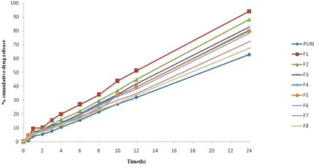
In vitro drug Permeation Study
The proniosomal gel formulations (F1 to F8) were characterized for their drug release and the results are reported in Table -3. The drug release was maximum from formulation containing Span 20 (F1)
among all tested formulations. From the permeation profile it was clear that the gel showed optimum drug release up to 24hrs. From the formulation F1 which was 106.64 ± 0.44% Figure 8. The rate of drug release was increased with the increasing HLB value of the surfactant added (Span 20 having high HLB value of 8.6 and greater transition temperature (TC) (Barry, 1994) Proniosomal gel prepared by using with lecithin exhibited better permeation, higher flux and optimum entrapment efficiency when compared with the formulations containing without lecithin. Hence the formulation F1 had selected as optimum formulation and stability testing
studies were carried out. These
observations were in accordance with earlier reports saying that incorporation of cholesterol was known to influence vesicle stability, permeability and entrapment efficiency. Cholesterol content resulted in a more intact and ordered lipid bilayer as a barrier for drug release and helped as a controlled release polymer and also decreased drug leakage by improving the fluidity of the bilayer membrane and reducing its permeability [14].
Ex vivo permeation study
Ex-vivo permeation studies were
conducted by using rat abdominal skin and the permeation results were shown in Fig- 9. It has been reported that the intercellular lipid barrier in the stratum corneum would
be dramatically losses and more
permeability follow by treatment with liposome and niosomes [15]. Fusion of niosomal vesicles to the skin surface results in higher flux due to the direct transfer of the drug from vesicles to skin.
Proniosomes has without lecithin content resulted in a lower flux Table-9,
although this lower flux was not
4603
Table 1: Composition of Valcyclovir proniosomal gel (f1 to f8)
Ingredients(mg) F1 F2 F3 F4 F5 F6 F7 F8
Valcyclovir 30 30 30 30 30 30 30 30
-
Span 20 200 - - - 360 - -
Span 40 - 200 - - - 360 - -
-
Span 60 - - 200 - - - 360
Span 80 - - - 200 - - - 360
Cholesterol 40 40 40 40 40 40 40 40
Lecithin 160 160 160 160 - - - -
Table 2: Percentage of drug content, entrapment efficiency and particle size of Valcyclovir proniosome gel
Formulation
Code % drug content
Entrapment Efficiency (%)
Particle Size (μm)
F1 94.66±3.73 96.44±1.33 20.02±0.45
F2 92.32±5.56 94.82±1.96 13.84±0.76
F3 87.33±4.54 91.66±3.55 12.79±0.44
F4 76.26±1.78 86.46±1.92 11.33±0.44
F5 88.70±5.23 93.80±6.89 13.24±0.72
F6 83.82±3.13 90.62±3.35 11.22±0.47
F7 77.4±4.35 90.04±2.42 10.14±0.37
F8 82.73±1.26 82.98±2.74 8.96±0.68
%- Percentage, μm- Micro meter, SD- Standard Deviation
Fig. 1: Entrapment efficiency of Valcyclovir in proniosomal gel for the formulation of F1 to F8
4604 Fig. 3: FTIR of Span 20
Fig. 4: FTIR spectrum of Cholesterol
Fig. 5: FTIR spectrum of lecithin
4605
Table 3: Release of Valcyclovir proniosome gel for the formulations F1 to F8 through
cellopheone membrane.
Formulation
Code Q24
Flux (μg/cm2/hr)
Permeability
Coefficient Enhancement Ratio
Valcyclovir
F1 F2 F3 F4 F5 F6 F7 F8
49.56 ± 2.97 106.64 ± 1.44
100.55± 2.21 97.03 ± 1.43 91.77 ±1.081 85.12 ± 4.72 81.83 ± 2.343
78.43 ± 1.41 73.94 ± 1.57
41.16 ± 1.37 103.99 ± 0.264
88.96 ± 0.133 76.98 ± 0.720 74.95 ± 0.532 81.68 ± 0.527 71.703 ± 0.551 65.073 ± 0.228 60.263 ± 0.628
1.740 ± 0.02 3.463 ± 0.08 2.965 ± 0.04 2.566 ± 0.24 2.498 ± 0.01 2.722 ± 0.01 2.390 ± 0.08 2.169 ± 0.01 2.008 ± 0.01
- 1.990 ± 0.005 1.703 ± 0.002 1.474± 0.013 1.434 ± 0.010 1.564 ± 0.010 1.373 ± 0.010 1.246 ± 0.004 1.154 ± 0.005 Q24- Cumulative amount drug permeated in 24 hours
Fig. 7: Release of Valcyclovir proniosome gel for the formulations F1 to F8 through cellopheone membrane
Table 4: Release of Valcyclovir proniosome gel for the formulations F1 to F8
through rat abdominal skin.
Formulation
Code Q24
Flux (μg/cm2/hr)
Permeability Coefficient
(Kp)
Enhancement Ratio (ER)
Valcyclovir F1 F2
F3 F4 F5 F6 F7 F8
45.67 ± 1.08 94.11 ± 1.81 88.18 ± 3.23 82.76 ± 5.47 80.56 ± 2.19 79.74 ± 0.83
78.18 ± 0.200 72.21 ± 1.39 67.35 ± 0.86
39.61 ± 2.351 95.93 ± 3.266 a,1,9,8,7,5,4,3
80.14 ± 1.32a,1,9,8,7,5,6
78.18 ± 3.122a,1,9,8,7,5
72.71 ± 1.701a,1,9,8
76.29 ± 1.783a,1,9,8,7,5
69.01 ± 3.246a,1,9,8
59.92 ± 0.92a,1,9
54.39 ± 1.043a,1
1.691 ± 0.013 3.197 ± 0.19 2.660 ± 0.010 2.606 ± 0.104 2.424 ± 0.056 2.543 ± 0.026 2.300 ± 0.048 1.986 ± 0.03 1.813 ± 0.014
- 1.890 ± 0.064 1.576 ± 0.061 1.540± 0.006 1.434 ± 0.033 1.503 ± 0.015 1.360 ± 0.005 1.174 ± 0.018 1.071 ± 0.008
4606
Fig. 9: Release of Valcyclovir proniosome gel for the formulations F1 to F8 through rat abdominal skin
These results suggest that inclusion of lecithin at a certain level may play an important role in drug permeation[7] .Therefore lower in its concentration in the formula resulted in slight decrease in drug permeation. Proniosomes prepared with span 80 showed a significantly lower enhancement effect then those prepared with span 20 (p<0.05). The rate of drug release was increases with the increasing HLB value of the surfactant added. Span 20 having high drug release this is may be due to its high HLB value and greater
transition temperature (TC) [16]. Whereas
span 40, 60 and 80 are having low HLB values compare to span 20.This was expected due to the larger size of the vesicles and the less lipophilic nature of the former, which makes it more difficult for these vesicles to penetrate or fuse with skin.
C
COONNCCLLUUSSIIOONN
The Transdermal Proniosomal Gels showed controlled drug release properties. The results of the present study indicated
that Valcyclovir proniosomal gel
containing with or without lecithin,
cholesterol and in combination of
surfactants like span 20, 40, 60, 80 shows sustained release of drug over a period of 24 hrs for the management of herpes simplex viruses. By carrying out different evaluation parameters finally F1 and F2 are found as the optimized formulations.
The percentage of drug content for formulation F1 was 94.66±3.73 and for F2 formulation is 97.32±5.56. The entrapment efficiency of F1 is 96.44±1.33and for F2 is 94.82±1.96. The particle size of F1is found to be 20.02±0.45and for F2 is 13.84±0.76. The ex-vivo drug release of F1 was founded as 94.11 ± 1.81 and F2 was 88.18 ± 3.23. The flux result of F1 formulation was 95.93 ± 3.266 and for F2 was 80.14 ± 1.32. The optimized F1 proniosomal gel system showed better ex-vivo permeation study and will be a great potential for delivery of anti viral drug Valcyclovir. Non-ionic surfactants with higher HLB values showed greater drug permeation. Finally, Valcyclovir proniosomal gel with lecithin and span-20 (F1) and Sapn-40 (F2) was optimized. The FTIR and SEM results are also supports the permeation of
Valcyclovir from non-ionic vesicles
containing lecithin..
A
ACCKKNNOOWWLLEEDDGGEEMMEENNTT
The authors acknowledge to the
Trinity College of pharmaceutical
sciences, peddapalli, karimnagar, for
providing the necessary instrumental
facilities and also guidance of faculty.
REFERENCES:
1. Naik, A., Kalia, Y.N., Guy, R.H.
Transdermal drug delivery:
4607
2. Fang, J.Y., Hong, C.T, Chiu, W.T.,
Wang, Y.Y. Effect of liposomes and niosomes on skin permeation
of enoxacin. Int Jour Pharm. 2001;
219, 61–72.
3. Almira, A., Blazek-Welsh., David,
G. Maltodextrin- Based
proniosomes. AAPS Pharmsci,
2001: 3:1.
4. Hu, C., Rhodes, D.G.
Proniosomes: A novel drug
carrier preparation, Int. J. Pharm,
1999; 185: 23-35.
5. Perrett, S., Golding, M.,
Williams, W.P. A simple method for the preparation of liposomes for pharmaceutical applications:
characterization of the
liposomes, Pharm. Pharmacol,
1 9 9 1 ; 4 3 : 154-161.
6. El-Laithy, H.M., Shoukry, O.,
Mahran, L.G. Novel sugar esters
proniosomes for transdermal
delivery of vinpocetine: Preclinical
and clinical studies, Eur Jour
Pharm Biopharm, 2011; 1: 43-55.
7. Alsarra, I.A., Bosela, A.A.,
Ahmed, S.M., Mahrous, G.M. Proniosomes as a drug carrier for transdermal delivery of ketorolac,
Eur Jour Pharm Biopharm, 2005;
59: 485-90.
8. Abd-Elbary, A., El-Laithy., H.M.,
Tadros, M.I. Sucrose stearate-based proniosomes-derived niosomes for
the nebulisable delivery of
cormolyn sodium, International Journal of Pharmaceutics, 2008; 357:189-198.
9. Chandra, A., Sharma P.K.
Proniosome based drug delivery of
poroxicam, African journal of
pharmacy and pharmacology,
2008; 2: 184-190
10.Sunil, T.N., Ramesh, P.,
Transdermal drug delivery of
zidovudin : effect of terpenes and
their mechanism of action. J.
Control. Rel, 2004; 95, 367-379.
11.Murdan, S., Van Den Bergh, B.,
Gregoriadis, G., Florence, A.T. Water in sorbitan monostearate
organogels (water in oil gels), Jour
Pharm Sci, 1999; 6: 615-619.
12.Vora, B., Khopade, A.J., Jain,
N.K. Proniosome based
transdermal delivery of
levonorgesterel for effective
contraception, Jour Control. Rel,
1998; 54: 149-165.
13.Prajapati, K.,Gupta, A.,
Balamurugan, M., Singh, M.
Design and development of a
proniosomal transdermal drug
delivery system for Flubiprofen,
Tropical Journal of
Pharmaceutical Research, 2007; 2:
687.
14.Hemant, N., Sharwaree, R.
Formulation development and
Evaluation of proniosomal gel of
Carvedilol, Intr Journal of pharm
research, 2012.
15.Sankar, V., Ruckmani, K., Durga,
S., Jailani, S. Proniosomes as drug
carrier, Pak. J. Pharm. Sci, 2010;
1: 103-107.
16.Thomas Litha, A., Jose Shoma, A.,
George Sachin John, A., Vidya Viswanad, A. Provesicular
Niosomes Gel: A Novel
Absorption Modulator for
Transdermal Delivery, Int Jour of
Drug Development & Research,