Available Online at www.ijpret.com 445
INTERNATIONAL JOURNAL OF PURE AND
APPLIED RESEARCH IN ENGINEERING AND
TECHNOLOGY
A PATH FOR HORIZING YOUR INNOVATIVE WORK
ECG ANALYSIS USING ARTIFIAL NEURAL NETWORK
ASST. PROF. RAJ CHALSE, MR. SURAJ A. KUBADE, MR.AKASH K. WAIRAGADE, MISS. VIBHA BALPANDE, MISS. RENKA KHONDE
Dept. of Information Technology, DMIETR, Wardha-442001.
Accepted Date: 15/03/2016; Published Date: 01/05/2016
\
Abstract: The performance of computer aided ECG analysis depends on the precise and
accurate delineation of QRS-complexes. In this we presents an application of Artificial Neural Network (ANN), Canny Edge Algorithm as a classifier for detection of QRS-complex in ECG. The proposed algorithm is evaluated on manually annotated standard databases such as MIT-BIH Arrhythmia database. For this work, we used Neural Network to reduce false detection caused by interference present in ECG signal and further noises and also inconsistent data in ECG image. Using the implemented Canny edge for features extraction and as an enhancement tool for ECG images. Although Back propagation algorithm builds a model that gives the correct information about the patient whether it is normal or abnormal. These results clearly establishes a system that help to easily identify whether the patient is normal or abnormal.
Keywords: ECG Analysis, Artifial Neural Network
. Corresponding Author: ASST. PROF. RAJ CHALSE
Access Online On:
www.ijpret.com
How to Cite This Article:
Raj Chalse, IJPRET, 2016; Volume 4 (9): 445-451
Available Online at www.ijpret.com 446 INTRODUCTION
A great deal of research has been conducted in the field of biomedical signal processing. A number of biomedical signals are recorded and used for patient monitoring or diagnostic purposes. The electrocardiogram (ECG) plays a key role in patient monitoring and diagnosis. An electrocardiogram is use to monitor your heart .Each beat of your heart is triggered by an electrical impulse normally generated from special cell in the upper right chamber of your heart. An electrocardiogram is also called as ECG or EKG record electrical signals as the travel through heart. Our project is basically design on the ECG on which we are perform the action such that to determine the given ECG image will be normal or abnormal. To recover such type of problem and to find the ECG of any type of person is normal or abnormal.
Artificial neural networks
Available Online at www.ijpret.com 447 The Back propagation Algorithm
The backpropagation algorithm (Rumelhart and McClelland, 1986) is used in layered feed-forward ANNs. This means that the artificial neurons are organized in layers, and send their signals “forward”, and then the errors are propagated backwards. The network receives inputs by neurons in the input layer, and the output of the network is given by the neurons on an output layer. There may be one or more intermediate hidden layers. The backpropagation algorithm uses supervised learning, which means that we provide the algorithm with examples of the inputs and outputs we want the network to compute, and then the error (difference between actual and expected results) is calculated. The idea of the backpropagation algorithm is to reduce this error, until the ANN learns the training data. The training begins with random weights, and the goal is to adjust them so that the error will be minimal. The activation function of the artificial neurons in ANNs implementing the i backpropagation algorithm is a weighted sum (the sum of the inputs x multiplied by their ji respective weights w):
Principal Component Analysis (PCA)
PCA is a statically procedure that use an orthogonal transformation to convert a set of observation possibly correlated variable into a set of values of linearly uncorrelated variable called principal component the no of principal component is less than the number of original variable [2].
Canny Edge Detector
The canny edge dector is an edge detection operator that uses a multistage algorithm to detect a wide range of edges in image.
II. PROBLEM RELATED TO ECG SIGNAL
Generally so many biomedical signal are weak signal and many type of disturbance appear due to interfaces of unwanted signal. This will lead to the in proper out of the machine. Although very few hospital contain the equipment to analysis the image. At some time the environment accomuted ECG signal.
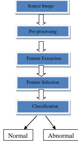
III. PROPOSED WORK
Available Online at www.ijpret.com 448 An application that give the directly give a result whether it is the normal or abnormal. And we also create Artificial neural network by using some algorithm that are disuss in introduction.
Fig: Basic design of proposed work
IV. IMPLEMENTED WORK
The described work is implemented with help of matlab. First we show the snapshot in which we browse the image after we click in select ecg image in that textbox we can add the name of
particular image
Fig 1: Browse the image Pre-processing
re
Feature Extraction
Feature Selection
Classification
Normal Abnormal
Available Online at www.ijpret.com 449 2. The next snapshot represent the selection of image from any file then click on the open and select the image from that particular file.
Fig 2: Select the image
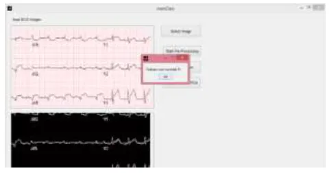
3. The next snap shot represent the processing page after clicking the start processing button we get image which we got in negative form.
Fig 3: Start Preprocessing
4. After start processing the feature extraction will be start in this we extract feture of image
after we click in the feature extraction ECG image should run successfully
Available Online at www.ijpret.com 450 5. After successful feature extraction we get popup window that window display the current message the message in the form of “feature successfully”. Then click on ok button this ecg image should be save in the db_feature. mat file of feature extraction now the database in the from of binary format
Fig 5: Feature Extracted Successfully
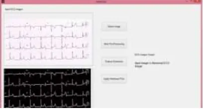
7. After applying the non collinear PCA we get the result whether the image is normal or abnormal.
Fig 6: Getting Result
CONCLUSION
The implementation of algorithm above will results a report including patient’s data and accurate heart rate calculation. In this section, the results of algorithm for each image processing techniques were shown. First of all, the selected ECG image was initially loaded into the software through computerized algorithms. An efficient method for extraction and digitization of ECG signal from various sources such as thermal ECG printouts, scanned ECG and captured ECG images from devices is proposed.
Available Online at www.ijpret.com 451 ACKNOWLEDGMENT
Author would like to give his sincere gratitude to guide Mr. Raj Chalse who encouraged and guided us throughout this paper.
REFERANCE
1. ECG ANALYSIS FOR ARRHYTHMIA DETECTION USING PCA AND ELMAN NEURAL NETWORK
harpreet kaur and rupinder kaur DIET, kharar, punjab, india.International journal of applied engineering and technology ISSN: 2277-212X (online) an open access, online international journal 2014.
2. DIGITIZATION OF ECG PAPER RECORDS USING MATLAB international journal of innovative
technology and exploring engineering (ijitee) issn: 2278-3075, volume-4 issue-6, november 2014.
3. APPLYING K-NEAREST NEIGHBOUR IN DIAGNOSING HEARTDISEASE PATIENTS. International
journal of information and education technology, vol. 2, no. 3, june 2012.
4. DATA CLASSIFICATION USING SUPPORT VECTOR MACHINE durgesh k. srivastava, lekha