Vol -2, Issue-2, February-April 2013
ISSN: 2278 – 7496
AJPER February-April 2013, Vol 2, Issue 2 (29-42)
RESEARCH
ARTICLE
Formulation and Optimization of Candesartan Loaded Eudragit Coated
Microspheres
Amit Sharma*1, Swati Dikshit1, Niraj Upamanyu1, Subhendu S. Mishra2, Ashish Pathak2
1. Department of Pharmaceutics, R.K.D.F College of Pharmacy, Bhopal, M.P., India.
2. Department of Pharmaceutics, Sapience Bioanalytical Research Laboratory, Bhopal, M.P.,
India.
Abstract:
The objective of present study is enhancement in the oral bioavailability of Candesartan using a lipid-based formulation i.e Eudragit S-100 microspheres. Candesartan is therapeutically a potent antihypertensive agent but it suffers a major drawback of poor oral bioavailability, which is estimated to be 15% due to low solubility in gastrointestinal fluid, hepatic first pass metabolism. The main emphasis of present study is on the use of new approach of lipid-based formulation to enhance the bioavailability of poorly water-soluble but highly permeable drug. The proposed formulation is aimed to enhance the bioavailability of Candesartan by using microspheres coated with eudragit S-100 polymer, which are transported through lymphatic system, thereby preventing hepatic first pass metabolism. Eudragit based microspheres were prepared by oil-in-oil solvent evaporation method using different drug- polymer ratios (1:1 to 1:4), stirring speeds (500-1500 rpm) and emulsifier concentrations (0.5%-1.25% wt/vol). Differential scanning calorimetry, study of the physical mixtures of drug and polymer revealed no drug-polymer interaction. All formulations were evaluated for particle size and shape, swell ability and percentage drug entrapment. The yield of preparation and the encapsulation efficiencies were high for all Eudragit microspheres. The in vitro drug release study of optimized formulation was also performed in simulated gastrointestinal fluids (SGF). The release profile of Candesartan from Eudragit microspheres was pH dependent.
Keywords: Eudragit S 100, Colon specific drug delivery, Microspheres, Candesartan.
Article Received on 4 March 2013.
Revised on 15 March 2013
Accepted on 18 March 2013
*Correspondence for
Author:
Mr. Amit Sharma
Dept. of Pharmaceutics
R.K.D.F. College of Pharmacy, N.H-12, Hoshangabad Road, Bhopal, M.P., India
Email:
AJPER February-April 2013, Vol 2, Issue 2 (29-42) INTRODUCTION:
Microspheres have been widely accepted as a means to achieve oral (1) and parenteral controlled
release (2) drug delivery system. The microsphere requires a polymeric substance as a carrier
and a core material. Among the various methods developed for formulation of microspheres, the
solvent evaporation method has gained much attention due to its ease of fabrication without
compromising the activity of drug. Eudragit S 100 is a water-insoluble polymer that is widely
used as a wall material for sustained release microsphere (3). This is due to its biocompatibility,
good stability, easy fabrication and low cost. In recent years, microsphere dosage forms have
gained increasing importance as oral controlled drug delivery systems. These systems present
several advantages in comparison to unit dosage forms such as more predictable gastric
emptying and less local irritation (4). Microsphere systems also minimize the possible intestinal
retention of undigested polymer materials in chronic dosing (5). The drug of choice,
Candesartan, is a potent antihypertensive agent belonging to the category of Angiotensine
receptor antagonist. It has a short biological (6) half-life of 5.1 ± 10.5 h & low bioavailability
15% The main emphasis of present study is on the use of new approach of lipid-based
formulation to enhance the bioavailability of poorly water-soluble4 but highly permeable drug by
using microspheres coated with eudragit S-100 polymer, which are transported through
lymphatic system, thereby preventing hepatic first pass metabolism also. In addition, the colon
has a long retention time and appears highly responsive to agents that enhance the absorption of
poorly absorbed drugs. The approaches used in the colonic delivery of drugs include the use of
prodrugs (7), pH-sensitive polymer coatings (8,9), time-dependent formulations (10), bacterial
degradable coatings (11), time pH-controlled deliveries (12), and intestinal luminal
pressure-controlled colon delivery capsules (13). The solvent evaporation method was selected as a
AJPER February-April 2013, Vol 2, Issue 2 (29-42)
methods allow for the use of different polymers and solvents, Emulsion evaporation permits
higher polymer concentration per batch production improving the microsphere yield, It can be
used for entrapment of hydrophobic and hydrophilic drugs, The hydrophobic drugs use oil in
water (o/w) emulsion technique, The hydrophilic drugs require the use of non-aqueous o/o
emulsion or double emulsion technique, The fast evaporation rate of the solvent permits a
reduction in the processing time. In addition, the colon has a long retention time and appears
highly responsive to agents that enhance the absorption of poorly absorbed drugs. The
approaches used in the colonic delivery of drugs include the use of prodrugs (1), pH-sensitive
polymer coatings (2, 3), time-dependent formulations (4), bacterialdegradable coatings (5), time
pH-controlled deliveries (6), and intestinal luminal pressure-controlled colon delivery capsules
(7).
MATERIALS AND METHOD
MATERIALS:
Candesartan was obtained as a gift sample from Matrix laboratories, Hyderabad, India. Eudragit L-100
was procured as a gift sample from Matrix laboratories, Hyderabad,India All other solvents and
reagents used were of analytical grade.
METHOD:
PREPARATION OF CORE CALCIUM ALGINATE BEADS
Different formulations of Calcium alginate beads were prepared using various drug: polymer ratio,
emulsifier concentration, and various stirring speed as shown in table 1.
Emulsion of drug and Span 80 was taken in a beaker and to it solution of sodium alginate was added
drop wise and stirrer well. This mixture of alginate and emulsion was then added to the solution of
AJPER February-April 2013, Vol 2, Issue 2 (29-42)
precipitate was formed by chemical reaction between sodium alginate and calcium chloride. The
prepared beads were left under continuous stirring at about 500 RPM for about 10 minutes and then
removed by filtration and washed with n- Hexane 4-5 times to remove the surface drug and then dried.
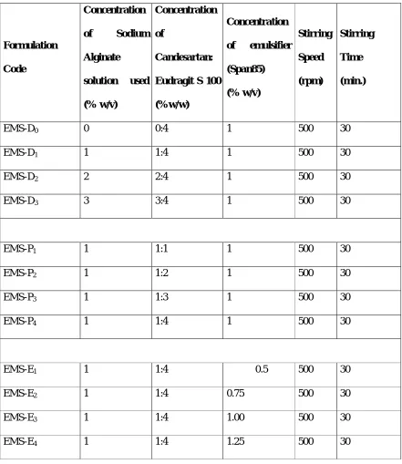
Tables:
Table 1: Composition of different Candesartan Microspheres
Formulation
Code
Concentration
of Sodium
Alginate
solution used
(% w/v)
Concentration
of
Candesartan:
Eudragit S 100
(%w/w)
Concentration
of emulsifier
(Span85) (% w/v) Stirring Speed (rpm) Stirring Time (min.)
EMS-D0 0 0:4 1 500 30
EMS-D1 1 1:4 1 500 30
EMS-D2 2 2:4 1 500 30
EMS-D3 3 3:4 1 500 30
EMS-P1 1 1:1 1 500 30
EMS-P2 1 1:2 1 500 30
EMS-P3 1 1:3 1 500 30
EMS-P4 1 1:4 1 500 30
EMS-E1 1 1:4 0.5 500 30
EMS-E2 1 1:4 0.75 500 30
EMS-E3 1 1:4 1.00 500 30
AJPER February-April 2013, Vol 2, Issue 2 (29-42)
EMS-R1 1 1:4 1 500 30
EMS-R2 1 1:4 1 750 30
EMS-R3 1 1:4 1 1000 30
EMS-R4 1 1:4 1 1500 30
E- Eudragit, MS- Microspheres, D1- Drug, P1- Polymer, E1- Emulsifier, R1- Stirring speed
COATING OF CORE CALCIUM ALGINATE SPHERES
The prepared optimized batch of alginate beads were coated with polymer Eudragit S 100 using
solvent evaporation method. In this method the beads were dispersed in different optimized batches of
Eudragit S 100 which were dissolved in acetone and methanol in 2:1 ratio and the solvent was
evaporated in a rotator evaporator using rotation of about 500 RPM and then dried.
Optimization
Various formulation variables e.g. drug concentration, polymer concentration, emulsifier concentration and
process variables viz. stirring speed, which could affect the preparation and properties of microspheres were
identified and studied. The compositions of formulation code of designed formulae of Candesartan
microspheres are given in table 1.
Optimization of formulation & process variables:
Various formulation variables were tried to prepare microspheres viz. drug concentration: 0%, 1%,
2% and 3%, Eudragit concentrations: 1%, 2%, 3%, 4% and emulsifier concentrations: 0.5%, 0.75%, 1.0%,
1.25%, were optimized. The effects of drug concentration, Eudragit concentration and emulsifier
concentration on the particle size, shape, size distribution and total drug loading efficiency are shown in
table 2 respectively.
AJPER February-April 2013, Vol 2, Issue 2 (29-42)
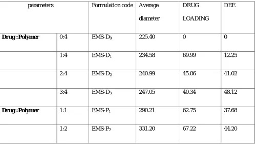
Optimization of drug ratio was the first step in the preparation of microspheres. Various ratios of drug
and polymers were taken i.e.0:1, 1:1, 2:1, 3:1. Keeping stirring speed (1000 rpm), emulsifier
concentration (1.25%v/v span 80), processing medium (light liquid paraffin oil, ethanol and acetone)
constant, microsphereswere prepared by the method and shown in table 2
From the above optimization parameters observed on the drug concentration it was observed that the
batch EMS-D3 is having the drug entrapment efficiency of 48.12 % and drug loaded in it is 40.34 %.
From the above graph it was concluded that as the drug concentration is increased the DEE is
increased whereas the drug loading decreases.
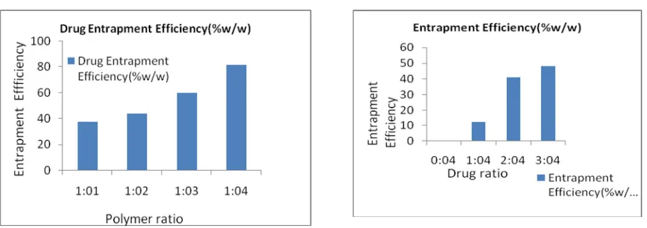
Optimization of drug polymer ratio
Optimization of drug polymer ratio was the first step in the preparation of microspheres. Various ratios
of drug and polymers were taken i.e. 1:1, 1:2, 1:3, 1:4. Keeping stirring speed (1000 rpm), emulsifier
concentration (1.25%v/v span 80), processing medium (light liquid paraffin oil, ethanol and acetone)
constant, microsphereswere prepared by the method and shown in table 2
Table 2: PREPARATION OF CORE CALCIUM ALGINATE BEADS
parameters Formulation code Average
diameter
DRUG
LOADING
DEE
Drug :Polymer 0:4 EMS-D0 225.40 0 0
1:4 EMS-D1 234.58 69.99 12.25
2:4 EMS-D2 240.99 45.86 41.02
3:4 EMS-D3 247.05 40.34 48.12
Drug :Polymer 1:1 EMS-P1 290.21 62.75 37.68
AJPER February-April 2013, Vol 2, Issue 2 (29-42)
1:3 EMS-P3 440.52 70.46 60.02
1:4 EMS-P4 579.32 84.64 81.78
Emulsifier
conc.(%v/v)
0.5 EMS-E1 580.24 70.24 80.01
0.75 EMS-E2 461.40 69.14 65.12
1.0 EMS-E3 391.52 50.20 49.60
1.25 EMS-E4 287.22 41.02 29.87
Stirring Speed (rpm) 500 EMS-R1 295.21 62.95 62.45
750 EMS-R2 263.24 64.74 69.22
1000 EMS-R3 231.02 73.31 76.85
1500 EMS-R4 182.12 58.72 57.20
Optimization of Emulsifier Concentration
Concentration of emulsifier is an important parameter which needs to be optimized because it is
responsible for optimum particle size and stability of the microsphere. Span 80 was selected as an
emulsifier and various concentrations of span 80 were taken. Microspheres were prepared by same
method as described a with optimized ratio of drug polymer (1:4), keeping stirring speed (1000 rpm),
and volume of processing medium constant as shown in table 2.
From the above optimization parameters observed on the emulsifier concentration it was observed that
the batch EMS-E3 is having the drug entrapment efficiency of 49.60 % and drug loaded in it is 50.20
AJPER February-April 2013, Vol 2, Issue 2 (29-42)
and loading is decreased. So the optimum emulsifier concentration recommended for best DEE and
loading is found to be 1%.
Optimization of stirring speed
Stirring speed plays an important role in the microspheres size distribution and drug loading.
Microspheres were prepared by same method as described above with optimized ratio of drug polymer
(1:4), keeping emulsifier concentration (1%v/v span 80) volume of processing medium constant, at
different speeds i.e.500, 750, 1000, 1500 rpm as shown in table 2
From the above optimization parameters observed by the change in stirring speed it was observed that
the batch EMS-R3 is having the drug entrapment efficiency of 76.85 % and drug loaded in it is 73.31 %
as shown in figure 4. From the above graph it was concluded that as the stirring speed is increased the
DEE and loading also increases but decreases after a certain point. So the optimum stirring speed
recommended for best DEE and loading is found to be 1000 RPM.
Drug Content
The amount of Candesartan associated with the microspheres was analyzed in terms of surface
adsorbed drug and entrapped drug.
Estimation of surface drug in microspheres:
100 mg of microspheres was dispersed in 10 ml of PBS (pH 7.4) and shaken vigorously for 10 minutes
and supernatant was kept aside. Similarly, the sediment was again treated in the same manner and
second supernatant was mixed with first supernatant and analyzed for candesartan content
spectrophotometrically as described previously. The amount of prednisolone in the mixed washings
gave the amount of drug adsorbed on the surface of the microspheres.
Estimation of entrapped drug in microspheres:
The amount of drug entrapped was estimated by crushing the microspheres and extracting with
aliquots of simulated gastric fluid pH 7.4 repeatedly. The extract was transferred to a 100 ml
AJPER February-April 2013, Vol 2, Issue 2 (29-42)
filtered and the absorbance was measured after suitable dilution spectrophotometrically (UV-1800,
Shimadzu) against appropriate blank. The amount of drug entrapped in the microspheres was
calculated by the following formula: D.E.E. = (Amount of drug actually present /Theoretical drug
load expected) × 100.
Fig 1 Optimization of Drug w.r.t. % D.E. Fig 2 Optimization of Drug Polymer Ratio w.r.t.% D.E.E
Fig 3 Optimization of Emulsifier Concentration Fig 4 Optimization of Stirring speed w.r.t
w.r.t. % D.E.E. % D.E.E
CHARACTERIZATION OF PREPARED MICROSPHERES
The prepared microspheres were characterized for shape , size and size distribution, percent drug loading
AJPER February-April 2013, Vol 2, Issue 2 (29-42) 1. Shape
Microspheres were suspended in water; a drop was placed on a glass slide, covered with a cover slip
and viewed under the optical microscope to examine their shape.
2. Scanning electron microscopy
Scanning electron microscopy (SEM) is one of the most commonly used method for
characterizing drug delivery systems, owing in large part of simplicity of sample preparation and
ease of operation. Scanning electron microscopy was carried out in order to characterize surface
morphology of the microspheres. In this study the morphological observations were carried out
to study the surface morphology of microspheres. SEM micrographs and typical surface
morphology of the microspheres are given in figures 3.
3. Swellability/Degree of Swelling
From the swelling study as shown in table 15 and figure 19 it was observed that as compared to
other polymer like pectin, guar gum etc which are having a swelling phenomena to a greater
AJPER February-April 2013, Vol 2, Issue 2 (29-42)
pH 7.4 Eudragit S 100 does not show a swelling phenomena to a greater extent as compared to
various gums.
4. In Vitro Drug Release Studies in Simulated Gastrointestinal Fluids of Different pH
In vitrodrug release studies were performed in simulated gastric fluid pH 1.2 for 2 h and then studied
in pH 4.5 i.e simulated gastric fluid and simulated intestinal fluid from 3rd to 6th hour and then finally
release observed in phosphate buffer pH 7.4. The comparison of different dissolution media are shown
in tables 6. The Percentage cumulative drug release was higher at phosphate buffer 7.4 and Acetate
buffer 4.5 when compared to 0.1N HCl because of the higher solubility of drug in between pH
4.0-7.0.The cumulative percentage drug released from the formulations was found to be 83.62% in
phosphate buffer 7.4. From the release profile it is clearly evident that the drug release increased as the
pH of the dissolution media increased.
The cumulative release of drug significantly Increased with increasing drug and polymer concentration
but after a certain extent release was found to decrease as shown in figures 6, 7. The increased density
of the polymer matrix at higher concentrations resulted in an increased diffusional pathlength. This
may decrease the overall drug release from the polymer matrix. Furthermore, smaller microspheres are
formed at a lower polymer concentration and have a larger surface area exposed to dissolution
medium, giving rise to faster drug release.
With increase in emulsifier concentration from 0.5 to 1.2 5%, drug release was also found to increase
from 75.14 to 82.22 %. Whereas as with the change in stirring speed the release was found to increase
but after a above 1000 rpm the release was found to decrease as shown in table and figure.
AJPER February-April 2013, Vol 2, Issue 2 (29-42) RESULT AND DISCUSSION:
Eudragit microspheres of candesartan were successfully prepared by solvent evaperation technique.The
microspheres produced were generally spherical, discrete upon dispersions in an aqueous medium and
having uniform size ranges from 25 to 32 µm.
The microspheres were coated with Eudragit S100 oil-in-oil solvent evaporation method. Different
core: coat ratios were taken to optimize the final formulation. The final product was finally dried for 24
hours.
The effect of various process variables viz.stirring speed and formulation variables e.g. drug
concentration, polymer concentration and emulsifier concentration were studied. The results suggested
that these variables influence the shape, size, size distribution, swellability, total drug loading
efficiency and in vitro drug release of the final preparation. Hence these parameters were optimized to
prepare microspheres of small size with narrow size distribution, good drug loading efficiency and
good drug release at colonic Ph. The amount of drug entrapped was estimated by crushing the
microspheres and extracting with aliquots of simulated gastric fluid pH 7.4 repeatedly. The extract was
transferred to a 100 ml volumetric flask and the volume was made up using simulated gastric fluid pH
7.4. The solution was filtered and the absorbance was measured after suitable dilution
spectrophotometrically (UV-1800, Shimadzu) against appropriate blank.
ACKNOWLEDGEMENT:
Authors are grateful to Dept of Pharmacy, Sapience Bioanalytical Research Lanoratory, Bhopal for
providing the necessary facilities to the authors.
REFERENCES:
1 Sahoo SK, Mallick AA, Barik BB, Senapati PC. Preparation and in vitro evaluation of ethyl cellulose
AJPER February-April 2013, Vol 2, Issue 2 (29-42)
2 Chowdary KPR, Koteshwara RN, and Malathi K.Ethyl cellulose microspheres of gliplizide:
characterization, in vitro and in vivo evaluation.Indian Journal of pharmaceutical sciences 2004; 66:
412- 416.
3 Sahoo SK, Mallick AA, Barik BB, Senapati PC. Formulation and in vitro Evaluation of Eudragit®
Microspheres of Stavudine. Tropical Journal of Pharmaceutical Research. 2005; 4: 369-375
4.M. Kilicarslan and T. Baykara, The Effect of the Drug/Polymer Ratio on the Properties of the
Verapamil HCl Loaded Microspheres Intended for Oral Administration,” Int. J. Pharm.2003; 252 (1–
2): 99–109.
5. J. Kramer and H. Blume, Biopharmaceutical Aspects of Multiparticulates,” in Multiparticulate Oral
Drug Delivery, Y. Ghebre-Sellasie, Ed. (Marcel Dekker, New York, NY,1994), pp. 307–332.
6. Goodman & Gilman's The Pharmacologic Basis of Therapeutics - 11th Ed.,2006, 458-461.
7. T.N. Tozer et al., Colon Specific Delivery of Dexamethasone from a Glucoside Prodrug in the
Guinea Pig,” Pharm. Res. 1991; 8(4): 445- 454.
8. M. Ashford et al., An In Vivo Investigation into the Suitability of pH Dependent Polymers for
Colonic Targeting, Int. J. Pharm. 1993; 95: 193–199.
9. M. Marvola et al., Enteric Polymers as Binders and Coating Materials in Multiple-Unit Site-Specific
Drug Delivery Systems, Eur. J. Pharm. Sci. 1999; 7 (3): 259–267.
10. C. Gazzaniga et al., Time Dependent Oral Delivery System for the Colon Targeting, S.T.P. Pharm.
Sci. 1995;5: 83–88.
11. L.F. Siew, A.W. Basit, and J.M.Newton, The Properties of Amylose-Ethylcellulose Films Cast
from Organic-Based Solvents as Potential Coatings for Colonic Drug Delivery, Eur. J. Pharm. Sci.
2000; 11(2), 133–139.
12. T. Ishibashi et al., Design and Evaluation of a New Capsule-Type Dosage Form for Colon Targeted
AJPER February-April 2013, Vol 2, Issue 2 (29-42)
13. Y. Yoshikawa et al., A Dissolution Test for a Pressure-Controlled Colon Delivery Capsule: