RESEARCH
Proteins adopt functionally active
conformations after type III secretion
Kevin James Metcalf
1,5, James Lea Bevington
1, Sandy Lisette Rosales
2, Lisa Ann Burdette
1,4, Elias Valdivia
3and Danielle Tullman‑Ercek
4*Abstract
Background: Bacterial production of natively folded heterologous proteins by secretion to the extracellular space can improve protein production by simplifying purification and enabling continuous processing. In a typical bacterial protein production process, the protein of interest accumulates in the cytoplasm of the cell, requiring cellular lysis and extensive purification to separate the desired protein from other cellular constituents. The type III secretion system of Gram‑negative bacteria is used to secrete proteins from the cytosol to the extracellular space in one step, but proteins must unfold during translocation, necessitating the folding of secreted proteins in the extracellular space for an efficient production process. We evaluated type III secretion as a protein production strategy by characterizing and quantifying the extent of correct folding after secretion.
Results: We probed correct folding by assaying the function after secretion of two enzymes—beta‑lactamase and alkaline phosphatase—and one single‑chain variable fragment of an antibody. Secreted proteins are correctly folded and functional after unfolding, secretion, and refolding in the extracellular space. Furthermore, structural and chemi‑ cal features required for protein function, such as multimerization and disulfide bond formation, are evident in the secreted protein samples. Finally, the concentration of NaCl in the culture media affects the folding efficiency of secreted proteins in a protein‑specific manner.
Conclusions: In the extracellular space, secreted proteins are able to fold to active conformations, which entails post‑translational modifications including: folding, multimerization, acquisition of metal ion cofactors, and formation of disulfide bonds. Further, different proteins have different propensities to refold in the extracellular space and are sensitive to the chemical environment in the extracellular space. Our results reveal strategies to control the secretion and correct folding of diverse target proteins during bacterial cell culture.
Keywords: Protein secretion, T3SS, Protein folding
© The Author(s) 2016. This article is distributed under the terms of the Creative Commons Attribution 4.0 International License (http://creativecommons.org/licenses/by/4.0/), which permits unrestricted use, distribution, and reproduction in any medium, provided you give appropriate credit to the original author(s) and the source, provide a link to the Creative Commons license, and indicate if changes were made. The Creative Commons Public Domain Dedication waiver (http://creativecommons.org/ publicdomain/zero/1.0/) applies to the data made available in this article, unless otherwise stated.
Background
Heterologous protein production is used to make protein products, such as therapeutics and industrial enzymes, and enables researchers to study proteins that would oth-erwise be difficult to isolate from their native source. In order for a protein to perform its function, the protein must adopt a three-dimensional structure that allows for proper function. When producing a heterologous protein, it is desired to maximize both product titer and
proper folding of the protein of interest. Secretion of het-erologous proteins to the extracellular space holds sev-eral advantages over intracellular production: proteins accumulate outside the cell, limiting cytotoxicity associ-ated with intracellular accumulation; secretion serves as a first step of purification, as the cell selectively secretes proteins to the extracellular space; and lysis of the pro-duction organism is not required, enabling continuous protein production [1, 2]. Cytosolic accumulation also may result in aggregation of the protein of interest into inclusion bodies. The insoluble inclusion body is then dissolved and refolded in dilute solution in vitro, a diffi-cult process that results in product losses [3]. Bacteria are
Open Access
*Correspondence: [email protected]
4 Department of Chemical and Biological Engineering, Northwestern University, Evanston, IL 60208, USA
often used as a cellular host for protein production due to their fast growth, high protein production capacity, and inexpensive culture cost. However, not all proteins are efficiently secreted by bacteria [1].
The type III secretion system (T3SS) is a protein secretion machine found in Gram-negative patho-genic bacteria. This multimeric heteroprotein structure is characterized by a long passageway that is 2–3 nm in internal diameter, termed the needle [4]. Given the diameter of a typical folded protein, considerable unfold-ing of the protein is required in order to fit through the needle. It is hypothesized that only secondary structures could exist in the secreted protein during translocation. Indeed, cryo-electron microscopy of secretion suggests that proteins are fully linearized before being ejected into the extracellular space [5]. Proteins secreted by a T3SS have been previously shown to adopt a native con-formation after secretion, both in the extracellular space and when delivered to the cytoplasm of a neighboring cell [6–8].
The constraints of this system present a unique condi-tion for protein folding. Proteins are secreted by the T3SS at a rate of 103–104 amino acids per second per appara-tus [9, 10] (about 1–10 proteins per second) and must be unfolded in order to pass through the T3SS [5]. Thus, proteins are released rapidly into the extracellular space in an unfolded and extended confirmation, in contrast to the mechanism of co-translational folding. Addition-ally, the extracellular space has a much lower macro-molecule concentration compared to inside the cell [11]. As a result, protein folding post-secretion may resem-ble in vitro refolding in dilute solution. By capitalizing on this feature of protein folding and coupling produc-tion with secreproduc-tion, this T3SS-based approach may hold advantages over industrial approaches that are based on inclusion body formation that requires a separate refold-ing step [12].
In this study, we tested the biochemical requirements for protein function to understand protein folding fol-lowing secretion by the T3SS. We used protein function (e.g., enzymatic activity or antigen binding) as a proxy for folding. We investigated the ability two enzymes (beta-lactamase and alkaline phosphatase) and one single-chain variable fragment (scFv) of an antibody to adopt an active conformation after secretion. We found in all cases that protein secretion to the extracellu-lar space allows the production of functional, correctly folded protein product. Moreover, we found that the concentration of sodium chloride in the culture medium could affect both secreted protein titer and the fraction of secreted proteins that are correctly folded, allowing for simultaneous optimization of both protein titer and folding.
Results
Secreted proteins are functional after secretion
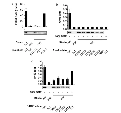
Beta-lactamase (EC:3.5.2.6, class A) is a monomeric enzyme that forms one intrachain disulfide bond, but is not required for activity [13]. No cofactors are required for activity [14]. We previously reported that the enzyme beta-lactamase adopts a catalytically active confor-mation after secretion by the T3SS [6]. We confirmed that beta-lactamase was indeed active in the extracel-lular space after secretion by the T3SS, and found that enzymatic activity in the extracellular space was both enzyme- and secretion-dependent (Fig. 1a). No secre-tion or activity was detected when secresecre-tion was pre-vented by deletion of the prgI gene, which codes for an essential component of the SPI-1 T3SS [15]. No activity was detected when the catalytic site of the enzyme was knocked out (ST71TS) [16], though the protein was still secreted. These results indicate that detected activity in the extracellular space was due to a catalytically active beta-lactamase. We mutated the two cysteine residues in beta-lactamase to serine to prevent disulfide bond for-mation. Both mutant enzymes were secreted, and the C121S mutation resulted in a catalytic activity similar to the wild type. Interestingly, the C75S mutation was not catalytically active, in contrast to previous reports in the literature [13]. Differences in N- and C-terminal modification may explain this difference—our secreted beta-lactamase bears a substantial N-terminal secretion signal and C-terminal epitopes that may affect the essen-tiality of Cys75.
The enzyme alkaline phosphatase (EC 3.1.3.1, isozyme 1) requires the acquisition of two Zn2+ and one Mg2+
A single-chain variable fragment (scFv) of an antibody is a monomeric protein that forms, but does not necessarily require, two intrachain disulfide bonds. 14B7* is an scFv of a mouse IgG antibody that binds to the protective anti-gen (PA) of the anthrax toxin [20, 21]. Binding of secreted 14B7* to PA was detected by enzyme-linked immuno-sorbent assay (ELISA) (Fig. 1c). No secretion or activ-ity was detected when secretion is prevented by deletion
of the prgI gene. Systematic mutation of each of the four cysteines to serine to prevent disulfide bond formation resulted in secretion and antigen binding, though each of the four mutants exhibited lower binding than wild type. Chemical reduction of the wild type 14B7* secreted sam-ple with 10% 2-mercaptoethanol did not affect binding activity, suggesting that disulfide bonds are not essential for binding activity after post-secretion folding.
0 20 40 60 80
a
b
0.1 0.2 0.3 0.4 0.5 0.6
WT WT S102
A
C168SC178SC268 S C336
S T60R
A405 (au)
WT prgI WT
PhoA allele Strain
WT
10% BME - - - +
WT WT ST71T
S
C75SC121S WT prgI WT
Bla allele Strain
A450 (au)
0.0 0.2 0.4 0.6 0.8 1.0 1.2 1.4
WT WTC40S
C105SC167SC241 S
WT prgI WT
14B7* allele Strain
WT 10% BME - - - +
c
Initial Rate (nM/s)
0.0
Secreted proteins form disulfide bonds
The presence of disulfide bonds in secreted proteins was confirmed by selective cysteine alkylation with the rea-gent 4′-acetamido-4′-maleimidylstilbene-2,2′-disulfonic acid (AMS). AMS selectively adds to free thiols, adding ~500 Da of mass with each addition. It will not cova-lently modify cysteines that participate in a disulfide bond. Reduction of the protein sample with tris(2-car-boxyethyl)phosphine (TCEP) will reduce disulfide bonds and convert all cysteines to the free thiol form. Thus, we can observe the disulfide bond state of a protein by detecting changes in molecular weight resulting from redox-dependent protein modification by AMS [22]. Greater cysteine modification will result in a protein that migrates more slowly in a denaturing polyacrylamide gel. For all proteins tested, the N-terminal SptP secre-tion signal sequence contains a cysteine residue at posi-tion 112 that is not expected to participate in a disulfide bond and is thus a free thiol. Indeed, a shift in migration was detected when all proteins are modified with AMS without TCEP pretreatment, indicating that the cysteine in the SptP secretion signal sequence is modified (Fig. 2, lane 3).
Disulfide bonds were detected in beta-lactamase (Fig. 2a). This protein contains one intrachain disulfide bond in the native protein, giving a total of three cysteine residues in the fusion protein. An increase in apparent molecular weight was observed when the protein was modified with AMS after TCEP pretreatment, indicat-ing that the protein contained a disulfide bond in the extracellular space. Disulfide bonds were also detected in both alkaline phosphatase and the 14B7* scFv (Fig. 2b, c). Both of these proteins contain two intrachain disulfide bonds in the native protein, giving a total of five cysteine residues in the fusion protein. When the sample was pretreated with TCEP before modification with AMS, a further increase in apparent molecular weight was observed, indicating that disulfide bonds were present in the secreted protein.
Specific activity of secreted enzymes is affected by salt concentration in growth medium
Activity of the secreted enzymes was compared with enzyme purified from the cytosol. While activity of the secreted enzymes was detected as shown in Fig. 1, it was not clear what fraction of the secreted enzymes were active. We define the parameter ffold as the fraction of
functional secreted protein, relative to the same protein fusion purified from the cytoplasm. Briefly, we assume that secreted enzymes that are folded are also active and catalyze reactions with rate kcat, while misfolded secreted
enzymes do not contribute to catalysis. The sample thus catalyzes reaction with an apparent rate constant, kcatapp,
that is less than or equal to kcat (see Additional file 1
sec-tion for a thorough descripsec-tion of the ffold parameter and
the apparent rate constant kcatapp).
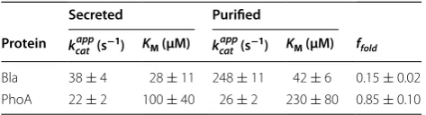
Beta-lactamase and alkaline phosphatase were purified from the cytosol and the enzyme concentration, [E]T, and the kinetic parameters KM, kcatapp, and Vmax were calculated
for each sample. In addition, the same kinetic parameters were calculated for secreted enzyme (Table 1). This anal-ysis was first performed in standard production media (Lysogeny Broth, Lennox; 5 g L−1 NaCl) [6].
a
TCEPAMS
+ +
-+ +
-Bla
72
55
c
TCEPAMS
+ +
-+ +
-14B7*
b
TCEP AMS
+ +
-+ +
-PhoA
55
43
72
55
The fraction of secreted beta-lactamase enzymes that are active was 15 ± 2%, relative to the purified form. The kinetic parameters KM and kcatapp of the purified beta-lacta-mase fusion compared well with published values for the wild type enzyme for the nitrocefin substrate (110 µM and 900 s−1, respectively) [23]. No statistically significant difference in the value of KM was found between puri-fied and secreted beta-lactamase fusion. However, the secreted and purified forms of beta-lactamase signifi-cantly differed in the calculated apparent rate constant,
kcatapp (p < 0.05), suggesting a folding defect in the secreted
enzyme, relative to the enzyme purified from the cell. The fraction of secreted alkaline phosphatase enzymes that are active was 85 ± 10%, relative to the purified form. Both kinetic parameters, KM and kcatapp, of the puri-fied alkaline phosphatase fusion were significantly differ-ent from the published values for the wild type enzyme for the para-nitrophenyl phosphate substrate (35 ± 5 µM and 176 ± 6 s−1, respectively) [24]. It should be noted that these reported kinetic parameters reported by Wojciechowski and Kantrowitz were calculated for reac-tions performed at 25 °C in a Tris-buffered solution, while all reactions with alkaline phosphatase in this study were conducted at 37 °C in LB media, prohibiting a direct comparison of the values. No statistically significant dif-ferences in the values of KM and kcatapp were found between purified and secreted alkaline phosphatase fusion. Thus, the activity of the alkaline phosphatase fusion studied in this work did not experience a significant folding defect after secretion, compared to protein purified from solu-ble cytosolic fraction. It should be noted that while the value of KM for the secreted and purified samples was not statistically significantly different, the large difference in KM between the samples may indicate that the folding of alkaline phosphatase is not well described by our simple two-state model (see Additional file 1 section for details on two-state model).
We attempted to increase the parameter ffold by
chang-ing culturchang-ing conditions. By changchang-ing the components in the growth medium, we hypothesized that the folding of
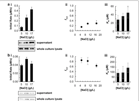
secreted protein could be modulated. The ionic strength of a solution is known to affect protein folding, likely through charge–charge interactions [25]. The concentra-tion of NaCl in the growth medium was varied between 5 and 17 g L−1 (0.09 and 0.3 M, respectively). The activ-ity was then calculated using activactiv-ity assays and quantita-tive western blotting, as above. Interestingly, the activity of beta-lactamase at saturating concentrations of sub-strate increased with NaCl concentration in the growth medium. This effect can be attributed in part to the fact that secreted protein titer increased monotonically with increasing [NaCl] (Fig. 3a.i). However, importantly, the increased activity in Fig. 3a.i was due to both increased enzyme concentration and increased ffold (Fig. 3a.ii).
Fur-ther, the calculated value of KM for secreted beta-lacta-mase was not significantly different between the three media conditions tested (Fig. 3a.iii).
The effect of NaCl in the growth media had an opposite effect on alkaline phosphatase. No change was observed in secreted protein titer or activity at saturating concen-trations of substrate (Fig. 3b.i). The value of ffold did not
change at low concentrations of NaCl but decreased at the highest salt concentration (Fig. 3b.ii). The calculated value of KM for secreted alkaline phosphatase was not significantly different between the three conditions tested (Fig. 3b.iii).
Discussion
Pure and correctly folded protein is desired when pro-ducing a heterologous protein. Production of heter-ologous proteins via secretion to the extracellular space holds many advantages over intracellular accumulation: purification is simplified; cytotoxicity is alleviated; and cell lysis is not required [1, 2]. The production and secre-tion of heterologous proteins can be achieved using the T3SS of various Gram-negative bacteria [7–9, 26, 27]. However, the mechanism of protein secretion requires an unfolding event during translocation [5]. For this produc-tion strategy to be effective, the protein should fold into a functional conformation after secretion. This event must occur in the extracellular space, a region of the cell cul-ture thought to be devoid of molecular chaperones that assist in protein folding. By testing for protein function, the folded state of the protein is probed, as only folded proteins are expected to be functionally active. The pro-tein function assays are sensitive enough to give infor-mation on the folded state of the protein of interest in a heterogeneous, dilute protein mixture.
Previous studies have demonstrated the ability of het-erologous proteins secreted by a T3SS to adopt active conformations [6–8]. In this study, we investigated the ability of secreted proteins with different chemical and structural requirements to adopt active conformations.
Table 1 Analysis of refolding efficiency of secreted enzyme in the culture supernatant, relative to purified, soluble cellular enzyme
Uncertainty is given as the standard error and is propagated for the ffold
calculation. All experiments were performed three times in biological replicate. Parameters for secreted samples were calculated for samples generated in LB media with 5 g L−1 NaCl
Secreted Purified
Protein kcatapp (s−1) KM (µM) k app
cat (s−1) KM (µM) ffold
The enzyme beta-lactamase folds into a catalytically active tertiary structure (Fig. 1a). The enzyme alkaline phosphatase adopts a catalytically active tertiary struc-ture, forms a dimer and two intrachain disulfide bonds, and acquires one Mg2+ and two Zn2+ ions per chain
(Fig. 1b). The scFv 14B7* folds into a tertiary structure that permits antigen binding (Fig. 1c). These data demon-strate that proteins can adopt active confirmations after secretion. Furthermore, in the extracellular space—an area thought to be devoid of folding chaperones—forma-tion of disulfide bonds, multimerizachaperones—forma-tion, and acquisichaperones—forma-tion of metal ion cofactors can still occur. We hypothesize that these interactions occur spontaneously in the extra-cellular space. It is unlikely that post-translational modi-fications are achieved inside the cell before secretion, owing to the limited conformations allowed by the type
III secretion system lumen. The cultures are grown aero-bically, and it is likely that the oxidizing environment of the culture medium allows for disulfide bond formation. It is not clear, however, what molecular species, if any, are involved in disulfide bond formation. Dissolved oxygen and/or other components of the growth media may be involved in disulfide bond formation. Further, the growth media is not chemically defined and likely contains trace metals for secreted proteins to sequester. Nonetheless, it is surprising that these proteins are able to adopt active conformations in the extracellular space after secretion.
A comparison of the specific activity of secreted and purified cellular enzyme was performed to compare the fraction of secreted proteins that adopt active confor-mations in the extracellular space. Secreted enzyme that is catalytically inactive will decrease the apparent rate
a
b
ii
ii
Initial Rate (μM/s)
0.0 0.1 0.2 0.3 0.4 0.5
supernatant
whole culture lysate
supernatant
whole culture lysate
0.00 0.02 0.04 0.06
0 20 40 60
KM
(μM
)
iii
[NaCl] (g/L)
5 10 17
Initial Rate (μM/s)
[NaCl] (g/L)
5 10 17
0.0 0.2 0.4 0.6 0.8 1.0
ffold
[NaCl] (g/L)
0.0 0.2 0.4 0.6 0.8 1.0
0 4 8 12 16
ffold
[NaCl] (g/L) *
*
[NaCl] (g/L)
5 10 17
0 50 150 250
KM
(μM
)
iii
[NaCl] (g/L)
5 10 17 200
100
20
0 4 8 12 16 20
i
i
Fig. 3 NaCl concentration in media affects secreted protein titer and folding efficiency. Both proteins studied are in the fusion format SptP‑POI‑ 2xFLAG‑6xHIS. For all plots, unless specified, the mean of three biological replicates is plotted, except where noted by the symbol * to indicate two biological replicates. Error bars represent one standard deviation, unless noted. a i Plot of raw activity of secreted Bla as a function of growth media with representative western blot of analyzed samples. ii Plot of ffold for secreted Bla as a function of growth media. iii Plot of KM for secreted Bla as
a function of growth media. Error bars represent the standard error. b. i Plot of raw activity of secreted PhoA as a function of growth media with representative western blot of analyzed samples. ii Plot of ffold for secreted PhoA as a function of growth media. iii Plot of KM for secreted PhoA as a
constant, kcatapp. Indeed, beta-lactamase had a lower value
of kcatapp in the secreted sample, compared to the
puri-fied protein (Table 1). This suggests that only a fraction of the secreted enzyme adopts an active conformation after secretion. Extending this reasoning, the percent-age of secreted beta-lactamase that is active is 15 ± 2% (Table 1). We speculate that this is due to misfolding or aggregation. Increasing the ionic strength of a solution has been shown to increase the thermodynamic stabil-ity of a folded monomeric protein [25], increasing the concentration of NaCl in the growth medium from 5 to 17 g L−1 increased f
fold of secreted beta-lactamase by
almost threefold (Fig. 3a.ii). This indicates that the abil-ity of the secreted protein to adopt an active conforma-tion after secreconforma-tion may be dependent on the chemical environment in which it folds. Secreted alkaline phos-phatase had a kcatapp value similar to the purified sample,
such that the percentage of folded enzyme is 85 ± 10% for the 5 g L−1 NaCl sample (Table 1). This indicates that the fraction of folding for secreted alkaline phosphatase is similar to the folding when the enzyme is purified from the soluble cellular fraction following cytosolic expres-sion. Increasing the concentration of NaCl from 5 to 10 g L−1 in the culture medium did not increase the value of ffold (Fig. 3b.ii). However, the value of ffold decreased at
the highest media NaCl concentration. This effect could be due to increased charge screening at high solution ionic strength, as charge–charge interactions are known to be important in dimerization of alkaline phosphatase [19, 28].
Understanding protein folding after secretion is impor-tant for improving protein production. Large-scale pro-tein production often involves production of the propro-tein of interest in inclusion bodies, which are then solubilized and refolded. Parallels exist between protein folding in the extracellular space and inclusion body solubiliza-tion followed by protein refolding in vitro. In an in vitro refolding procedure, the inclusion body is solubilized in a high concentration solution of a chaotrope, such as guanidinium chloride. This also unfolds the proteins in the inclusion body. The solution is then diluted to allow for proteins to fold into a native conformation. Dilution decreases the concentration of protein in solution in addition to decreasing the concentration of chaotrope. The lower concentration of protein in solution improves protein refolding, as each chain is less likely to form inter-chain aggregates. Secretion of protein to the extracellular space mimics this process, as the extracellular space also has a lower protein concentration, relative to inside the bacterial cell. The needle structure of the T3SS extends ~50 nm from the outer membrane [29], potentially beyond any extracellular cellular structures, such as the lipopolysaccharide layer. Thus, we speculate that proteins
secreted by the T3SS to the extracellular space will expe-rience a folding environment that, as in in vitro refold-ing, can be tailored to increase the folding of the secreted protein. In addition to allowing the facile production of correctly folded heterologous protein in the supernatant of bacterial culture, protein secretion by the T3SS offers a unique condition to study protein folding. Refolding of proteins after secretion may access different folding tra-jectories than found in co-translational or in vitro folding conditions. Given the rate and directionality of secretion, proteins may fold in a unique vectorial folding pathway. Further, the unfolded conformations that are accessible to the protein during secretion are likely constrained, which in turn may change the conformations that are accessible after secretion. We anticipate that this system will pro-vide a unique folding environment for future study.
Conclusions
Production of heterologous proteins in bacteria is improved using a secretion strategy. In this study, the T3SS of Salmonella enterica was used to secrete pro-teins to the extracellular space. A folded protein product in the extracellular space is desired, but the T3SS secre-tion mechanism requires proteins to be unfolded during translocation. We characterized folding of secreted pro-teins by quantifying protein function, either by enzymatic activity or antigen binding. Indeed, the three secreted proteins in this study adopt a functional confirmations after secretion to the extracellular space. However, only a fraction of secreted proteins are functional, between 10 and 85%. Media composition was used to modify the chemical environment in the extracellular space. Increas-ing the concentration of NaCl in the culture medium had a protein-specific effect on the ability of a secreted protein to adopt an active conformation in the extracel-lular space. Further understanding of how chemical and genetic variables control the folding of secreted proteins will be important for process development of bacterial strains for production of secreted proteins. An improved production process achieves secretion a protein at high titer to the extracellular space and that spontaneously adopts a functional conformation. This would simplify purification and not require refolding unit operations.
Methods
Strains and growth conditions
(34 µg mL−1 chloramphenicol and/or 50 µg mL−1 kana-mycin) for 12–16 h at 37 °C and 225 rpm in an orbital shaker overnight. Overnight cultures were subcultured into fresh LB-L media supplemented with 100 µM iso-propyl β-d-1-thiogalactopyranoside (IPTG) and the appropriate antibiotics. All culturing steps were per-formed in 24-well blocks (Axygen).
Salmonella enterica strains were transformed with the plasmids listed in Additional file 1: Table S1 using electroporation. All experiments were performed using a two-plasmid system, with the upregulation vector (PlacUV5hilA), as reported by Metcalf et al. [6], in addition to the export vector that carried the gene coding for the protein of interest [26]. Controlled overexpression of hilA allows for controlled expression of genes coding for both the secretion apparatus and the protein of interest [6].
DNA manipulations
PCR was performed with Pfu DNA polymerase and the primers listed in Additional file 1: Table S2. Restric-tion enzymes and ligase (NEB) were used according to the manufacturer’s instructions. For all cloning, E. coli DH10B cells were used. Mutations to bla and phoA are specified with respect to the full-length mature wild type protein. Mutations to 14b7* are specified with respect to the order of cysteines, from amino to carboxy termi-nus, in the antibody fusion gene and does not include the cysteine in the N-terminal SptP signal sequence. The bla gene was amplified from the plasmid pTrc99A [31]. The phoA gene was amplified from E. coli MG1655. The 14b7* gene was amplified from the plasmid pFLAG-APEx 14B7* (gift from the Georgiou lab).
Protein separation and western blotting
Samples were separated by SDS-PAGE. Proteins were transferred to a polyvinylidene fluoride (Millipore) membrane for chemiluminescence detection, using the TransBlot SD unit (Bio-Rad). Membranes were interro-gated with Mouse anti-FLAG antibodies per manufac-turer’s instructions (Sigma). A secondary labeling step was carried out with Goat anti-Mouse IgG (H + L), HRP conjugate antibodies, per manufacturer’s instructions (Thermo). Bands were visualized with pico or west-femto chemiluminescent substrate (Thermo) and imaged with a ChemiDoc XRS + unit (Bio-Rad).
Protein purification
Culture homogenate was purified using a His Gravi-Trap column (GE Healthcare # 11-0033-99). Eluted pro-tein sample was separated by SDS-PAGE, stained with Coomassie G-250, and quantified using densitometry relative to a bovine serum albumin standard (Thermo).
Purified protein samples were diluted in LB-L and stored at 4 °C for enzyme activity assays.
Protein quantification
Supernatant samples were harvested 8 h after subculture. Supernatant samples were harvested from the cell cul-ture by two sequential centrifugation steps of 2272×g for 10 min, 4× Laemmli buffer was added to each sample for a final concentration of Laemmli buffer of 1×, and boiled for 95 °C for 5 min. Purified protein samples were used to create four standards that were included with each blot to construct a standard curve. Samples were separated by SDS-PAGE and transferred to a polyvinylidene fluo-ride (Millipore). A linear least-squares regression of the standard samples were used to calculate the concentra-tion of each supernatant sample.
Beta‑lactamase activity assay
Samples were grown overnight in LB-L media, then sub-cultured 1:100 in LB-L media and grown for 8 h at 37 °C and 225 rpm. The cultures were pelleted by one centrifu-gation step of 2272×g for 10 min and the supernatant was passed through a 0.45 μm filter. Samples were then sub-jected to a nitrocefin hydrolysis assay, per the substrate vendor (Sigma). 100 μL of reaction buffer (0.1 M phos-phate, 1 mM ethylenediaminetetraacetic acid, 50 g mL−1 nitrocefin (EMD Millipore), 0.5% dimethyl sulfoxide, pH 7) was mixed with 10 μL culture supernatant and the absorbance at 486 nm was observed over time. The reaction was performed in a disposable UV-Transparent cuvette (BrandTech, part# 759215) without stirring at 37 °C. The absorbance at 486 nm was measured every 5 s for 60 s with a UV–Vis spectrophotometer (Nanodrop, part# 2000c). The reaction was linear for the first 40 s of all reactions tested and the change in the absorbance as a function of time was determined using a linear least-squares fit. The initial reaction velocity was calculated using an extinction coefficient of 20,500 M−1 cm−1, as specified by the vendor. The experiment was performed on different days in biological triplicate. Error bars repre-sent one standard deviation.
Alkaline phosphatase activity assay
phosphate. For endpoint assays, multiple equal-volume reactions were performed simultaneously in a 96-well microtiter plate and incubated at 37 °C without shaking for at least 1 h. The absorbance at 405 nm of each well was measured using a Synergy HTX Multi-Mode Reader spectrophotometer (Bio-Tek). For kinetic assays, the reaction was performed in a disposible UV-Transparent cuvette (BrandTech, part# 759215) without stirring at 37 °C. The absorbance at 405 nm was measured every 5 s for 60 s with a UV–Vis spectrophotometer (Nanodrop, part# 2000c). The reaction was linear for the first 40 s of all reactions tested and the change in the absorbance as a function of time was determined using a linear least-squares fit. The initial reaction velocity was calculated using an extinction coefficient of 18,000 M−1 cm−1, as specified by the vendor. The experiment was performed on different days in biological triplicate. Error bars repre-sent one standard deviation.
Enzyme‑linked immunosorbent assay (ELISA)
Samples were grown overnight in LB-L media, then subcultured 1:100 in LB-L media and grown for 8 h at 37 °C and 225 rpm. The cultures were pelleted by one centrifugation step of 2272×g for 10 min and the super-natant was passed through a 0.45 μm filter. The wells of a 96-well microtiter plate (Santa Cruz Biotechnol-ogy, Inc., part# sc-204463) was coated with 100 µL of a solution of 4 µg mL−1 protective antigen (PA) of the anthrax toxin (List Biological Laboratories, part# 171E) in 5 mM HEPES, 50 mM NaCl, pH 7.5 covered at 4 °C overnight. Liquid was removed by inversion and wells were incubated with a 200 µL blocking solution (200 µL 2% milk, 0.05% TBST) at room temperature for 1 h. Liquid was removed by inversion and 100 µL filtered culture supernatant was added to the wells. Samples were incubated at room temperature for 2 h. Liquid was removed by inversion and wells were rinsed with 0.05% TBST three times. Next, 100 µL of primary labeling solution (1:10,000 dilution of Mouse FLAG anti-body (Sigma) diluted in 0.05% TBST) was added to the wells and incubated 1 h at room temperature. Liquid was removed by inversion and wells were rinsed with 0.05% TBST five times. Then, 100 µL of secondary labe-ling solution (1:5000 dilution of Goat anti-Mouse IgG (H + L), HRP conjugate antibodies (Thermo) diluted in 0.05% TBST) was added to the wells and incubated 1 h at room temperature. Liquid was removed by inver-sion and wells were rinsed with 0.05% TBST five times. 100 µL of 3,3′,5,5′-Tetramethylbenzidine (TMB) Liq-uid Substrate (Thermo) was added to the wells and incubated for approximately 20 min at room tem-perature. The reaction was quenched with 100 µL 2 M H2SO4. Absorbance at 450 nm was measured using a
Synergy HTX Multi-Mode Reader spectrophotometer (Bio-Tek).
Cysteine alkylation
Filtered supernatant samples were precipitated with trichloroacetic acid (20% w/w final concentration) over-night at 4 °C, washed twice with cold acetone, and dried by heating. Dried samples were resuspended in 50 µL resuspension buffer (1 M Tris (base), pH 7.5, 3% w/w sodium dodecyl sulfate). Resuspension buffer was sup-plemented with 10 mM tris(2-carboxyethyl)phosphine (TCEP) for reduced samples, as necessary. Samples were incubated at room temperature for 10 min. Next, 8.8 µL of 100 mM 4-acetamido-4′-maleimidylstilbene-2,2′ -disulfonic acid (AMS) was added to samples (final con-centration is 15 mM), as necessary. Samples to which AMS was not added were diluted with equal volume distilled water. Samples were incubated at room tem-perature for 2 h, protected from light. Samples were then mixed with Laemmli buffer, boiled, and separated by SDS-PAGE, and bands were detected by a western blot, as specified above.
Error estimation of Michaelis–Menten model
Error estimation was conducted using Matlab (Math-works, R2014a). Biological replicates were treated as independent samples. Three independent replicates were then analyzed for both purified and secreted samples. Least-squares minimization was used to fit a modified Michaelis-Menten model (Eq. 1) to the measured values and determine the parameters kcatapp and KM.
where V0 is the initial reaction rate, [E]T is the total enzyme concentration, [S] is the substrate concentration, and kcatapp and KM are fitting parameters. To compare the
parameters from different treatments, a t test (Eq. 2) was used where the degrees of freedom was determined by the pooled sample size.
where t is the t statistic, β is the fitting parameter of inter-est (i.e., kcatapp and KM), and Se is the standard error
calcu-lated from the nonlinear regression. A p value of 0.05 was used to define significance.
(1)
V0
[E]T =
kcatapp·[S] KM+[S]
(2)
t= β1−β2
Se21−Se22
Additional file
Abbreviations
AMS: 4′‑acetamido‑4′‑maleimidylstilbene‑2,2′‑disulfonic acid; ELISA: enzyme‑ linked immunosorbent assay; LB: lysogeny broth; PA: protective antigen of anthrax toxin; scFv: single‑chain variable fragment; TCEP: tris(2‑carboxyethyl) phosphine; T3SS: type III secretion system.
Authors’ contributions
KJM and DTE designed research, analyzed data, and wrote the paper. KJM, SLR, LAB, and EV performed research. JLB and KJM performed statistical analysis. All authors read and approved the final manuscript.
Author details
1 Department of Chemical and Biomolecular Engineering, University of California, Berkeley, CA 94720, USA. 2 Department of Nutritional Science and Toxicology, University of California, Berkeley, CA 94720, USA. 3 Department of Plant and Microbial Biology, University of California, Berkeley, CA 94720, USA. 4 Department of Chemical and Biological Engineering, Northwestern Uni‑ versity, Evanston, IL 60208, USA. 5 Present Address: Department of Biomedical Engineering, Northwestern University, Evanston, IL 60208, USA.
Acknowledgements
We thank George Georgiou (University of Texas at Austin) for providing the plasmid pFLAG‑APEx 14B7*, the laboratory of Michelle Chang (University of California, Berkeley) for providing use of laboratory equipment, and members of the Danielle Tullman‑Ercek lab for helpful discussions, experimental advice, and criticisms of this manuscript.
Competing interests
The authors declare that they have no competing interests.
Availability of data and materials
All data generated during and/or analysed during this study are included in the published article and its supplementary information files.
Funding
KJM was supported by a National Science Foundation Graduate Research Fellowship and a University of California, Berkeley Chancellor’s Fellowship, SLR was supported by the Biology Fellows Program (University of California, Berkeley) and the Synthetic Biology Engineering Research Center (SynBERC) Scholar program, LAB was supported by a National Science Foundation Graduate Research Fellowship, and EV was supported by the Pre‑Initiative for Maximizing Student Development (National Institute of Health and University of California, Berkeley) and the SynBERC Scholar program.
Received: 18 August 2016 Accepted: 25 November 2016
References
1. Georgiou G, Segatori L. Preparative expression of secreted proteins in bacteria: status report and future prospects. Curr Opin Biotechnol. 2005;16:538–45.
2. Stader JA, Silhavy TJ. Engineering Escherichia coli to secrete heterologous gene products. Methods Enzymol. 1990;185:166–87.
3. Swartz JR. Advances in Escherichia coli production of therapeutic proteins. Curr Opin Biotechnol. 2001;12:195–201.
4. Cornelis GR. The type III secretion injectisome. Nat Rev Microbiol. 2006;4:811–25.
5. Radics J, Königsmaier L, Marlovits TC. Structure of a pathogenic type 3 secretion system in action. Nat Struct Mol Biol. 2013;21:82–7. 6. Metcalf KJ, Finnerty C, Azam A, Valdivia E, Tullman‑Ercek D. Using tran‑
scriptional control to increase titers of secreted heterologous proteins by the type III secretion system. Appl Environ Microbiol. 2014;80:5927–34. 7. Derouazi M, Toussaint B, Quénée L, Epaulard O, Guillaume M, Marlu R,
et al. High‑yield production of secreted active proteins by the pseu‑ domonas aeruginosa type III secretion system. Appl Environ Microbiol. 2008;74:3601–4.
8. Majander K, Anton L, Antikainen J, Lång H, Brummer M, Korhonen TK, et al. Extracellular secretion of polypeptides using a modified Escherichia coli flagellar secretion apparatus. Nat Biotechnol. 2005;23:475–81.
9. Singer HM, Erhardt M, Steiner AM, Zhang M‑M, Yoshikami D, Bulaj G, et al. Selective purification of recombinant neuroactive peptides using the flagellar type III secretion system. mBio. 2012. doi:10.1128/mBio.00115‑12. 10. Schlumberger MC, Müller AJ, Ehrbar K, Winnen B, Duss I, Stecher B, et al.
Real‑time imaging of type III secretion: Salmonella SipA injection into host cells. Proc Natl Acad Sci USA. 2005;102:12548–53.
11. Hingorani KS, Gierasch LM. Comparing protein folding in vitro and in vivo: foldability meets the fitness challenge. Curr Opin Struct Biol. 2014;24:81–90.
12. Clark EDB. Protein refolding for industrial processes. Curr Opin Biotechnol. 2001;12:202–7.
13. Schultz SC, Dalbadie‑McFarland G, Neitzel JJ, Richards JH. Stability of wild‑ type and mutant RTEM‑1 beta‑lactamases: effect of the disulfide bond. Proteins. 1987;2:290–7.
14. Enzyme Nomenclature Database. Swiss Institute of Bioinformatics. http:// enzyme.expasy.org/EC/3.5.2.6. Accessed 8 Aug 2016.
15. Kimbrough TG, Miller SI. Contribution of Salmonella typhimurium type III secretion components to needle complex formation. Proc Natl Acad Sci USA. 2000;97:11008–13.
16. Dalbadie‑McFarland G, Neitzel JJ, Richards JH. Active‑site mutants of beta‑lactamase: use of an inactive double mutant to study requirements for catalysis. Biochemistry. 1986;25:332–8.
17. Stec B, Holtz KM, Kantrowitz ER. A revised mechanism for the alka‑ line phosphatase reaction involving three metal ions. J Mol Biol. 2000;299:1303–11.
18. Butler‑Ransohoff JE, Rokita SE, Kendall DA, Banzon JA, Carano KS, Kaiser ET, et al. Active‑site mutagenesis of E. coli alkaline phosphatase: replace‑ ment of serine‑102 with nonnucleophilic amino acids. J Org Chem. 1992;57:142–5.
19. Boulanger RR, Kantrowitz ER. Characterization of a monomeric Escherichia coli alkaline phosphatase formed upon a single amino acid substitution. J Biol Chem. 2003;278:23497–501.
20. Leysath CE, Monzingo AF, Maynard JA, Barnett J, Georgiou G, Iverson BL, et al. Crystal structure of the engineered neutralizing antibody M18 complexed to domain 4 of the anthrax protective antigen. J Mol Biol. 2009;387:680–93.
21. Little SF, Leppla SH, Cora E. Production and characterization of monoclo‑ nal antibodies to the protective antigen component of Bacillus anthracis toxin. Infect Immun. 1988;56:1807–13.
22. Sechi S, Chait BT. Modification of cysteine residues by alkylation: a tool in peptide mapping and protein identification. Anal Chem. 1998;70:5150–8. 23. Sigal IS, DeGrado WF, Thomas BJ, Petteway SR. Purification and properties
of thiol beta‑lactamase: a mutant of pBR322 beta‑lactamase in which the active site serine has been replaced with cysteine. J Biol Chem. 1984;259:5327–32.
24. Wojciechowski CL, Kantrowitz ER. Altering of the metal specificity of Escherichia coli alkaline phosphatase. J Biol Chem. 2002;277:50476–81. 25. Song B, Cho J‑H, Raleigh DP. Ionic‑strength‑dependent effects in protein
folding: analysis of rate equilibrium free‑energy relationships and their interpretation. Biochemistry. 2007;46:14206–14.
26. Widmaier DM, Tullman‑Ercek D, Mirsky EA, Hill R, Govindarajan S, Minshull J, et al. Engineering the Salmonella type III secretion system to export spider silk monomers. Mol Syst Biol. 2009;5:309.
27. Azam A, Li C, Metcalf KJ, Tullman‑Ercek D. Type III secretion as a gener‑ alizable strategy for the production of full‑length biopolymer‑forming proteins. Biotechnol Bioeng. 2015. doi:10.1002/bit.25656.
28. Torriani A. Alkaline phosphatase subunits and their dimerization in vivo. J Bacteriol. 1968;96:1200–7.
29. Kubori T, Matsushima Y, Nakamura D, Uralil J, Lara‑Tejero M, Sukhan A, et al. Supramolecular structure of the Salmonella typhimurium type III protein secretion system. Science. 1998;280:602–5.
30. Hoiseth SK, Stocker BAD. Aromatic‑dependent Salmonella typhimurium are non‑virulent and effective as live vaccines. Nature. 1981;291:238–9. 31. Amann E, Ochs B, Abel K‑J. Tightly regulated tac promoter vectors useful
for the expression of unfused and fused proteins in Escherichia coli. Gene. 1988;69:301–15.