R E V I E W
Open Access
Expression and function of
cartilage-derived pluripotent cells in joint
development and repair
Zhou Jiang
1†, Sijing Yu
1†, Hengyi Lin
1and Ruiye Bi
2*Abstract
Cartilage-derived pluripotent cells reside in hyaline cartilage and fibrocartilage. These cells have the potential for multidirectional differentiation; can undergo adipogenesis, osteogenesis, and chondrogenesis; and have been classified as mesenchymal stem cells (MSCs) conforming to the minimal criteria of the International Society for Cellular Therapy. Cartilage tissue is prone to injury and is difficult to repair. As cartilage-derived pluripotent cells are the closest cell source to cartilage tissue, they are expected to have the strongest ability to differentiate into cartilage compared to other MSCs. This review focuses on the organizational distribution, expression, and function of cartilage-derived pluripotent cells in joint development and repair to help explore the therapeutic potential of in situ cartilage-derived pluripotent cells for joint cartilage repair.
Keywords:Cartilage-derived pluripotent cell, Chondroprogenitor cell, Stem cell-based therapy, Cartilage repair, Stem cell transplantation
Background
Articular cartilage in long bones is made up of hyaline cartilage. The condylar cartilage (CC) located in the temporomandibular joint (TMJ) is generally considered fibrocartilage. The articular disc, including the meniscus and the TMJ disc, is also composed of fibrocartilage. Due to the lack of nerves, blood vessels, and lymphatic vessels and the effect of its weight-bearing role, cartilage tissue shows difficulty repairing itself when injured.
With the rise of regenerative medicine and tissue en-gineering, cell-based approaches have been successfully used in cartilage repair. Both autologous chondrocytes and mesenchymal stem cells (MSCs) are currently used as seed cells for repairing cartilage injury. However, the amount of healthy cartilage available for chondrocyte harvesting is often limited during autologous chondro-cyte transplantation. Chondrochondro-cyte phenotypes are diffi-cult to maintain during diffi-culture expansion, and these cells are prone to dedifferentiating and losing their
capacity to form cartilage. Instead, MSCs are considered a preferable cell source for cartilage repair because they are easy to isolate, retain some stem cell properties during in vitro expansion, and can differentiate into chondrocytes.
MSCs can be isolated from the bone marrow [1], peri-osteum [2], synovium [3], and adipose tissue [4]. Gener-ally, the closer the cell source is to the injured cartilage tissue, the more effective the differentiation into carti-lage tissue is [5]. Therefore, if MSCs are also present in the articular surface, they are expected to have the strongest ability to differentiate into cartilage and repair injured cartilage tissue.
Recent studies have found that articular cartilage con-tains pluripotent cell populations that can undergo chondrogenic, osteogenic, and adipogenic differentiation. These cells have been classified as MSCs conforming to the minimal criteria of the International Society for Cellular Therapy, which include being plastic-adherent, showing multipotentiality, and expressing an MSC marker phenotype [6, 7]. Therefore, these populations are expected to be potential cell sources for cartilage repair, and in-depth and comprehensive studies on their function in joint development and repair can help us
© The Author(s). 2020Open AccessThis article is distributed under the terms of the Creative Commons Attribution 4.0 International License (http://creativecommons.org/licenses/by/4.0/), which permits unrestricted use, distribution, and reproduction in any medium, provided you give appropriate credit to the original author(s) and the source, provide a link to the Creative Commons license, and indicate if changes were made. The Creative Commons Public Domain Dedication waiver (http://creativecommons.org/publicdomain/zero/1.0/) applies to the data made available in this article, unless otherwise stated. * Correspondence:[email protected]
†Zhou Jiang and Sijing Yu contributed equally to this work.
2Department of Orthognathic and TMJ Surgery, State Key Laboratory of Oral Diseases, National Clinical Research Center for Oral Diseases, West China Hospital of Stomatology, Sichuan University, Chengdu, China
explore ideal stem cell-based therapies for cartilage repair. Since these cells had various names in different studies, we named these cells cartilage-derived pluripo-tent cells in our study.
Organizational distribution of cartilage-derived pluripotent cells
In long bones In hyaline cartilage
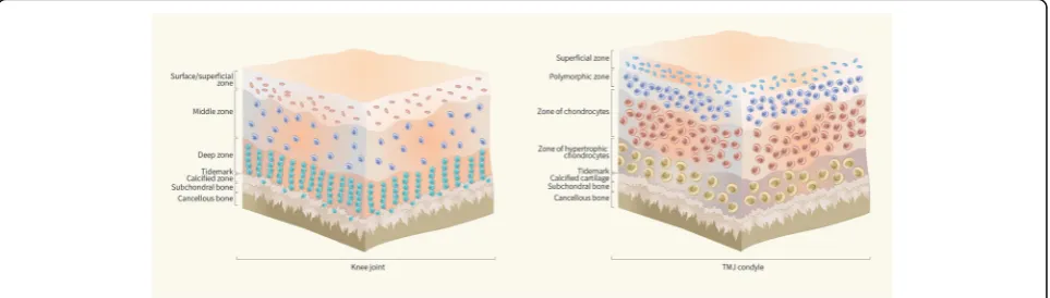
Hyaline cartilage is compartmentalized into the surface zone, middle zone, deep zone, and calcified zone (Fig. 1a), with biochemical and morphological variations existing at different depths [8]. Multiple studies have confirmed the presence of pluripotent cells with stem cell characteristics in hyaline cartilage [6, 9,10], and the surface zone of the cartilage tissue, including the articu-lar surface, is a relatively abundant source of these pluri-potent cells. In the development of articular cartilage, Hayes et al. [11] found that articular surface zone cells from animal knee joints had a longer cell cycle than the underlying transitional zone cells, and Hunziker et al. [12] found that the superficial zone (SZ) consisted of slowly dividing stem cells, which suggested the presence of a chondroprogenitor or stem cell population in the articular cartilage surface. Further, Dowthwaite et al. [8] and Hattori et al. [9] both successfully isolated stem/pro-genitor cells from the surface zone of calf/bovine articu-lar cartilage, and the latter study reported that these progenitors make up approximately 0.1% of all cells that can be extracted from the surface zone of the articular cartilage tissue. Grogan et al. [13] found that the fre-quency of progenitor cells in full-thickness human ar-ticular cartilage was 0.14%, and no difference was found between the control and osteoarthritis (OA) groups. Interestingly, Pretzel et al.’s [14] study indicated a much higher percentage of CD105+/CD166+ progenitors in OA (16.7%) cartilage compared to normal (15.3%) cartilage,
and the CD166+ cells were almost exclusively located in the superficial and middle cartilage zones. A recent study demonstrated that high-efficiency colony-forming cells (HCCs) can also be isolated from the deep zone of bovine articular cartilage, although the SZ has significantly more progenitor cells than the deep zone [15].
In meniscus
A recent study showed that multipotent stem cells are present in the human meniscus and are phenotypically similar to MSCs [16]. Shen et al. [17] identified and characterized a population of meniscus-derived stem cells (MeSCs) that displayed low immunogenicity and possessed immunosuppressive functions. Then, this team found that human meniscus stem/progenitor cells (hMeSPCs) displayed both MSC characteristics and high expression levels of type II collagen [18]. Similarly, another study showed that MSCs isolated from rabbit menisci have universal stem cell characteristics, includ-ing clonogenicity, multipotency, self-renewal capacity, and expression of stem cell markers, and a pronounced tendency to chondrogenic differentiation appeared both in vivo and in vitro compared to that of bone marrow-derived stem cells (BMSCs) [19]. Gamer et al. [20] isolated and localized stem/progenitor cells from murine menisci grown in explant culture, and localization studies suggested that endogenous progenitor cells may reside in the superficial and outer regions of the meniscus in vivo.
In TMJ fibrocartilage
Unlike hyaline cartilage, fibrocartilage in the TMJ con-dyle consists of various proportions of both fibrous and cartilaginous tissue and is divided into four distinct zones (Fig. 1b): the fibrous SZ, a polymorphic zone, a zone of chondrocytes, and a zone of hypertrophic
chondrocytes [21]. In the fibrocartilage of the TMJ, Embree et al. [22] first showed that the fibrous SZ tissue in the TMJ condyle is a niche that harbors fibrocartilage stem cells (FCSCs). The skeletal stem/progenitor cell marker αSMA was traced in transgenic mice [23] by pedigree tracking studies, and the researchers found that the mature col2a1+ chondrocyte progeny located in CC tissues were differentiated from undifferentiated αSMA+ cells in the fibrous SZ, indicating that the fibrocartilage stem cells in the SZ can produce mature chondrocytes (Fig.2).
Cytological features of cartilage-derived pluripotent cells
Although cartilage-derived pluripotent cells currently lack definitive biomarkers, these cells are generally char-acterized according to several cytological features, such as increased chondrogenic protein expression, specific cell surface markers, a high colony-forming efficiency, a pluripotent differentiation capacity, and migratory ability.
Increased chondrogenic protein expression during cartilage development
Cartilage development is a continuous dynamic process of cell differentiation and protein expression. In hyaline cartilage, pluripotent cells at different stages of differen-tiation exhibit expression of different genes, including fibronectin, hyaluronan, tenascin, type I collagen, type II collagen, minor type IX and XI collagens, proteoglycans, matrilins, and cartilage oligomeric protein (COMP) [24–27], which participate in the formation of abundant extracellular matrix (ECM). In this process, undifferen-tiated mesenchymal cells eventually differentiate into
chondrocytes. In mature articular cartilage, the cells of the surface zone produce high quantities of SZ protein (SZP/ proteoglycan 4), encoding lubricin for surface lubrication [28,29]. In the middle and deep zones, the cartilage ECM is composed mainly of type II collagen, aggrecan, and hya-luronan [30]. Assays of these chondrogenic proteins and their corresponding mRNA/gene levels, especially those of type II collagen, aggrecan, and COMP, can be used to measure the chondrogenic differentiation potential of specific cell populations.
Surface markers
Cartilage-derived pluripotent cells are generally charac-terized according to MSC-related surface markers, which can effectively distinguish them from chondrocytes. These pluripotent cells express the classical MSC markers CD105, CD73, and CD90 and lack expression of CD45, CD34, CD14 or CD11b, CD79a, or CD19, ac-cording to the International Society for Cellular Therapy [7]. CD10 and CD166 (ALCAM) are also regarded as distinctive markers for MSCs [31, 32]. In addition, these pluripotent cells express stem cell markers, such as Stro-1, NotchStro-1, VCAM-1 (CD106), and Integrin β1 (CD29) [8, 10, 13,33], and chondrogenic markers, such as Sox9 [6, 10]. These surface markers were also expressed in combinations, including CD9+/CD90+/CD166+ for OA [34], CD166+/CD90+ for normal [35], and CD105+/ CD166+ for both normal and OA samples from human articular cartilage [14].
Recent studies have shown that cartilage-derived pluri-potent cells also express specific surface markers. Gro-gan et al.’s [36] study found that in normal human knee joints, chondrocytes with a high chondrogenic capacity
expressed increased levels of CD44, CD49c, CD49f, and CD151. CD90 and CD166 were also highly expressed, which suggests that these highly chondrogenic subpopu-lations might correspond to those with progenitor characteristics. Williams et al. [6] found that CD49e might be utilized as a specific marker for the cartilage progenitor cell population in normal human articular cartilage from femoral condyles. In knee joints from late-stage OA, CD146 might be a new cell surface marker for the cartilage progenitor cell population [37]. Another study compared protein changes between human bone marrow MSCs and chondrogenic progeni-tor cells (CPCs) from knee articular cartilage from OA samples, and 4 cell surface proteins were found with significantly increased expression in the CPCs: AMPN (CD13), CD109 antigen, CADM1, and CD49b [38].
However, as these studies examined cartilage-derived pluripotent cells from different species and with a differ-ent cell origin and cartilage status (normal or OA), the surface markers currently used for identification are not unified. Therefore, the identification of specific markers to effectively distinguish cells that can differentiate into cartilage is needed.
Colony formation
Colony-forming ability is a well-recognized trait of stem/ progenitor cells that has been used extensively to perform quantitative and functional analysis of clonal populations of progenitors [39], and cartilage-derived pluripotent cells have a strong clonogenic potency.
In hyaline cartilage, Dowthwaite et al. [8] defined a colony as more than four cells and found that articular surface cells with high affinity for fibronectin showed a significantly enhanced colony-forming efficiency (CFE) relative to all other cohorts. Compared to nonclonal dedifferentiated chondrocytes, immature bovine chon-droprogenitor cells from the metacarpophalangeal (MCP) joints showed 2.6-fold greater telomerase activity and significantly longer telomere lengths of chromo-somes during long-term clonal expansion in a mono-layer culture [40]. Williams et al. [6] found that clonal cartilage progenitor cells isolated from human articular cartilage could proliferate to over 60 population doublings (PD) cultured in monolayers, taking over 200 days. Similarly, in the TMJ condyle, FCSCs formed sixfold more colonies than CC cells [22].
Interestingly, CPCs from articular cartilage of the later stages of human OA were also found to undergo 60 PD in monolayer culture, with incrementally increasing time for each doubling event [10]. Another study showed that articular cartilage-derived CPCs from cartilage from OA samples can be subdivided into two populations: an early senescent population (ES-OA-CPCs) that underwent replicative exhaustion by 30 PD and a late senescent
population (LS-OA-CPCs) that was capable of prolonged expansion and displayed similar growth rates compared to stem cells from normal cartilage [39]. These findings suggested that although early senescence is an inherent property of a subset of activated progenitors, there is also a pool of progenitors with extended viability and regenerative potential residing within cartilage from OA samples.
Pluripotent differentiation
The potential for chondrogenic, osteogenic, and adipo-genic differentiation has become a defining feature of cartilage-derived pluripotent cells that distinguishes them from mature chondrocytes. Progenitors from the articular cartilage surface treated with bone morpho-genic protein-7 (BMP-7) showed robust chondrogenesis and produced ECM for cartilage [9]. Studies have also found that CPCs from both normal articular cartilage and OA-derived stem cell populations demonstrate trili-neage differentiation into adipogenic, osteogenic, and chondrogenic [6, 41] lineages, suggesting that stem cells from human OA cartilage also have the potential for cartilage repair. Koelling et al. [10] observed that CPCs from knee joints from late-stage OA regained a round chondrocyte-like phenotype and exhibited collagen type II mRNA expression as well as collagen type II protein expression 3 weeks after transfer to a 3D-alginate culture without any chondrogenic supplementation. High mRNA levels of sox-9 and collagen type II and low levels of runx-2 and collagen type I were also identified in these cells. Moreover, CPCs adhere to and are influ-enced by ECM components, and downregulation of runx-2 enhances their chondrogenic potential. Subse-quently, another study showed that CD146+ chondro-progenitors from knee joints of late-stage OA showed a lower potential for adipogenesis and osteogenesis but a much higher potential for chondrogenesis compared to unsorted chondrocytes and adipose-derived MSCs, as these CD146+ cell subpopulations showed increased type II collagen, aggrecan, and Sox9 expression [37]. In TMJ condyles, FCSCs underwent adipogenesis, chondrogenesis, and osteogenesis, and their individual colonies showed heterogeneous differentiation poten-tial (22.5% trilineage, 64.5% bilineage, and 12.9% single lineage) [22].
Migratory ability
scratching in healthy cartilage explants from mature cattle, and these migrating cells were highly clonogenic and multipotent and expressed CPC-associated markers, including CD105, CD73, CD90, CD29, CD44, Notch-1, and Sox9. The latter study also suggested that migratory cartilage-derived pluripotent cells might exist in healthy cartilage.
In situ cartilage-derived pluripotent cell-based therapies for joint cartilage repair
Articular cartilage is a type of tissue that is easily dam-aged and difficult to repair. Currently, MSC-based ther-apies, mainly BMSCs, are widely used to effectively repair cartilage defects [43]. In recent years, researchers have discovered a special population of stem cells, in situ cartilage-derived pluripotent cells, which reside in both healthy and injured joint cartilage/fibrocartilage tissues and exhibit strong repair capabilities. There are two main strategies for in situ cartilage-derived pluripotent cell-based therapies for joint cartilage repair. One is to transplant cartilage-derived pluripotent cell-containing grafts into the cartilage defect, and the other is the intra-articular injection of pluripotent cells (Table 1). Never-theless, in situ cartilage-derived pluripotent cells display different reparative characteristics in hyaline cartilage, meniscus, and TMJ condyle.
When hyaline cartilage-derived pluripotent cells are transplanted into artificially constructed cartilage defects of the femur, high expression of type II collagen, and cartilage-like repair tissue formation were observed, and these results have been successfully verified in equine [44] and goat models [6]. However, this conclusion may not hold in some cases [45,46]. Marcus et al. [46] dem-onstrated that when bovine articular cartilage-derived pluripotent cells were intramuscularly injected into a severe-combined immunodeficient (SCID) mouse, these cells could survive within the muscle mass but failed to produce cartilage-like tissue despite expressing Sox9 and type II collagen. These findings suggest that the cells may require further signals and a more favorable envir-onment for chondrogenic differentiation.
In culture of man-made injured meniscus explants in vitro, meniscus-derived pluripotent cells could bridge and reintegrate torn meniscal fibrocartilage along the tear channel, as evidenced by the migratory ability in response to the chemokine signaling stromal-derived factor-1/stromal-derived factor-1 receptor (SDF-1/ CXCR4) axis, a pronounced tendency toward chondro-genic differentiation, a greater than 100% increase in fibrochondrocyte proliferation, the elevated expression of Sox9 and decreased expression of type X collagen, and the resistance to cellular hypertrophy and terminal differentiation during the tissue repair process in a rat [47] and a rabbit [19] model. On this basis, Jayasuriya
et al. [47] proposed that the initiation of the observed meniscal tissue repair is possible without first forming a blood clot, provided that an influx of stem cells is readily available near the damage site. Furthermore, the intra-articular injection of meniscus-derived pluripotent cells can enhance the regeneration of the injured meniscus at an early stage of OA, promoting neotissue formation with an improved shape and increased mature ECM and resulting in reduced expression of OA markers such as type I collagen, type X collagen, and hypoxia-inducible factor 2a (HIF-2a) but increased expression of collagen II [17,18]. Notably, Jiang et al. [48] discovered a class of human chondrocyte-derived progenitor cells (CDPCs) and transplanted them into patients with large knee car-tilage defects, leading to reduced knee pain and swelling and eliminating the locking sensation, which supported the possibility of cartilage-derived pluripotent cell-based therapies for human joint cartilage repair.
In addition to transplantation and intra-articular injec-tion of cartilage-derived pluripotent cells, endogenous CPCs can be exploited to initiate cartilage regeneration and repair with drugs. Embree et al. [22] demonstrated the therapeutic application of sclerostin (SOST) in a rabbit model of injury to the TMJ disc and secondary OA; this molecule is an exogeneous canonical Wnt inhibitor, as canonical Wnt signals may be enhanced in diseased human TMJ condylar fibrocartilage.
However, there are some drawbacks of the current studies on cartilage-derived pluripotent cell-based ther-apies for joint cartilage repair. One is the use of stable cell lines because these cell lines may deviate from the primary cells that were used to generate them in vitro. The other is that animal cartilage is different from human cartilage in terms of its main structural features, including cellular distribution, vascularity, and collagen structure. Therefore, future research should focus on the application of cartilage-derived pluripotent cells in the treatment of human joint cartilage diseases.
Conclusions
Table 1 Cartilage-derived pluripotent cell-based therapies for joint cartilage repair Cartil age Cell Source Species Mod el Mod e o f action Stud y/ev aluatio n
In vivo/ in
vitro Resu lt Conclus ion Year Auth or Hyaline carti lage Autolog ous chond roprogenitor cells Articular cartilage Equine 15 mm cartilage defec ts on the medi al troch lear ridge of the fem ur A graft : autologous chon droprogenit or cell s trans planted in a fibri n mat rix Lam ene ss (pain), arthr osc opic, radio graph ic, gross, histo logic, and im mun ohistoc hemical an alyses In vivo Improve d the amou nt of type II collagen an d decreased centr al osteop hyte formation Had significan tly better rep air tissue 2015 Frisb ie et al. [ 44 ] Chon droprogenito rs Articular cartilage Goat A circular 6 mm defec t in the lateral fem oral cond yle A graft : Chondro -Gi de® me mbrane se eded with goat chon droprogenit ors Im mun ohistolog ical and poly merase chain react ion (PCR ) analyse s, routine hist ology and im mun ocytoche mistry an alyses, repair tissue grad ing In vivo Positive collagen type II and aggrecan lab eling, repair scores for chond roprogenitors ranged from 7 (abn ormal) to 10 (nearly normal ) Forme d a cartilage-like re-pair tissue 2010 Wi lliams et al. [ 6 ] Chon droprogenito r cells Hyaline cartilage Sheep Grow th pla te defec ts at the mar gin of the medi al asp ect of the proxi mal tibia e A graft : end ochon drally ossi fiying cartilage from the pe ripheral mar gin of the se condary center of ossi fication and the adj acent zone of Ranvi er tissu e Rad iolog ical ass essment of lon gitudinal growth, hist ological analy sis In vivo End ochond ral ossi fication continu ed and no short ening no de formity resul ted Survived and persisted as cartilaginous tissue but was unable to restore, rep air or fu nction as a growth plate 1994 Wi rth et al. [ 45 ] Chon droprogenito r cells Articular cartilage Bovine Thi gh mu scle of severe-com bined immu nodefi cient (SCI D) mice Intr amuscular inje ction Cry osection ing and PCR an alyses In vivo Expre ssed sox9 an d type II collagen Survived but failed to create a robust cartilage pellet 2014 Mar cus et al. [ 46 ] Menis cus Cartil age-derived progen itor cells (C -PCs)
Knee articular cartilage
Human A radial tear in the inner anterior hor n of the rat men iscus An exp lant organ cult ure Ti ssue im mun ohistoc hemis try an d st aining, messeng er RN A exp ression, cell sur face mar ker, stem ce ll diff erentiat ion and we stern blot an alyses In vitro Eleva ted sox9 exp ressi on, main tained lowe r expre ssion of type X collagen, resis ted cell ular hypert rophy and terminal differ entiation , mob ilized in respon se to chemok ine signali ng SDF-1/CXCR4 axis Had the reparative ability to bridge and rei ntegrate torn me niscal fibrocart ilage 2019 Jayasuriya et al. [ 47 ] Menis cus-derived mesen chym al stem cells (MMSC s)
Medial and lateral menis
ci Rabbit A wound with 1 mm dia meter in the cente r of each men iscus An exp lant organ cult ure and a graft : the Matri gel with cell s used for im plantation into nude rat skin His tochem istry, im mun ocytoche mistry, real-ti me quantitative polymer-ase chai n reaction (RT-qPC R) an d we stern blot ting an alyses In vivo and in vitro A pronounc ed tenden cy to chond roge nic differ entiation , homing trait s, more format ion of carti lage-related prot eins Served as an alternative cell therapy in repairin g damaged menis cus 2015 Ding and
Huang [19
Table 1 Cartilage-derived pluripotent cell-based therapies for joint cartilage repair (Continued) Cartil age Cell Source Species Mod el Mod e o f action Stud y/ev aluatio n
In vivo/ in
vitro Resu lt Conclus ion Year Auth or lab eling and de tecti on, hist-olo gy, trans mission el ectron mi croscopy and immun o-st aining analyse s mar kers such as col lagen I, collagen X, an d hyp oxia-inducible fact or 2a (HIF -2a) but increased exp ression o f collagen II homing via the SDF-1/CXCR4 chemoki ne axis Alloge nous menis cus-derived stem cells (MeSC s) Meniscu s Rabbit The remo val of the anterior half of the rabb it medi al me niscus and a rabb it early exper iment al OA mod el Intr a-articular inje ction Cel l labeling and detec tion, radi ograp hic evaluation, hist ology, im mun ohistoc hemis try, trans missio n electro n mi croscopy, real -time PCR, biom echanic al eva luation In vivo Did not elicit immu nologi cal rejection , but promo ted neo-t issue format ion with better -defin ed shape and more matured extrace llular matrix, furt her prot ected joint surface cartilage and main tained joint spa ce Evoked a new strategy for
articular cartilage protect
ion and menis cus regenerat ion 2013 She n et al. [ 17 ] Chon drocyte -derived progen itor cells (CDPC s)
Knee articular cartilage
Although many studies have demonstrated the joint car-tilage reparative capability of in situ carcar-tilage-derived pluripotent cells residing in hyaline cartilage, meniscus, and TMJ condyle, evidence from clinical trials is lacking. Hence, the effectiveness and mechanisms of cartilage-derived pluripotent cell-based therapies for human joint cartilage repair remain to be further elucidated.
Abbreviations
BMP-7:Bone morphogenic protein-7; BMSCs: Bone marrow-derived stem cells; CC: Condylar cartilage; CCs: Cartilage cells; CDPCs: Chondrocyte-derived progenitor cells; CFE: Colony-forming efficiency; COMP: Cartilage oligomeric protein; CPCs: Chondrogenic progenitor cells/Chondroprogenitor cells; ECM: Extracellular matrix; ES: Early senescent; FCSCs: Fibrocartilage stem cells; HCCs: High-efficiency colony-forming cells; HIF-2a: Hypoxia-inducible factor-2a; hMeSPCs: Human meniscus stem/progenitor cells; LS: Late senescent; MCP: Metacarpophalangeal; MeSCs: Meniscus-derived stem cells;
MSCs: Mesenchymal stem cells; OA: Osteoarthritis; PD: Population doublings; SCID: Severe-combined immunodeficient; SDF-1: Stromal derived factor-1; SOST: Sclerostin; SZ: Superficial zone; SZP: Superficial zone protein; TMJ: Temporomandibular joint
Acknowledgements Not applicable
Authors’contributions
ZJ and SY wrote the manuscript. HL collected the related references and edited the manuscript. RB provided guidance and revised this manuscript. All authors read and approved the final manuscript.
Funding
This study was supported by National Natural Science Foundation of China (NSFC) No. 81801003 and No. 81771097.
Availability of data and materials
The datasets generated and analyzed during the current study are available
in the PubMed repository,www.ncbi.nlm.nih.gov/pubmed.
Ethics approval and consent to participate Not applicable
Consent for publication Not applicable
Competing interests
The authors declare that they have no competing interests.
Author details
1State Key Laboratory of Oral Diseases, National Clinical Research Center for Oral Diseases, West China Hospital of Stomatology, Sichuan University, Chengdu, China.2Department of Orthognathic and TMJ Surgery, State Key Laboratory of Oral Diseases, National Clinical Research Center for Oral Diseases, West China Hospital of Stomatology, Sichuan University, Chengdu, China.
Received: 24 September 2019 Revised: 11 February 2020 Accepted: 14 February 2020
References
1. Prockop DJ. Marrow stromal cells as stem cells for nonhematopoietic
tissues. Science. 1997;276:71–4.
2. De Bari C, Dell'Accio F, Vanlauwe J, Eyckmans J, Khan IM, Archer CW, et al.
Mesenchymal multipotency of adult human periosteal cells demonstrated
by single-cell lineage analysis. Arthritis Rheum. 2006;54:1209–21.
3. De Bari C, Dell'Accio F, Tylzanowski P, Luyten FP. Multipotent mesenchymal
stem cells from adult human synovial membrane. Arthritis Rheum. 2001;44:
1928–42.
4. Zuk PA, Zhu M, Ashjian P, De Ugarte DA, Huang JI, Mizuno H, et al. Human
adipose tissue is a source of multipotent stem cells. Mol Biol Cell. 2002;13:
4279–95.
5. Centeno CJ. Clinical challenges and opportunities of mesenchymal stem
cells in musculoskeletal medicine. PM R. 2014;6:70–7.
6. Williams R, Khan IM, Richardson K, Nelson L, McCarthy HE, Analbelsi T, et al.
Identification and clonal characterisation of a progenitor cell sub-population in normal human articular cartilage. PLoS One. 2010;5:e13246.
7. Dominici M, Le Blanc K, Mueller I, Slaper-Cortenbach I, Marini F, Krause D,
et al. Minimal criteria for defining multipotent mesenchymal stromal cells. The International Society for Cellular Therapy position statement.
Cytotherapy. 2006;8:315–7.
8. Dowthwaite GP, Bishop JC, Redman SN, Khan IM, Rooney P, Evans DJ, et al.
The surface of articular cartilage contains a progenitor cell population. J Cell
Sci. 2004;117:889–97.
9. Hattori S, Oxford C, Reddi AH. Identification of superficial zone articular
chondrocyte stem/progenitor cells. Biochem Biophys Res Commun. 2007;
358:99–103.
10. Koelling S, Kruegel J, Irmer M, Path JR, Sadowski B, Miro X, et al. Migratory
chondrogenic progenitor cells from repair tissue during the later stages of
human osteoarthritis. Cell Stem Cell. 2009;4:324–35.
11. Hayes AJ, MacPherson S, Morrison H, Dowthwaite G, Archer CW. The
development of articular cartilage: evidence for an appositional growth
mechanism. Anat Embryol (Berl). 2001;203:469–79.
12. Hunziker EB, Kapfinger E, Geiss J. The structural architecture of adult
mammalian articular cartilage evolves by a synchronized process of tissue resorption and neoformation during postnatal development. Osteoarthr
Cartil. 2007;15:403–13.
13. Grogan SP, Miyaki S, Asahara H, D'Lima DD, Lotz MK. Mesenchymal
progenitor cell markers in human articular cartilage: normal distribution and changes in osteoarthritis. Arthritis Res Ther. 2009;11:R85.
14. Pretzel D, Linss S, Rochler S, Endres M, Kaps C, Alsalameh S, et al. Relative
percentage and zonal distribution of mesenchymal progenitor cells in human osteoarthritic and normal cartilage. Arthritis Res Ther. 2011;13:R64.
15. Yu Y, Zheng H, Buckwalter JA, Martin JA. Single cell sorting identifies
progenitor cell population from full thickness bovine articular cartilage.
Osteoarthr Cartil. 2014;22:1318–26.
16. Segawa Y, Muneta T, Makino H, Nimura A, Mochizuki T, Ju YJ, et al.
Mesenchymal stem cells derived from synovium, meniscus, anterior cruciate ligament, and articular chondrocytes share similar gene expression profiles.
J Orthop Res. 2009;27:435–41.
17. Shen W, Chen J, Zhu T, Yin Z, Chen X, Chen L, et al. Osteoarthritis
prevention through meniscal regeneration induced by intra-articular
injection of meniscus stem cells. Stem Cells Dev. 2013;22:2071–82.
18. Shen W, Chen J, Zhu T, Chen L, Zhang W, Fang Z, et al. Intra-articular
injection of human meniscus stem/progenitor cells promotes meniscus regeneration and ameliorates osteoarthritis through stromal cell-derived
factor-1/CXCR4-mediated homing. Stem Cells Transl Med. 2014;3:387–94.
19. Ding Z, Huang H. Mesenchymal stem cells in rabbit meniscus and bone
marrow exhibit a similar feature but a heterogeneous multi-differentiation potential: superiority of meniscus as a cell source for meniscus repair. BMC Musculoskelet Disord. 2015;16:65.
20. Gamer LW, Shi RR, Gendelman A, Mathewson D, Gamer J, Rosen V.
Identification and characterization of adult mouse meniscus stem/
progenitor cells. Connect Tissue Res. 2017;58:238–45.
21. Shibukawa Y, Young B, Wu C, Yamada S, Long F, Pacifici M, et al.
Temporomandibular joint formation and condyle growth require Indian
hedgehog signaling. Dev Dyn. 2007;236:426–34.
22. Embree MC, Chen M, Pylawka S, Kong D, Iwaoka GM, Kalajzic I, et al.
Exploiting endogenous fibrocartilage stem cells to regenerate cartilage and repair joint injury. Nat Commun. 2016;7:13073.
23. Grcevic D, Pejda S, Matthews BG, Repic D, Wang L, Li H, et al.In vivofate
mapping identifies mesenchymal progenitor cells. Stem Cells. 2012;30:187–96.
24. Sandell LJ, Nalin AM, Reife RA. Alternative splice form of type II procollagen
mRNA (IIA) is predominant in skeletal precursors and non-cartilaginous
tissues during early mouse development. Dev Dyn. 1994;199:129–40.
25. Krug D, Klinger M, Haller R, Hargus G, Buning J, Rohwedel J, et al. Minor
cartilage collagens type IX and XI are expressed during embryonic stem
cell-derivedin vitrochondrogenesis. Ann Anat. 2013;195:88–97.
26. Zhang Y, Chen Q. Changes of matrilin forms during endochondral ossification.
27. Geng H, Carlsen S, Nandakumar KS, Holmdahl R, Aspberg A, Oldberg A, et al. Cartilage oligomeric matrix protein deficiency promotes early onset and the chronic development of collagen-induced arthritis. Arthritis Res Ther. 2008;10:R134.
28. Amanatullah DF, Yamane S, Reddi AH. Distinct patterns of gene expression
in the superficial, middle and deep zones of bovine articular cartilage. J
Tissue Eng Regen Med. 2014;8:505–14.
29. Mori Y, Chung UI, Tanaka S, Saito T. Determination of differential gene
expression profiles in superficial and deeper zones of mature rat articular cartilage using RNA sequencing of laser microdissected tissue specimens.
Biomed Res. 2014;35:263–70.
30. Jiang Y, Tuan RS. Origin and function of cartilage stem/progenitor cells in
osteoarthritis. Nat Rev Rheumatol. 2015;11:206–12.
31. Bruder SP, Ricalton NS, Boynton RE, Connolly TJ, Jaiswal N, Zaia J, et al.
Mesenchymal stem cell surface antigen SB-10 corresponds to activated leukocyte cell adhesion molecule and is involved in osteogenic
differentiation. J Bone Miner Res. 1998;13:655–63.
32. Jones EA, Kinsey SE, English A, Jones RA, Straszynski L, Meredith DM, et al.
Isolation and characterization of bone marrow multipotential mesenchymal
progenitor cells. Arthritis Rheum. 2002;46:3349–60.
33. Li S, Sengers BG, Oreffo RO, Tare RS. Chondrogenic potential of human
articular chondrocytes and skeletal stem cells: a comparative study. J
Biomater Appl. 2015;29:824–36.
34. Fickert S, Fiedler J, Brenner RE. Identification of subpopulations with
characteristics of mesenchymal progenitor cells from human osteoarthritic cartilage using triple staining for cell surface markers. Arthritis Res Ther.
2004;6:R422–32.
35. Ozbey O, Sahin Z, Acar N, Ozcelik FT, Ozenci AM, Koksoy S, et al.
Characterization of colony-forming cells in adult human articular cartilage.
Acta Histochem. 2014;116:763–70.
36. Grogan SP, Barbero A, Diaz-Romero J, Cleton-Jansen AM, Soeder S,
Whiteside R, et al. Identification of markers to characterize and sort human
articular chondrocytes with enhancedin vitrochondrogenic capacity.
Arthritis Rheum. 2007;56:586–95.
37. Su X, Zuo W, Wu Z, Chen J, Wu N, Ma P, et al. CD146 as a new marker for
an increased chondroprogenitor cell sub-population in the later stages of
osteoarthritis. J Orthop Res. 2015;33:84–91.
38. Matta C, Boocock DJ, Fellows CR, Miosge N, Dixon JE, Liddell S, et al.
Molecular phenotyping of the surfaceome of migratory chondroprogenitors and mesenchymal stem cells using biotinylation, glycocapture and quantitative LC-MS/MS proteomic analysis. Sci Rep. 2019;9:9018.
39. Fellows CR, Williams R, Davies IR, Gohil K, Baird DM, Fairclough J, et al.
Characterisation of a divergent progenitor cell sub-populations in human osteoarthritic cartilage: the role of telomere erosion and replicative senescence. Sci Rep. 2017;7:41421.
40. Khan IM, Bishop JC, Gilbert S, Archer CW. Clonal chondroprogenitors
maintain telomerase activity and Sox9 expression during extended monolayer culture and retain chondrogenic potential. Osteoarthr Cartil.
2009;17:518–28.
41. Nelson L, McCarthy HE, Fairclough J, Williams R, Archer CW. Evidence of a
viable pool of stem cells within human osteoarthritic cartilage. Cartilage.
2014;5:203–14.
42. Seol D, McCabe DJ, Choe H, Zheng H, Yu Y, Jang K, et al. Chondrogenic
progenitor cells respond to cartilage injury. Arthritis Rheum. 2012;64:3626–37.
43. O'Sullivan J, D'Arcy S, Barry FP, Murphy JM, Coleman CM. Mesenchymal
chondroprogenitor cell origin and therapeutic potential. Stem Cell Res Ther. 2011;2:8.
44. Frisbie DD, McCarthy HE, Archer CW, Barrett MF, McIlwraith CW. Evaluation
of articular cartilage progenitor cells for the repair of articular defects in an
equine model. J Bone Joint Surg Am. 2015;97:484–93.
45. Wirth T, Byers S, Byard RW, Hopwood JJ, Foster BK. The implantation of
cartilaginous and periosteal tissue into growth-plate defects. Int Orthop.
1994;18:220–8.
46. Marcus P, De Bari C, Dell'Accio F, Archer CW. Articular
chondroprogenitor cells maintain chondrogenic potential but fail to form a functional matrix when implanted into muscles
of SCID mice. Cartilage. 2014;5:231–40.
47. Jayasuriya CT, Twomey-Kozak J, Newberry J, Desai S, Feltman P, Franco JR,
et al. Human cartilage-derived progenitors resist terminal differentiation and
require CXCR4 activation to successfully bridge meniscus tissue tears. Stem
Cells. 2019;37:102–14.
48. Jiang Y, Cai Y, Zhang W, Yin Z, Hu C, Tong T, et al. Human cartilage-derived
progenitor cells from committed chondrocytes for efficient cartilage repair
and regeneration. Stem Cells Transl Med. 2016;5:733–44.
Publisher’s Note