Original article
†the two first authors made equal contributions (Askari and Nikpoor are first co-Authors).
1: Department of Biotechnology, Pasteur Institute of Iran, Tehran, Iran
2: Department of Immunology, School of Medicine, Mashhad University of Medical Sciences, Mashhad, Iran 3: Physiology Research Center, Faculty of Medicine, Iran University of Medical Sciences, Tehran, Iran 4: Biochemistry and Nutrition Research Center, Mashhad University of Medical Sciences, Mashhad, Iran 5: National Institute for Genetic Engineering and Biotechnology, Tehran, Iran
6: Fazeli-Sanati Genetic Laboratory, Tehran, Iran
7: Eye Research Center, Rassoul Akram Hospital, Iran University of Medical Sciences, Tehran, Iran
www.RBMB.net
Association of Htra1 Gene Polymorphisms
with the Risk of Developing AMD
in Iranian Population
Mohammad Askari
†1, Amin Reza Nikpoor
†2, Fazel Gorjipour
3,
Mohsen Mazidi
4, Mohammad Hosein Sanati
5, Hajar Aryan
6, Alireza Irani
7,
Khalil Ghasemi Falavarjani
7, Hossein Nazari
7, Kazem Mousavizadeh*
8Abstract
Background:Half of the cases of vision loss in people under 60 years of age have been attributed to
age-related macular degeneration (AMD). This is a multifactorial disease with late onset. It has been demonstrated that many different genetic loci are implicated in the risk of developing AMD in different populations. In the current study, we investigated the association of high-temperature requirement A-1 (HTRA1) gene polymorphisms with the risk of developing AMD in the Iranian population.
Methods: Genomic DNA samples were extracted from 120 patients with AMD and 120 healthy age-
and sex-matched controls. A 385 base-pair fragment of the HTRA1 gene promoter region was amplified using the polymerase chain reaction (PCR) technique and sequenced. The frequencies of the alleles were calculated and statistical analysis was performed using SPSS software.
Results: Our study demonstrated that the rate of polymorphisms rs11200638 -625 G>A and
rs2672598 -487T>C were significantly greater in AMD patients than in healthy controls from the Iranian population.
Conclusions: The results of our study indicate that HTRA1 gene promoter region polymorphisms
are associated with the risk of developing AMD in the Iranian population.
Keywords: HTRA1, Iran, Macular Degeneration, Single Nucleotide Polymorphisms
Introduction
Age-related macular degeneration (AMD) is a neurodegenerative disease that affects vision in the elderly in the developed countries and also causes about half of the cases of vision loss
in people under 60 (1). The onset of the disease and its primary stages in adults and the elderly is subclinical and emerges gradually (2). Chronic degeneration of light receptors in the retinal
pigment epithelium (RPE) results in vision impairment in these patients (3). The primary stage of the disease, called age-related maculopathy (ARM), is due to drusen (cellular debris) associated with hyperpigmented or depigmented regions. Advanced states of the disease are divided into two main types: dry, also called atrophic or non-exudative AMD, and wet, also called neovascular or exudative AMD (4).
Presence of drusen and/or abnormal pigmentation in the RPE and geographic atrophy (GA) are common characteristics of the dry form, while the presence of serous detachments in the RPE, or chondroidal neovascularization (CNV), and are common characteristics of the wet form. The cause of the wet form of the disease is excessive sub- or intra-RPE ingrowth of blood vessels resulting in hemorrhage and the formation of scar tissue in the neovascularization site. Exudative GA-associated forms of the disease could result in severe vision impairment or loss of vision (4-6).
Epidemiologic studies indicate that about 50 million people suffer from AMD worldwide, with eight million in the United States. The rate of disease is increasing in the elderly (7, 8). There is currently no estimation about the rate of the disease in Iran. Although no distinct causes have been described for developing AMD, studies have demonstrated that numerous genetic and environmental factors, including ageing, are included in the pathogenesis of the disease (9). Familial history of AMD, smoking, alcohol consumption, nutrition, and specific drugs have been implicated in the risk of developing AMD (7, 10-12). Furthermore, numerous studies have discovered the roles of numerous genes in the pathogenesis of the disease. These genes include the factor H complement gene located in the 1q32 region, LOC387715 (ARMS2)/HTRA1 locus in 10q26 region, and genes of the B, C2, and C3 complement systems (6, 13-17). High-temperature requirement A-1 (HTRA1) is a member of heat shock proteins and serine protease family expressed in human and mouse retinas. Ageing is associated with the increase of the expression of this gene in the retina (18, 19). Currently, several polymorphisms on the HTRA1 locus have been identified and their associations with AMD studied. Among these
expression and the risk of developing AMD in different populations (15, 20-26). As yet, a potential association between variations in the HTR1 gene and risk of developing AMD has not been studied in the Iranian population. In the current study we analyzed the allele frequency in patients and a control group from the Iranian population and the association of these alleles with the risk of developing AMD.
Materials and Methods
Study designIn this cross-sectional case-control study 120 AMD patients and 120 age- and sex-matched healthy controls were recruited. Patients were from a mixed Iranian population who were admitted to the Fazeli-Sanati Medical Genetics Laboratory in Tehran. All study protocols complied with the Helsinki Declaration for studies including human subjects and were approved by the medical ethics committee of Tehran University of Medical Sciences.
Demographic information of all patients and controls was recorded. Patients of 55 years of age or more who were suffering from exudative or non-exudative AMD in at least one eye and did not suffer from other non-AMD related complications, such as diabetic retinopathies or degenerative myopathy, were included in this study. The control group included age- and sex-matched people who were free of primary or advanced AMD and other ocular diseases, such as cataracts and macular hemorrhages.
To diagnose AMD, patients and controls were examined by an ophthalmologist in the Fazeli-Sanati Medical Genetics Laboratory in Tehran. The examinations included visual acuity measurements, fundus examinations, retinal photographs, and fluorescein angiography. Finally, the disease was diagnosed and classified according to the Age-related Eye Disease Study Research Group (AREDS) (27).
Genotyping
Five ml of venous blood were collected from each subject and DNA was extracted using a GeneJET DNA Extraction kit (Fermentas, Lithuania). DNA samples were stored at -80 ̊C.
were used to amplify 385 base pairs of the HTRA1 gene promoter region according to previous studies (24). PCR products were sequenced using an ABI 3130 sequencer (Applied Biosystems, USA). Sequencing results were compared with dbSNP database to determine the presence of polymorphisms in subjects.
Statistical analysis
Data were analyzed using SPSS software version 16 (IBM, USA). Descriptive data were represented as mean ± standard deviation and percent. Descriptive statistics, correlation, logistic regression, chi-square and independent t-test were used for data analysis. P-values less than 0.05 were considered significant. The Hardy-Weinberg test was used to examine the frequency of alleles in the study population.
Result
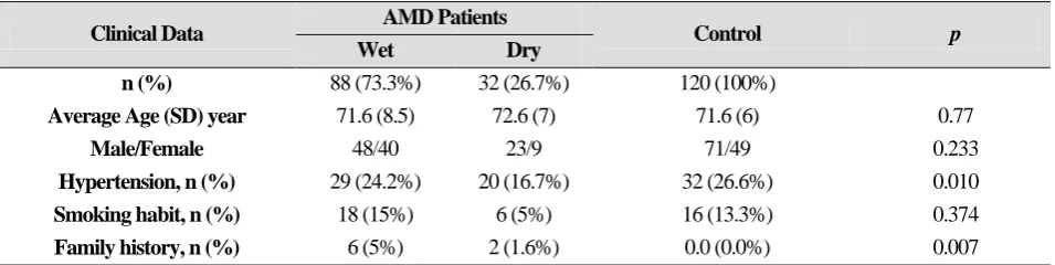
To study the polymorphisms of the HTRA1 gene promoter region, 120 patients 72 ± 8.11 years of age and 120 healthy controls 71.5 ± 6 years of age were recruited. Patients had a higher rate of hypertension (p = 0.01) and more family histories of AMD (p = 0.007) than control group subjects (Table 1).
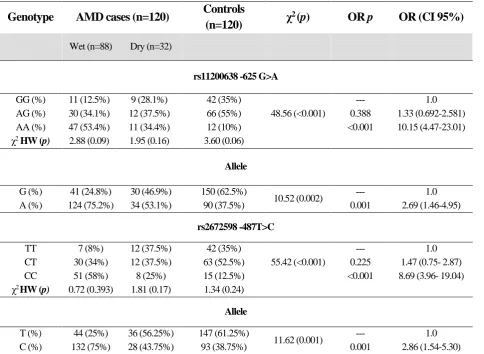
Genotyping data showed that allelic frequencies complied with Hardy-Weinberg equilibrium. Polymorphisms rs11200638 -625 G>A (p = 0.001, χ2 = 48.56) and rs2672598 -487 T>C (p = 0.001, χ2 = 64.17) were significantly higher in AMD patients than in controls. There was a significant linkage
disequilibrium between these two
polymorphisms (X2 p < 0.001, D': 0.93, r2: 0.845) (Tables 2 and 3).
Allele A of polymorphism rs11200638 -625 G>A was significantly greater in the patients with wet AMD than controls and associated with the risk of developing AMD (p = 0.001, OR (95% CI) = 2.69 (1.46-4.95). Allele C of polymorphism rs2672598 -487T>C was also significantly higher in patients with wet AMD than controls and associated with increased risk of developing AMD (p = 0.001, OR (95% CI) = 2.68 (1.54-5.30)). Genotype AA of polymorphism rs11200638 -625 G>A (p < 0.001, OR (95%CI) = 10.15(4.47-23.01)) and genotype CC of polymorphism rs2672598 -487T>C (p < 0.001, OR (95% CI) = 8.69 (3.96- 19.04)) were associated with greater risk of developing AMD (Table 3).
Table 1. Clinical and demographic data of study population
Table 2. Linkage disequilibrium of polymorphisms rs2672598 -487T>C and rs11200638 -625 G>A
r2
D' p χ2
Total rs2672598 -487T>C T>C
rs11200638 -625 G>A
CC CT
TT
0.845 0.93
<0.001 62
4 0
58
GG
108 4
104 0
AG
70 66
1 3
AA
240 74
105 61
Total
p
Control AMD Patients
Clinical Data
Dry Wet
120 (100%) 32 (26.7%)
88 (73.3%)
n (%)
0.77 71.6 (6)
72.6 (7) 71.6 (8.5)
Average Age (SD) year
0.233 71/49
23/9 48/40
Male/Female
0.010 32 (26.6%)
20 (16.7%) 29 (24.2%)
Hypertension, n (%)
0.374 16 (13.3%)
6 (5%) 18 (15%)
Smoking habit, n (%)
0.007 0.0 (0.0%)
2 (1.6%) 6 (5%)
Family history, n (%)
Table 3. Frequency of genotypes and alleles of polymorphisms rs2672598 -487T>C and rs11200638 -625 G>A
Discussion
According to the results of the current study, the frequencies of polymorphisms rs2672598 -487T>C and rs11200638 -625 G>A in the promoter region of the HTRA1 gene were significantly higher in AMD patients than controls. Heterozygotes bearing the AG genotype and AA homozygotes of polymorphism rs11200638 -625 G>A were most frequent among wet AMD patients and showed prevalences of 34.1 and 53.4%, respectively. The frequencies were similar to those with the dry form of the disease, while AG heterozygotes and GG wild-type homozygotes were the dominant genowild-types of controls, with 55 and 35% prevalences, respectively.
In the analysis of the allele frequencies in different groups we found that the A allele was significantly higher in patients than controls and
increased the risk of developing AMD. We observed a high association of this allele with the wet form of the disease (OR (CI 95%): 2.69 (1.46-4.95)). Due to the tight linkage disequilibrium of these polymorphisms in the subjects (χ2 p < 0.001, D':0.93, r2: 0.845), results showed similar frequencies of polymorphism rs2672598 -487T>C in the study population. Also, other studies have noted linkage disequilibrium between polymorphisms rs2672598 -487T>C and rs11200638 -625 G>A (24, 28).
While the T allele was more frequent in patients with dry than with wet AMD, the frequencies of C alleles of polymorphism rs2672598 -487T>C in patients with wet and dry forms of the disease were 75 and 43.7%, respectively. According to the results of the
OR (CI 95%) OR p
χ2 (p)
Controls (n=120) AMD cases (n=120)
Genotype
Dry (n=32) Wet (n=88)
rs11200638 -625 G>A
1.0 ---
48.56 (<0.001) 42 (35%)
9 (28.1%) 11 (12.5%)
GG (%)
1.33 (0.692-2.581) 0.388
66 (55%) 12 (37.5%)
30 (34.1%) AG (%)
10.15 (4.47-23.01) <0.001
12 (10%) 11 (34.4%)
47 (53.4%) AA (%)
3.60 (0.06) 1.95 (0.16)
2.88 (0.09) χ2 HW (p)
Allele
1.0 ---
10.52 (0.002) 150 (62.5%)
30 (46.9%) 41 (24.8%)
G (%)
2.69 (1.46-4.95) 0.001
90 (37.5%) 34 (53.1%)
124 (75.2%) A (%)
rs2672598 -487T>C
1.0 ---
55.42 (<0.001) 42 (35%)
12 (37.5%) 7 (8%)
TT
1.47 (0.75- 2.87) 0.225
63 (52.5%) 12 (37.5%)
30 (34%) CT
8.69 (3.96- 19.04) <0.001
15 (12.5%) 8 (25%)
51 (58%) CC
1.34 (0.24) 1.81 (0.17)
0.72 (0.393) χ2 HW (p)
Allele
1.0 ---
11.62 (0.001) 147 (61.25%)
36 (56.25%) 44 (25%)
T (%)
2.86 (1.54-5.30) 0.001
93 (38.75%) 28 (43.75%)
132 (75%) C (%)
C allele of polymorphism rs2672598 -487T>C with the risk of developing AMD was greater than with polymorphism rs11200638 -625 G>A. Higher prevalences of the A and C mutations of polymorphisms rs11200638 -625 G>A and rs2672598 -487T>C are indicative of associations of these polymorphisms with the risk of developing wet AMD. Numerous studies have shown that polymorphisms rs2672598 -487T>C and rs11200638 -625 G>A in the HTRA1 gene promoter region are associated with increased risk of developing AMD in different populations. According to a study by Yoshida et al., the rs11200638 -625 G>A mutation is associated with an increased risk of developing wet AMD in the Japanese population (29). Other studies in Asian societies found similar results (15, 30, 31). Also many studies have found a relationship between the A allele of polymorphism rs11200638 -625 G>A and the risk of developing wet AMD in European and American populations (15, 17). Also, in the case of the rs2672598 -487 T>C polymorphism, our results replicated the results from previous studies. These studies have shown that rs2672598 -487T>C is associated with increased risk of developing wet AMD (25, 32).
Our study is the first to show that rs2672598 and rs11200638 are associated with AMD in the Iranian population. In a recent study in Iran, other polymorphic regions, including CFH Y402H and HTRA1 LOC387715, in AMD were studied (33). Studies in Turkey and Egypt found associations between polymorphisms CFH Y402H and LOC387715 A69S and the risk of AMD (34-36). Based on our results, individuals with family histories of AMD were more likely to develop the disease than those without. Similarly, in the family-based studies, the positive family history for AMD has been reported. These results indicate a familial factor in the pathogenesis of AMD (2, 37, 38). No significant relationship between cigarette smoking and AMD was seen (p = 0.374), although patients with the wet form of the disease had higher levels of cigarette smoking than those with the dry form. Other studies have reported a cumulative effect of smoking and
polymorphism rs11200638 -625 G>A on the risk of developing AMD (32, 39).
The role of the A allele of polymorphism rs11200638 -625 G>A on the increased risk of developing AMD could be attributed to changes in the transcriptional factor binding sites in the HTRA1 gene promoter region for adaptor related protein complex 2α (AP2α) and serum response factor (SRF) (15). Increased HTRA1 expression can contribute to pathogenesis and augment AMD injuries by increasing the cell death signals through serine protease pathways (40). AMD patients bearing an allele of polymorphism rs11200638 -625 G>A express more HTRA1 than normal individuals, which could be related to the increased risk of developing the disease (22, 31). The rs2672598 -487T>C mutation is also located in the HTRA1 gene promoter region bound by the ELK-1 transcription factor (25); however, no clear relationship between this polymorphism and an elevated risk of developing AMD has been described.
It appears that genetic changes in the HTRA1 gene promoter in the 10q26 chromosomal region are related to the pathogenesis of AMD. Polymorphisms rs2672598 -487T>C and rs11200638 -625 G>A in this region have been demonstrated to be associated with an increased risk of developing AMD in our study and other studies in different populations. This association has also been observed to be related to the more advanced form of wet AMD in the same populations. To further clarify genetic polymorphisms associated with risk of developing AMD other regions of the HTRA1 gene should be investigated. This will be help to identify other genetic risk factors associated with the disease.
Acknowledgments
This study is funded by Cellular and Molecular Research Center (CMRC), Tehran University of Medical Sciences.
We would like to appreciate the head and all staff of CMRC for their support during current project. We also thank Dr. Saeid Kargozar for his useful comments on this manuscript.
References
1. Bandello F, Lafuma A, Berdeaux G. Public health impact of neovascular age-related macular degeneration treatments extrapolated from visual acuity. Investigative ophthalmology & visual science. 2007;48(1):96-103.
2. Coleman HR, Chan CC, Ferris FL, Chew EY. Age-related macular degeneration. The Lancet. 2008;372(9652):1835-45.
3. Lu F, Zhao P, Fan Y, Tang S, Hu J, Liu X, et al. An association study of SERPING1 gene and age-related macular degeneration in a Han Chinese population. Molecular vision. 2010;16:1.
4. Xu Y, Guan N, Xu J, Yang X, Ma K, Zhou H, et al. Association of CFH, LOC387715, and HTRA1 polymorphisms with exudative age-related macular degeneration in a northern Chinese population. Molecular vision. 2008;14:1373.
5. de Jong PTVM. Age-related macular degeneration. New England Journal of Medicine. 2006;355(14):1474-85.
6. Klein RJ, Zeiss C, Chew EY, Tsai JY, Sackler RS, Haynes C, et al. Complement factor H polymorphism in age-related macular degeneration. Science. 2005;308(5720):385-9.
7. Klein R, Peto T, Bird A, Vannewkirk MR. The epidemiology of age-related macular degeneration. American journal of ophthalmology. 2004;137(3):486-95.
8. Klein R. Overview of progress in the epidemiology of age-related macular degeneration. Ophthalmic epidemiology. 2007;14(4):184-7.
9. Van Newkirk MR, Nanjan MB, Wang JJ, Mitchell P, Taylor HR, McCarty CA. The prevalence of age-related maculopathy: the visual impairment project. Ophthalmology. 2000;107(8):1593.
10. Rakic J. Multifactorial influences on age-related macular degeneration. Bulletin de la Société belge d'ophtalmologie. 2006(301):9.
11. Francis PJ, George S, Schultz DW, Rosner B, Hamon S, Ott J, et al. The< i> LOC387715</i> Gene, Smoking, Body Mass Index, Environmental Associations with Advanced Age-Related Macular Degeneration. Human heredity. 2007;63(3-4):212-8. 12. Chong EWT, Kreis AJ, Wong TY, Simpson JA,
and meta-analysis. American journal of ophthalmology. 2008;145(4):707-15.
13. Haines JL, Hauser MA, Schmidt S, Scott WK, Olson LM, Gallins P, et al. Complement factor H variant increases the risk of age-related macular degeneration. Science. 2005;308(5720):419-21. 14. Jakobsdottir J, Conley YP, Weeks DE, Mah TS, Ferrell RE, Gorin MB. Susceptibility genes for age-related maculopathy on chromosome 10q26. The American Journal of Human Genetics. 2005;77(3):389-407.
15. DeWan A, Liu M, Hartman S, Zhang SSM, Liu DTL, Zhao C, et al. HTRA1 promoter polymorphism in wet age-related macular degeneration. Science. 2006;314(5801):989-92.
16. Zerbib J, Richard F, Puche N, Leveziel N, Cohen SY, Korobelnik JF, et al. R102G polymorphism of the C3 gene associated with exudative age-related macular degeneration in a French population. Molecular vision. 2010;16:1324.
17. Weger M, Renner W, Steinbrugger I, Köfer K, Wedrich A, Groselj-Strele A, et al. Association of the HTRA1− 625G> A promoter gene polymorphism with exudative age-related macular degeneration in a Central European population. Mol Vis. 2007;13:1274-9.
18. Tocharus J, Tsuchiya A, Kajikawa M, Ueta Y, Oka C, Kawaichi M. Developmentally regulated expression of mouse HtrA3 and its role as an inhibitor of TGF‐β signaling. Development, growth & differentiation. 2004;46(3):257-74.
19. De Luca A, De Falco M, Severino A, Campioni M, Santini D, Baldi F, et al. Distribution of the serine protease HtrA1 in normal human tissues. Journal of Histochemistry & Cytochemistry. 2003;51(10):1279-84.
20. Chu J, Zhou C, Lu N, Zhang X, Dong F. Genetic variants in three genes and smoking show strong associations with susceptibility to exudative age-related macular degeneration in a Chinese population. Chinese Medical Journal (English Edition). 2008;121(24):2525. 21. Francis PJ, Zhang H, DeWan A, Hoh J, Klein ML. Joint effects of polymorphisms in the HTRA1, LOC387715/ARMS2, and CFH genes on AMD in a Caucasian population. Molecular vision.
22. Cameron DJ, Yang Z, Gibbs D, Chen H, Kaminoh Y, Jorgensen A, et al. HTRA1 variant confers similar risks to geographic atrophy and neovascular age-related macular degeneration. Cell Cycle. 2007;6(9):1122-5.
23. Chan CC, Shen D, Zhou M, Ross RJ, Ding X, Zhang K, et al. Human HtrA1 in the archived eyes with age-related macular degeneration. Transactions of the American Ophthalmological Society. 2007;105:92. 24. Chen H, Yang Z, Gibbs D, Yang X, Hau V, Zhao P, et al. Association of HTRA1 polymorphism and bilaterality in advanced age-related macular degeneration. Vision research. 2008;48(5):690-4. 25. DeAngelis MM, Ji F, Adams S, Morrison MA, Harring AJ, Sweeney MO, et al. Alleles in the HtrA serine peptidase 1 gene alter the risk of neovascular age-related macular degeneration. Ophthalmology. 2008;115(7):1209-15. e7.
26. Gotoh N, Yamada R, Nakanishi H, Saito M, Iida T, Matsuda F, et al. Correlation between CFH Y402H and HTRA1 rs11200638 genotype to typical exudative age‐related macular degeneration and polypoidal choroidal vasculopathy phenotype in the Japanese population. Clinical & experimental ophthalmology. 2008;36(5):437-42.
27. Goldberg J, Flowerdew G, Smith E, Brody JA, Tso MO. Factors associated with age-related macular degeneration an analysis of data from the fi1r8t national health and nutrition examination survey. American Journal of Epidemiology. 1988;128(4):700-10.
28. Gibbs D, Yang Z, Constantine R, Ma X, Camp NJ, Yang X, et al. Further mapping of 10q26 supports strong association of< i> HTRA1</i> polymorphisms with age-related macular degeneration. Vision research. 2008;48(5):685-9.
29. Yoshida T, DeWan A, Zhang H, Sakamoto R, Okamoto H, Minami M, et al. HTRA1 promoter polymorphism predisposes Japanese to age-related macular degeneration. Molecular vision. 2007;13:545. 30. Mori K, Horie-Inoue K, Kohda M, Kawasaki I, Gehlbach PL, Awata T, et al. Association of the HTRA1 gene variant with age-related macular degeneration in the Japanese population. Journal of human genetics. 2007;52(7):636-41.
31. Yang Z, Camp NJ, Sun H, Tong Z, Gibbs D,
Cameron DJ, et al. A variant of the HTRA1 gene increases susceptibility to age-related macular degeneration. Science. 2006;314(5801):992-3.
32. Tam POS, Ng TK, Liu DTL, Chan WM, Chiang SWY, Chen LJ, et al. HTRA1 variants in exudative age-related macular degeneration and interactions with smoking and CFH. Investigative ophthalmology & visual science. 2008;49(6):2357-65.
33. Nazari Khanamiri H, Ghasemi Falavarjani K, Hossein Sanati M, Aryan H, Irani A, Hashemi M, et al. Complement Factor H Y402H and LOC387715 A69S Polymorphisms in Association with Age-Related Macular Degeneration in Iran. Journal of Ophthalmic & Vision Research. 2014;9(2):181-7. 34. Soysal Y, Inan ÜÜ, Küsbeci T, Imirzalioğlu N. Age-related macular degeneration and association of CFH Y402H and LOC387715 A69S polymorphisms in a Turkish population. DNA and cell biology. 2012;31(3):323-30.
35. Yücel D, Yilmaz M, Durukan AH, Özgül RK. Association of CFH Y402H polymorphism with both forms of advanced age-related macular degeneration in Turkish patients. Ophthalmic genetics. 2012;33(3):144-9.
36. Abbas RO, Azzazy HM. Association of single nucleotide polymorphisms in CFH, ARMS2 and HTRA1 genes with risk of age-related macular degeneration in Egyptian patients. Ophthalmic genetics. 2013;34(4):209-16.
37. Heiba IM, Elston RC, Klein BE, Klein R. Sibling correlations and segregation analysis of age‐related maculopathy: The beaver dam eye study. Genetic epidemiology. 1994;11(1):51-67.
38. Gorin MB. Genetic insights into age-related macular degeneration: controversies addressing risk, causality, and therapeutics. Molecular aspects of medicine. 2012;33(4):467-86.
39. Tuo J, Ross RJ, Reed GF, Yan Q, Wang JJ, Bojanowski CM, et al. The HtrA1 promotor polymorphism, smoking, and age-related macular degeneration in multiple case-control samples. Ophthalmology. 2008;115(11):1891.
40. Chien J, Staub J, Hu SI, Erickson-Johnson MR, Couch FJ, Smith DI, et al. A candidate tumor suppressor HtrA1 is downregulated in ovarian cancer. Oncogene. 2004;23(8):1636-44.