Complete genome sequence of Meiothermus ruber
type strain
(21
T)
Brian J Tindall1, Johannes Sikorski1, Susan Lucas2, Eugene Goltsman2, Alex
Copeland2, Tijana Glavina Del Rio2, Matt Nolan2, Hope Tice2, Jan-Fang Cheng2, Cliff Han2,3, Sam Pitluck2, Konstantinos Liolios2, Natalia Ivanova2, Konstantinos Mavromatis2, Galina Ovchinnikova2, Amrita Pati2, Regine Fähnrich1, Lynne Goodwin2,3, Amy Chen4, Krishna Palaniappan4, Miriam Land2,5, Loren Hauser2,5, Yun-Juan Chang2,5, Cynthia D. Jeffries2,5, Manfred Rohde6, Markus Göker1, Tanja Woyke2, James Bristow2, Jonathan A. Eisen2,7, Victor Markowitz4, Philip Hugenholtz2, Nikos C. Kyrpides2, Hans-Peter Klenk1, and Alla Lapidus2*
1 DSMZ - German Collection of Microorganisms and Cell Cultures GmbH, Braunschweig, Germany
2 DOE Joint Genome Institute, Walnut Creek, California, USA
3 Los Alamos National Laboratory, Bioscience Division, Los Alamos, New Mexico, USA
4 Biological Data Management and Technology Center, Lawrence Berkeley National Laboratory, Berkeley, California, USA
5 Oak Ridge National Laboratory, Oak Ridge, Tennessee, USA
6 HZI – Helmholtz Centre for Infection Research, Braunschweig, Germany 7 University of California Davis Genome Center, Davis, California, USA
*Corresponding author: Alla Lapidus
Keywords: thermophilic, aerobic, non-motile, free-living, Gram-negative, Thermales, Deino-cocci, GEBA
Meiothermus ruber (Loginova et al. 1984) Nobre et al. 1996 is the type species of the genus
Meiothermus. This thermophilic genus is of special interest, as its members share relatively low degrees of 16S rRNA gene sequence similarity and constitute a separate evolutionary li-neage from members of the genus Thermus, from which they can generally be distinguished by their slightly lower temperature optima. The temperature related split is in accordance with the chemotaxonomic feature of the polar lipids. M. ruber is a representative of the low-temperature group. This is the first completed genome sequence of the genus Meiothermus
and only the third genome sequence to be published from a member of the family Therma-ceae. The 3,097,457 bp long genome with its 3,052 protein-coding and 53 RNA genes is a part of the GenomicEncyclopedia ofBacteria andArchaea project.
Introduction
Strain 21
T(= DSM 1279 = ATCC 35948 = VKM
B-1258) is the type strain of the species
Meiother-mus ruber
, which is the type species of the genus
Meiothermus
[1]. Strain 21
Twas first described as
a member of the genus
Thermus
by Loginova and
Egorova in 1975 [2], but the species name to
which it was assigned was not included on the
Approved Lists of Bacterial Names [3].
Conse-quently
Thermus ruber
was revived, according to
Rule 28a of the International Code of
Nomencla-ture of Bacteria [4] in 1984 [5]. It received its
cur-rent name in 1996 when transferred from the
North-Eastern China [10], Northern Taiwan [11] and
Iceland [12]. Interestingly, the genus
Meiothermus
is heterogeneous with respect to pigmentation.
The yellow pigmented species also form a distinct
group on the basis of the 16S rRNA gene sequence
similarity, with the red/orange pigmented strains
forming two groups, one comprising
M. silvanus
and the other the remaining species [9,10]. Like all
members of the
Deinococci
the lipid composition
of the cell membrane of members of the genus
Meiothermus
is based on unusual and
characteris-tic structures. Here we present a summary
classi-fication and a set of features for
M. ruber
21
T,
to-gether with the description of the complete
ge-nomic sequencing and annotation.
Classification and features
The 16S rRNA genes of the seven other type
strains in the genus
Meiothermus
share between
88.7% (
M. silvanus
) [13] and 98.8% (
M.
taiwanen-sis
) [14] sequence identity with strain 21
T,
whe-reas the other type strains from the family
Ther-maceae
share 84.5 to 87.6% sequence identity
[15].
Thermus
sp. R55-10 from the Great Artesian
Basin of Australia (AF407749), as well as other
reference strains,
e.g
. 16105 and 17106 [12], and
the uncultured bacterial clone 53-ORF05 from an
aerobic sequencing batch reactor (DQ376569)
show full length 16S rRNA sequences identical to
that of strain 21
T. A rather large number of
iso-lates with almost identical 16S rRNA gene
se-quences originates from the Great Artesian Basin
of Australia, clone R03 (AF407684), and various
hot springs in Hyogo, Japan (strain H328;
AB442017), Liaoning Province, China (strain
L462; EU418906, and others), Thailand (strain
O1DQU (EU376397), a Finnish paper production
facility (strain L-s-R2A-3B.2; AM229096) and
oth-ers), but also the not validly published ‘
M.
rosa-ceus
’ (99.9%) [16] from Tengchong hot spring in
Yunnan (China). Environmental samples and
me-tagenomic surveys do not surpass 81-82%
se-quence similarity to the 16S rRNA gene sese-quence
of strain 21
T, indicating a rather mixed impression
about the environmental importance of strains
belonging to the species
M. ruber
, as occurring
on-ly in very restricted extreme habitats (status
Au-gust 2009).
Figure 2 shows the phylogenetic neighborhood of
M. ruber
21
Tin a 16S rRNA based tree. The
se-quences of the two 16S rRNA gene copies in the
genome are identical and differ by only one
nuc-leotide from the previously published sequence
generated from ATCC 35948 (Z15059).
A detailed physiological description based on five
strains has been given by Loginova
et al.
[5]. The cells
are described as Gram-negative nonmotile rods that
are 3 to 6 by 0.5 to 0.8 µm (Table 1), have rounded
ends, and are nonsporeforming [5]. In
potato-peptone-yeast extract broth incubated at 60°C,
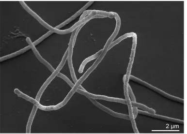
fila-mentous forms (20 to 40 µm in length) are observed
along with shorter rods (Figure 1) [5]. No filamentous
forms are observed after 16 h of incubation.
M. ruber
is obligately thermophilic [5]. On
potato-peptone-yeast extract medium, the temperature range for
growth is approx. 35-70°C, with an optimum
temper-ature at 60°C (the generation time is then 60 min) [5].
A bright red intracellular carotenoid pigment is
pro-duced, which resembles retro-dehydro-γ-carotene
(neo A, neo B) in its spectral properties [2]. The
ab-sorption spectra of acetone, methanol-acetone (l:l),
and hexane extracts show three maxima at 455, 483,
and 513 nm. The major carotenoid has since been
iden-tified as a
1‘-β-glucopyranosyl-3,4,3‘,4‘-tetradehydro-1‘,2‘-dihydro-β,ψ-caroten-2-one, with the glucose
ace-tylated at position 6 [30]. One strain (strain INMI-a)
contains a bright yellow pigment resembling
neuros-poraxanthine in its spectral properties [5], although it
may well have been misidentified, since other species
within the genus
Meiothermus
are yellow pigmented
[8,9].
M. ruber
is obligately aerobic [5]. It grows in
mi-nimal medium supplemented with 0.15% (wt/vol)
peptone as an N source, 0.05% (wt/vol) yeast extract,
and one of the following carbon sources at a
concen-tration of 0.25% (wt/vol):
D-glucose, sucrose,
mal-tose,
D-galactose,
D-mannose, rhamnose,
D-cellobiose,
glycerol,
D-mannitol, acetate, pyruvate, succinate,
fu-marate, or
DL-malate (sodium salts). No growth
oc-curs if the concentration of
D-glucose in the medium
is raised to 0.5% (wt/vol) [5]. Only moderate growth
occurs when ammonium phosphate (0.1%, wt/vol) is
substituted for peptone as the N source. No growth
occurs in the control medium without a carbon
source. No growth occurs on minimal medium
sup-plemented with 0.25% (wt/vol)
D-glucose, 0.05%
(wt/vol) yeast extract and one of the following
nitro-gen sources at a concentration of 0.1% (wt/vol):
alanine, glycine, asparagine, tyrosine,
L-glutamate, ammonium sulfate, nitrate, or urea.
Fur-ther lists of carbon source utilization, which differ in
part from the above list, are published elsewhere
[7,10-12]. Nitrates are not reduced and milk is not
peptonized [5], but
M. ruber
strain 21
Tis positive for
catalase and oxidase [10]. The most comprehensive
and updated list of physiological properties is
proba-bly given by Albuquerque et al [7].
Figure 2. Phylogenetic tree highlighting the position of M. ruber 21T relative to the type strains of the
Table 1. Classification and general features of M. ruber 21T according to the MIGS recommendations [17]
MIGS ID Property Term Evidence code
Current classification
Domain Bacteria TAS [18]
Phylum Deinococcus -Thermus TAS [1, 19-23]
Class Deinococci TAS [24,25]
Order Thermales TAS [25,26]
Family Thermaceae TAS [25,27]
Genus Meiothermus TAS [1]
Species Meiothermus ruber TAS [1,5]
Type strain 21 TAS [5]
Gram stain negative TAS [5]
Cell shape rod TAS [5]
Motility non motile TAS [5]
Sporulation not reported TAS [5]
Temperature range 35°C–70°C TAS [5]
Optimum temperature 60°C TAS [5]
Salinity growth with 1% NaCl TAS [7]
MIGS-22 Oxygen requirement obligately aerobic TAS [5]
Carbon source a diverse set of sugars TAS [5]
Energy source carbohydrates TAS [5]
MIGS-6 Habitat hot springs TAS [5]
MIGS-15 Biotic relationship free-living TAS [5]
MIGS-14 Pathogenicity not reported
Biosafety level 1 TAS [28]
Isolation hot spring TAS [5]
MIGS-4 Geographic location Kamchatka Peninsula, Russia TAS [5]
MIGS-5 Sample collection time 1973 or before TAS [2]
MIGS-4.1
MIGS-4.2 Latitude Longitude unknown unknown
MIGS-4.3 Depth unknown
MIGS-4.4 Altitude unknown
Evidence codes - IDA: Inferred from Direct Assay (first time in publication); TAS: Traceable Author Statement (i.e., a direct report exists in the literature); NAS: Non-traceable Author Statement (i.e., not directly observed for the living, isolated sample, but based on a generally accepted property for the species, or anecdotal evidence). These evidence codes are from of the Gene Ontology project [
29
]. If the evidence code is IDA, then the property was directly observed by one of the authors or an expert mentioned in the acknowledgementsChemotaxonomy
Initial reports on the polar lipids of
M. ruber
indi-cated that they consist of two major glycolipids GL
1a(~ 42%) and GL
1b(~ 57%) and one major
phospho-lipid PL
2(~ 93%), with small amounts of two other
phospholipids PL
1and PL
3[37]. Detailed work
indi-cates that in strains of
Thermus oshimai
,
T.
thermo-philus
,
M. ruber
, and
M. taiwanensis
the major
phos-pholipid is a 2’-O-(1, 2-diacyl-
sn
-glycero-3-phospho)
–3’-O-(α-N-acetyl-glucosaminyl)-N-glyceroyl
alky-lamine [38]. This compound is related to the major
2-OH iso-17:0) and a mixture of two compounds that
cannot be fully resolved by thin layer
chromatogra-phy carrying either 3-hydroxylated fatty acids or
unsubstituted fatty acids. The basic glycolipid
struc-ture dihexosyl – N-acyl-hexosaminyl – hexosyl –
di-acylglycerol is a feature common to all members of
the genera
Thermus
and
Meiothermus
examined to
date. There is currently no evidence that members of
the family
Thermaceae
(as currently defined)
pro-duce significant amounts of polar lipids containing
only two aliphatic side chains. The consequences of
having polar lipids containing three aliphatic side
chains on membrane structure has yet to be
ex-amined. Such peculiarities also indicate the value of
membrane composition in helping to unravel
evolu-tion at a cellular level. The major fatty acids of the
polar lipids are iso-C15:0 (30-40%) and iso-C17:0
(13-17%), followed by anteiso-C
15:0, C
16:0, iso-C
16:0,
anteiso-C
17:0, iso-C
17:0-2OH, and, at least in some
stu-dies, iso-C
17:1ω9c
(the values range from 3-10%).
Other fatty acid values are below 2%, including 3-OH
branched chain fatty acids. The values vary slightly
between the different studies [7,9,11,12,37].
De-tailed structural studies suggest that long chain diols
may be present in small amounts, substituting for
the 1-acyl-
sn
-glycerol [38]. Although not routinely
reported the presence of alkylamines (amide linked
to the glyceric acid of the major phospholipid) can
be deduced from detailed structural studies of the
major phospholipid [38]. Menaquinone 8 is the
ma-jor respiratory quinone, although it is not clear
which pathway is used for the synthesis of the
naph-thoquinone ring nucleus [41]. Ornithine is the major
diamino acid of the peptidoglycan in the genus
Meio-thermus
[1].
Genome sequencing and annotation
Genome project history
This organism was selected for sequencing on the
basis of its phylogenetic position [42], and is part
of the
Genomic
Encyclopedia of
Bacteria and
Arc-haea
project [43]. The genome project is
depo-sited in the Genome OnLine Database [36] and the
complete genome sequence is deposited in
Gen-Bank. Sequencing, finishing and annotation were
performed by the DOE Joint Genome Institute
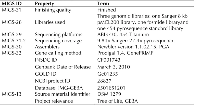
(JGI). A summary of the project information is
shown in Table 2.
Table 2. Genome sequencing project information
MIGS ID Property Term
MIGS-31 Finishing quality Finished
MIGS-28 Libraries used Three genomic libraries: one Sanger 8 kb pMCL200 library, one fosmide libraryand one 454 pyrosequence standard library MIGS-29 Sequencing platforms ABI3730, 454 Titanium
MIGS-31.2 Sequencing coverage 9.84× Sanger; 27.4× pyrosequence MIGS-30 Assemblers Newbler version 1.1.02.15, PGA MIGS-32 Gene calling method Prodigal 1.4, GenePRIMP
INSDC ID CP001743
Genbank Date of Release March 3, 2010
GOLD ID Gc01235
NCBI project ID 28827 Database: IMG-GEBA 2501651201 MIGS-13 Source material identifier DSM 1279
Project relevance Tree of Life, GEBA
Growth conditions and DNA isolation
M. ruber
21
T, DSM 1279, was grown in DSMZ
me-dium 256 (Nutrient Agar) [44] at 50°C. DNA was
isolated from 0.5-1 g of cell paste using Qiagen
Genomic 500 DNA Kit (Qiagen, Hilden, Germany)
following the standard protocol as recommended
by the manufacturer, with modification L for cell
lysis as described in Wu
et al.
[43].
Genome sequencing and assembly
The genome was sequenced using a combination
of Sanger and 454 sequencing platforms. All
gen-eral aspects of library construction and
sequenc-ing can be found at th
fragments of 1,000 bp and entered into assembly
as pseudo-reads. The sequences were assigned
quality scores based on Newbler consensus
q-scores with modifications to account for overlap
redundancy and adjust inflated q-scores. A hybrid
454/Sanger assembly was made using PGA
as-sembler. Possible misassemblies were corrected
and gaps between contgis were closed by primer
walks off Sanger clones and bridging PCR
frag-ments and by editing in Consed. A total of 431
Sanger finishing reads were produced to close
gaps, to resolve repetitive regions, and to raise the
quality of the finished sequence. Illumina reads
were used to improve the final consensus quality
using an in-house developed tool (the Polisher
[45]). The error rate of the completed genome
se-quence is less than 1 in 100,000. Together, the
combination of the Sanger and 454 sequencing
platforms provided 37.24× coverage of the
ge-nome. The final assembly contains 30,479 Sanger
reads and 371,362 pyrosequencing reads.
Genome annotation
Genes were identified using Prodigal [46] as part
of the Oak Ridge National Laboratory genome
an-notation pipeline, followed by a round of manual
curation using the JGI GenePRIMP pipeline [47].
The predicted CDSs were translated and used to
search the National Center for Biotechnology
In-formation (NCBI) nonredundant database,
Uni-Prot, TIGRFam, Pfam, PRIAM, KEGG, COG, and
In-terPro databases. Additional gene prediction
anal-ysis and functional annotation was performed
within the Integrated Microbial Genomes - Expert
Review (IMG-ER) platform [48].
Genome properties
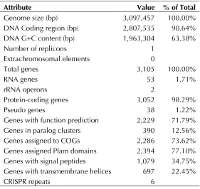
The genome consists of a 3,097,457 bp long
chro-mosome with a 63.4% GC content (Table 3 and
Figure 3). Of the 3,105 genes predicted, 3,052
were protein-coding genes, and 53 RNAs; thirty
eight pseudogenes were also identified. The
ma-jority of the protein-coding genes (71.8%) were
assigned with a putative function while the
re-maining ones were annotated as hypothetical
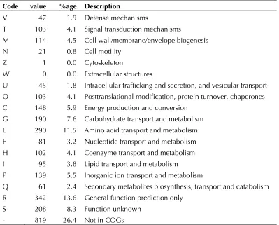
pro-teins. The distribution of genes into COGs
func-tional categories is presented in Table 4.
Table 3. Genome Statistics
Attribute Value % of Total
Genome size (bp) 3,097,457 100.00%
DNA Coding region (bp) 2,807,535 90.64% DNA G+C content (bp) 1,963,304 63.38%
Number of replicons 1
Extrachromosomal elements 0
Total genes 3,105 100.00%
RNA genes 53 1.71%
rRNA operons 2
Protein-coding genes 3,052 98.29%
Pseudo genes 38 1.22%
Genes with function prediction 2,229 71.79%
Genes in paralog clusters 390 12.56%
Genes assigned to COGs 2,286 73.62%
Genes assigned Pfam domains 2,394 77.10% Genes with signal peptides 1,079 34.75% Genes with transmembrane helices 697 22.45%
Figure 3. Graphical circular map of the genome. From outside to the center: Genes on forward strand (color by COG categories), Genes on reverse strand (color by COG categories), RNA genes (tRNAs green, rRNAs red, other RNAs black), GC content, GC skew.
Table 4. Number of genes associated with the general COG functional categories Code value %age Description
J 146 5.8 Translation, ribosomal structure and biogenesis A 0 0.0 RNA processing and modification
K 131 5.2 Transcription
L 117 4.7 Replication, recombination and repair B 2 0.1 Chromatin structure and dynamics
Table 4 (cont.) Number of genes associated with the general COG functional categories Code value %age Description
V 47 1.9 Defense mechanisms
T 103 4.1 Signal transduction mechanisms
M 114 4.5 Cell wall/membrane/envelope biogenesis N 21 0.8 Cell motility
Z 1 0.0 Cytoskeleton
W 0 0.0 Extracellular structures
U 45 1.8 Intracellular trafficking and secretion, and vesicular transport O 103 4.1 Posttranslational modification, protein turnover, chaperones C 148 5.9 Energy production and conversion
G 190 7.6 Carbohydrate transport and metabolism E 290 11.5 Amino acid transport and metabolism F 81 3.2 Nucleotide transport and metabolism H 102 4.1 Coenzyme transport and metabolism I 95 3.8 Lipid transport and metabolism
P 139 5.5 Inorganic ion transport and metabolism
Q 61 2.4 Secondary metabolites biosynthesis, transport and catabolism R 342 13.6 General function prediction only
S 208 8.3 Function unknown - 819 26.4 Not in COGs
Acknowledgements
We would like to gratefully acknowledge the help of Susanne Schneider (DSMZ) for DNA extraction and quality analysis. This work was performed under the auspices of the US Department of Energy Office of Science, Biological and Environmental Research Pro-gram, and by the University of California, Lawrence Berkeley National Laboratory under contract No.
DE-AC02-05CH11231, Lawrence Livermore National La-boratory under Contract No. DE-AC52-07NA27344, and Los Alamos National Laboratory under contract No. DE-AC02-06NA25396, UT-Battelle, and Oak Ridge National Laboratory under contract DE-AC05-00OR22725, as well as German Research Foundation (DFG) INST 599/1-2 and SI 1352/1-2.
References
1. Nobre MF, Trüper HG, Da Costa MS. Transfer of
Thermus ruber (Loginova et al. 1984), Thermus silvanus (Tenreiro et al. 1995), and Thermus chlia-rophilus (Tenreiro et al. 1995) to Meiothermus
gen. nov. as Meiothermus ruber comb. nov., Mei-othermus silvanus comb. nov., and Meiothermus chliarophilus comb. nov., respectively, and emendation of the genus Thermus. Int J Syst Bac-teriol 1996; 46:604-606
2. Loginova LG, Egorova LA. Obligate thermophilic-bacterium Thermus ruber in hot springs of Kam-chatka. Mikrobiologiya 1975; 44:661-665. 3. Skerman VBD, McGowan V, Sneath PHA.
Ap-proved Lists of Bacterial Names. Int J Syst
Bacte-riol 1980; 30:225-420
4. Lapage SP, Sneath PHA, Lessel EF, Skerman VBD, Seeliger HPR, Clark WA. 1992. International Code of Nomenclature of Bacteria (1990 Revi-sion). Bacteriological Code. American Society for Microbiology, Washington, DC.
5. Loginova LG, Egorova LA, Golovacheva RS, Sere-gina LM. Thermus ruber sp. nov., nom. rev. Int J Syst Bacteriol 1984; 34:498-499.
6. Euzéby JP. List of bacterial names with standing in nomenclature: A folder available on the Internet.
7. Albuquerque L, Ferreira C, Tomaz D, Tiago I, Veríssimo A, da Costa MS, Nobre MF. Meiother-mus rufus sp. nov., a new slightly thermophilic red-pigmented species and emended description of the genus Meiothermus. Syst Appl Microbiol
2009; 32:306-313
8. Tenreiro S, Nobre MF, da Costa MS. Thermus silvanus sp. nov. and Thermus chliarophilus sp. nov., two new species related to Thermus ruber
but with lower growth temperatures. Int J Syst Bacteriol 1995; 45:633-639
9. Pires AL, Albuquerque L, Tiago I, Nobre MF, Em-padinhas N, Veríssimo A, da Costa MS. Meio-thermus timidus sp. nov., a new slightly thermo-philic yellow-pigmented species. FEMS Microbiol Lett 2005; 245:39-45
10. Zhang XQ, Zhang WJ, Wei BP, Xu XW, Zhu XF, Wu M. Meiothermus cateniformans sp. nov., a slightly thermophilic species from north-eastern China. Int J Syst Evol Microbiol 2010; 60 :840-844 11. Chen MY, Lin GH, Lin YT, Tsay SS. Meiothermus
taiwanensis sp. nov., a novel filamentous, ther-mophilic species isolated in Taiwan. Int J Syst Evol Microbiol 2002; 52:1647-1654
12. Chung AP, Rainey F, Nobre MF, Burghardt J, Cos-ta MSD. Meiothermus cerbereus sp. nov., a new slightly thermophilic species with high levels of 3-hydroxy fatty acids. Int J Syst Bacteriol 1997;
47:1225-1230
13. Nobre MF, Trüper HG. Da Costa. Transfer of
Thermus ruber (Loginova et al. 1984), Thermus silvanus (Tenreiro et al. 1995), and Thermus chlia-rophilus (Tenreiro et al. 1995) to Meiothermus
gen. nov. as Meiothermus ruber comb. nov., Mei-othermus silvanus comb. nov., and Meiothermus chliarophilus comb. nov., respectively, and emendation of the genus Thermus. Int J Syst Bac-teriol 1996; 46:604-606
14. Chen MY, Lin GH, Lin YT, Tsay SS. Meiothermus taiwanensis sp. nov., a novel filamentous, ther-mophilic species isolated in Taiwan. Int J Syst Evol Microbiol 2002; 52:1647-1654
15. Chun J, Lee JH, Jung Y, Kim M, Kim S, Kim BK, Lim YW. EzTaxon: a web-based tool for the
iden-tification of prokaryotes based on 16S ribosomal RNA gene sequences. Int J Syst Evol Microbiol
2007; 57:2259-2261
16. Chen C, Lin L, Peng Q, Ben K, Zhou Z. Meio-thermus rosaceus sp. nov. isolated from Teng-chong hot spring in Yunnan, China. FEMS Micro-biol Lett 2002; 216:263-268
17. Field D, Garrity G, Gray T, Morrison N, Selengut
J, Sterk P, Tatusova T, Thomson N, Allen MJ, An-giuoli SV, et al. The minimum information about a genome sequence (MIGS) specification. Nat Biotechnol 2008; 26:541-547
18. Woese CR, Kandler O, Wheelis ML. Towards a natural system of organisms: proposal for the do-mains Archaea, Bacteria, and Eucarya. Proc Natl Acad Sci USA 1990; 87:4576-4579
19. Garrity GM, Lilburn TG, Cole JR, Harrison SH, Euzéby J, Tindall BJ. Taxonomic outline of the
Bacteria and Archaea, Release 7.7 March 6, 2007. Part 2 - The Bacteria: Phyla "Aquificae", "Thermotogae", "Thermodesulfobacteria", " Deino-coccus-Thermus", "Chrysiogenetes", "Chloroflexi", "Thermomicrobia", "Nitrospira", "Deferribacteres", "Cyanobacteria", and "Chlorobi".
20. Weisburg WG, Giovannoni SJ, Woese CR. The
Deinococcus-Thermus phylum and the effect of rRNA composition on phylogenetic tree construc-tion. Syst Appl Microbiol 1989; 11:128-134
21. Brooks BW, Murray RGE. Nomenclature for Mi-crococcus radiodurans and other radiation resis-tant cocci: Deinococcaceae fam. nov. and Dei-nococcus gen. nov., including five species. Int J Syst Bacteriol 1981; 31:353-360.
22. Rainey FA, Nobre MF, Schumman P, Stacke-brandt E, da Costa MS. Phylogenetic diversity of the deinococci as determined by 16S ribosomal DNA sequence comparison. Int J Syst Bacteriol
1997; 47:510-514
23. Brock TD, Freeze H. Thermus aquaticus gen. n. and sp. n., a nonsporulating extreme thermophile.
J Bacteriol 1969; 98:289-297
Bacteriolo-gy, Second Edition, Volume 1, Springer, New York, 2001, p. 395.
25. List Editor. Validation List no. 85. Validation of publication of new names and new combinations previously effectively published outside the IJ-SEM. Int J Syst Evol Microbiol 2002; 52:685-690
26. Rainey FA, da Costa MS. Order II. Thermales ord. nov. In: Garrity GM, Boone DR, Castenholz RW (eds), Bergey's Manual of Systematic Bacteriolo-gy, Second Edition, Volume 1, Springer, New York, 2001, p. 403.
27. da Costa MS, Rainey FA. Family I. Thermaceae
fam. nov. In: Garrity GM, Boone DR, Castenholz RW (eds), Bergey's Manual of Systematic Bacteri-ology, Second Edition, Volume 1, Springer, New York, 2001, p. 403-404.
28. Classification of bacteria and archaea in risk groups. www.baua.de TRBA 466.
29. Ashburner M, Ball CA, Blake JA, Botstein D, But-ler H, Cherry JM, Davis AP, Dolinski K, Dwight SS, Eppig JT, et al. Gene Ontology: tool for the unification of biology. Nat Genet 2000; 25:25-29
30. Burgess ML, Barrow KD, Gao C, Heard GM, Glenn D. Carotenoid glycoside esters from the thermophilic bacterium Meiothermus ruber.J Nat Prod 1999; 62:859-863
31. Castresana J. Selection of conserved blocks from multiple alignments for their use in phylogenetic analysis. Mol Biol Evol 2000; 17:540-552
32. Lee C, Grasso C, Sharlow MF. Multiple sequence alignment using partial order graphs. Bioinformat-ics 2002; 18:452-464
33. Stamatakis A, Hoover P, Rougemont J. A Rapid Bootstrap Algorithm for the RAxML Web Servers.
Syst Biol 2008; 57:758-771
34. Yarza P, Richter M, Peplies J, Euzeby JP, Amann R, Schleifer KH, Ludwig W, Glöckner FO, Rossel-lo-Mora R. The All-Species Living Tree project: A 16S rRNA-based phylogenetic tree of all se-quenced type strains. Syst Appl Microbiol 2008;
31:241-250
35. Pattengale ND, Alipour M, Bininda-Emonds ORP, Moret BME, Stamatakis A. How many bootstrap replicates are necessary? Lect Notes Comput Sci
2009; 5541:184-200
36. Liolios K, Chen IM, Mavromatis K, Tavernarakis N, Hugenholtz P, Markowitz VM, Kyrpides NC. The Genomes On Line Database (GOLD) in 2009: status of genomic and metagenomic projects and their associated metadata. Nucleic Acids Res 2010; 38:D346-D354
37. Donato ME, Seleiro EA, da Costa MS. Polar lipid and fatty acid composition of strains of Thermus ruber.Syst Appl Microbiol 1991; 14:234-239. 38. Yang YL, Yang FL, Jao SC, Chen MY, Tsay SS, Zou
W, Wu SH. Structural elucidation of phosphogly-colipids from strains of the bacterial thermophiles
Thermus and Meiothermus. J Lipid Res 2006;
47:1823-1832
39. Anderson R, Hansen K. Structure of a novel phos-phoglycolipid from Deinococcus radiodurans.J Biol Chem 1985; 260:12219-12223 40. Ferreira AM, Wait R, Nobre MF, da Costa MS.
Characterization of glycolipids from Meiothermus
spp. Microbiology 1999; 145:1191-1199 41. Hiratsuka T, Furihata K, Ishikawa J, Yamashita H,
Itoh N, Seto H, Dairi T. An alternative menaqui-none biosynthetic pathway operating in microor-ganisms. Science 2008; 321:1670-1673
42. Klenk HP, Göker M. En route to a genome-based classification of Archaea and Bacteria? Syst Appl Microbiol 2010; 33:175-182
43. Wu D, Hugenholtz P, Mavromatis K, Pukall R, Dalin E, Ivanova NN, Kunin V, Goodwin L, Wu M, Tindall BJ, et al. A phylogeny-driven genomic encyclopaedia of Bacteria and Archaea. Nature
2009; 462:1056-1060
44. List of growth media used at DSMZ:
45. Lapidus A, LaButti K, Foster B, Lowry S, Trong S, Goltsman E. POLISHER: An effective tool for us-ing ultra short reads in microbial genome assem-bly and finishing. AGBT, Marco Island, FL, 2008. 46. Hyatt D, Chen GL, Locascio PF, Land ML,
Bioinformatics 2010; 11:119
47. Pati A, Ivanova N, Mikhailova N, Ovchinikova G, Hooper SD, Lykidis A, Kyrpides NC. GenePRIMP: A gene prediction improvement ipeline for mi-crobial genomes. Nat Methods 2010; 7:455-457

![Table 1. Classification and general features of M. ruber 21T according to the MIGS recommendations [17]](https://thumb-us.123doks.com/thumbv2/123dok_us/673671.2066104/4.612.97.524.74.506/table-classification-general-features-ruber-according-migs-recommendations.webp)