Serkan Güler
1, A–C, F, Alparslan Apan
2, A–D, F, Nuray Bayar Muluk
3, A–F,
Bilgehan Budak
4, A, C, F, Goksen Öz
1, B, C, Emine A. Kose
1, B, CSevoflurane vs. TIVA in Terms
of Middle Ear Pressure During Laparoscopic Surgery
1 Department of Anesthesiology, Faculty of Medicine, Kırıkkale University, Turkey2 Department of Anesthesiology, Faculty of Medicine, Giresun University, Turkey 3 ENT Department, Faculty of Medicine, Kırıkkale University, Turkey
4 ENT Department, Audiology Division, Faculty of Medicine, Hacettepe University, Turkey
A – research concept and design; B – collection and/or assembly of data; C – data analysis and interpretation;
D – writing the article; E – critical revision of the article; F – final approval of article; G – other
Abstract
Objectives. The aim of this study was to investigate the effects of CO2 insufflation on the pressure of the middle
ear cavity (PMEC) during laparoscopic surgery under total intravenous anesthesia (TIVA) with propofol or sevo-flurane as an inhalational anesthetic maintenance.
Material and Methods. Sixty patients who underwent laparoscopic/or non-laparoscopic surgery under general anesthesia were included in the study. For anesthetic maintenance with inhalation agents, 20 non-laparoscopic surgery patients in Group 1 were applied sevoflurane (2–2.5%). Forty patients who underwent laparoscopic surgery were randomized into two groups. Anesthesia was maintained with sevoflurane (2–2.5%) in twenty patients in Group 2 and the TIVA technique in 20 patients in Group 3. In Group 1, PMEC was measured before anesthesia, 10 and 30 min after endotracheal intubation, 10 min before extubation, and 15, 30, 60 min and 6 hours in the post-operative period. In Group 2 and 3, PMEC was measured before the anesthesia, 10 min after intubation, 10 and 30 min after CO2 insufflation, just before the CO2 elimination, 10 min before the extubation, and 15, 30, 60 min
and 6 hours after extubation in the postoperative period.
Results. PMEC was significantly increased in Group 1 at 10 min after intubation, at 30 min of the operation, before extubation, and at postoperative 15 and 30 min (p < 0.05). In Group 3, differences between PMECs were detected at the 30th min of insufflation (p = 0.005), and during elimination (p = 0.035) compared to the initial measurement.
Generally, the values remained positive in Group 1 and negative in Group 3. There was a significant difference between Group 1 and Group 3 at 10 min after the induction (p = 0.001). There was no statistically significant dif-ference in PMECs between Group 2 and 3 patients undergoing laparoscopic surgery.
Conclusions. Our results indicate that, in laparoscopic surgery, TIVA used for the maintenance of anesthesia did not increase the PMEC and the changes caused by sevoflurane were also in the normal range of middle ear pressures. In patients with previous ear surgery, if there is a need of classical surgical procedures in the future, sevoflurane anesthesia should not be the first choice due to its effects on PMEC, which cause it to be increased over 50 daPa, especially at 30 min after intubation. Patient characteristics including previous ear surgery should be considered in selecting the optimum anesthetic agents and technique (Adv Clin Exp Med 2014, 23, 3, 447–454).
Key words: pressure of the middle ear cavity (PMEC),laparoscopy, sevoflurane, TIVA, CO2 insufflation. Adv Clin Exp Med 2014, 23, 3, 447–454
ISSN 1899–5276
ORIGINAL PAPERS
© Copyright by Wroclaw Medical University
The effects of volatile anesthetics on the pres-sure of the middle ear cavity (PMEC) have been clearly defined. Nitrous oxide has been demonstrat-ed to cause a time-relatdemonstrat-ed increase in the pressure with accumulation in a closed environment, and this effect is also valid for the middle ear [1, 2].
Increase in PMEC may result in membrane rup-ture due to the increase in intra-tympanic pressure,
The aim of this study was to determine the effects of CO2 insufflation on PMEC during
lap-aroscopic surgery under TIVA versus sevoflu-rane maintenance and to compare the results with age- and gender-matched healthy patients under-going non-laparoscopic surgery with sevoflurane anesthesia.
Material and Methods
This prospectivecohort study was undertak-en after obtaining the approval of the Local Ethics Committee of Kırıkkale University School of Med-icine (Date: 2009, Number: 014) and written in-formed consent from all patients. The study was conducted in the Anesthesiology and ENT Depart-ments of the Kırıkkale School of Medicine, and Hacettepe University School of Medicine, Audiol-ogy Division of the ENT Department from April 2009 to July 2010.
Patients
Sixty American Society of Anesthesiologists (ASA) physical status I or II patients undergoing laparoscopic/or non-laparoscopic surgery were included in the study. For inhalational anesthet-ic maintenance, 20 non-laparoscopanesthet-ic surgery pa-tients in Group 1 were given sevoflurane (2–2.5%). Forty patients who underwent laparoscopic sur-gery were divided into two groups by sealed enve-lope technique. Twenty patients in Group 2 were given sevoflurane (2–2.5%), and the TIVA tech-nique was applied for the 20 patients in Group 3 for anesthesia maintenance.
In all groups, the ears of the patients were ex-amined using a hand-held device (Heine Beta 100 Otoscope, Herrsching, Germany) and PMECs were measured by the same author.
Patients with ear wax, perforation of the ear drum, a history of previous surgery on the external or middle ear, previous surgery including cranio-facial operations that preclude reaching the exter-nal auricular meatus, and laparoscopy procedures under other positions except supine were excluded from the study.
Measurement of the PMEC
PMEC and other variables (compliance, gra-dient) were measured by a hand-held device (In-teracoustics A/S Assens DK-5610 Model MT10, Golden Triangle, MN, USA) in each ear of all pa-tients. In Group 1 patients, PMEC was measured before anesthesia, 10 and 30 min after endotrache-al intubation, 10 min before extubation, and 15,
30, 60 min and 6 h after extubation in the postop-erative period. In the patients of Group 2 and 3, PMEC was measured before the anesthesia, 10 min after intubation, 10 and 30 min after CO2
insuffla-tion, just before CO2 elimination, 10 min before
extubation, and 15, 30, 60 min and 6 h after extu-bation in the postoperative period.
Anesthetic Procedure
Patients were monitored according to routine anesthetic care, including EKG attached to lead II, noninvasive arterial blood pressure, oxygen satu-ration, end-tidal CO2, and temperature
(Datex-Ohmeda, Cardiocap 5 Monitor, Helsinki, Finland), and measurements were recorded every 5 min. In-travenous access was achieved on the non-domi-nant hand with a 20 G cannula, and hydration was performed at a rate of 10 mL/kg/h.
Induction of anesthesia: Propofol 2–2.5 mg/kg (Propofol® 1%, Fresenius Kabi AB, Sweden),
ro-curonium bromide 0.6 mg/kg (Esmeron®,
Orga-non, Holland) and fentanyl 1 µg/kg(Fentanyl®,
Abbott, Ireland).
Maintenance of the anesthesia: Sevoflurane (Sevorane® liquid, Abbott, Ireland) end-tidal
con-centration 2–2.5% and air-oxygen (FiO2: 35%)
mix-ture was adjusted for the maintenance anesthesia after intubation in the control (Group 1, n = 20) and laparoscopic surgery (Group 2, n = 20) groups. In Group 3 (n = 20), TIVA was administered using propofol at a rate of 12 mg/kg/h for the first 20– 30 min, then infusion was decreased to 9 mg/kg/h for 20–30 min, and finally infusion was followed with 6 mg/kg/h. Fentanyl 1–2 µg/kg was given for analgesia every 30–45 min and for supplementa-tion in all patients. When an increase in heart rate or mean blood pressure of more than 15% from baseline measurements was observed, a 0.3 mg/kg dose of additional rocuronium was applied ac-cording to the clinical requirements. Mechanical ventilation was set at a rate of 4 Lt.min–1 and FiO
2:
35% during the maintenance with an air/oxygen mixture. Tidal volume was set at 8–10 mL/kg, and respiratory frequency was adjusted according to the end-tidal CO2 value, which was maintained at
about 40–45 mm Hg (Dräger, Julian, Lübeck, Ger-many). In order to determine the effect of only CO2 and to accurately interpret the results, we did
not administer N2O to our patients.
Laparoscop-ic surgery was performed using a Storz unit (Karl Storz-Endoskope, Tuttlingen, Germany). Intra-ab-dominal pressure was increased 1–2 mm Hg/min and reached 15 mm Hg at maximum [7].
performed when ventilatory parameters (tidal vol-ume > 5–7 mL/kg, frequency > 10 per min) reached adequate levels.
Approach to the patients in the recovery room: patients were transferred to the recovery room when patient cooperation was obtained, and they stayed there for at least 1 h in order for the vital signs to be evaluated and ear measurements be made. Pain management was mainly performed in the surgical ward. In the recovery room, fentanyl 25–50 µg or petidine 20–25 mg i.v. was adminis-tered and repeated as required. Tramadol (Contra-mal, Abdi İbrahim, Turkey) or petidine (Aldolan, Gerot Pharmazeutika GmbH, Austria) 400 mg maximum dose was administered per day and in the case of uncontrolled pain, lornoxicam (Xefo, Nycomed GmbH, Austria) was added every 8 h. Side effects including dizziness, headache, nausea and vomiting were determined and recorded.
This study was conducted in accordance with Helsinki Declaration [8] and Good Clinical Perfor-mance Guidelines [9]. Data from the Consultancy Thesis of Dr. Serkan Güler was used in the prepa-ration of this study [10].
Statistical Analysis
Statistical analysis was performed using SPSS version 15.0 for Windows. Demographic variables including age, weight and hemodynamic variables were evaluated using ANOVA and post hoc Bon-ferroni correction. Categorical variables includ-ing ASA physical status, gender and distribution
of surgeries were determined with a chi-square test. Variables obtained from ear measurements were analyzed between the groups using ANOVA.
Post hoc Bonferroni, Mann-Whitney-U tests, and Friedman and Wilcoxon Signed Ranks tests were used for intra group analysis.
A p value < 0.05 was considered as statistical-ly significant.
Results
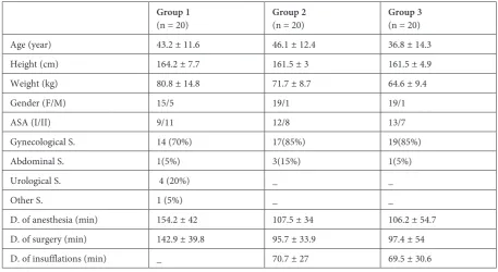
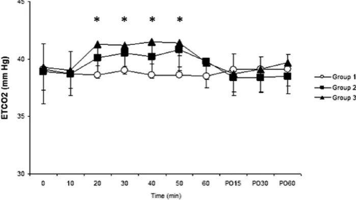
There was no significant difference between de-mographic variables including age, height, weight, distribution of gender, and ASA physical status in Groups 1–3. There was also no significant differ-ence between groups in terms of duration of anes-thesia, surgery and insufflation (Table 1). Hemody-namic changes shown in Fig. 1–3 were also similar between the groups. Heart rates were significantly different between Group 1 and 2 at 20 min. There was also a significant difference between Group 1 and 3 in terms of mean arterial blood pressures at 15 and 30 min after the operation. The changes in end-tidal CO2 values were significantly higher in Groups 2 and 3 at 20, 30, 40 and 50 min (p < 0.05) during the operation (Fig. 3).
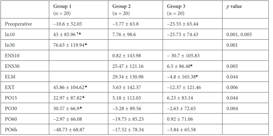
The PMEC values of the groups are demon-strated in Table 2. When compared with initial values, PMECs increased in Group 1 at the 10th
(p = 0.005) and 30th (p = 0.001) min of the
opera-tion, after extubation (p = 0.006), and at the post-operative periods of the 15th (p = 0.044) and 30th
Table 1. Patient characteristics, ASA physical status, types and durations (D) of surgery (S), anesthesia and insufflation (Data is given as mean ± SD or n (%)
Group 1
(n = 20) Group 2 (n = 20) Group 3 (n = 20)
Age (year) 43.2 ± 11.6 46.1 ± 12.4 36.8 ± 14.3
Height (cm) 164.2 ± 7.7 161.5 ± 3 161.5 ± 4.9
Weight (kg) 80.8 ± 14.8 71.7 ± 8.7 64.6 ± 9.4
Gender (F/M) 15/5 19/1 19/1
ASA (I/II) 9/11 12/8 13/7
Gynecological S. 14 (70%) 17(85%) 19(85%)
Abdominal S. 1(5%) 3(15%) 1(5%)
Urological S. 4 (20%) _ _
Other S. 1 (5%) _ _
D. of anesthesia (min) 154.2 ± 42 107.5 ± 34 106.2 ± 54.7
D. of surgery (min) 142.9 ± 39.8 95.7 ± 33.9 97.4 ± 54
Fig. 1. Heart rate (HR) changes between study groups. * – heart rates were significantly different between Group 1 and 2 at 20 min. (p < 0.05)
Fig. 2. Mean arterial pressure (MAP) changes between groups. * – there was significant dif-ference between Group 1 and 3 in terms of mean arterial blood pressures at 15 and 30 min after operation (p < 0.05)
Fig. 3. End-tidal CO2 changes between groups. * – the changes in end-tidal CO2 values
(p = 0.004) mins. In Group 3, a difference between PMECs at the 30th min of insufflation (p = 0.005),
and during elimination (p = 0.035) were detect-ed, according to the initial measurement. There was a significant difference between Group 1 and Group 3 at 10 min after the induction (p = 0.001). Generally, the values remained positive in Group 1 and negative in Group 3.
PMEC was generally measured under the base-line limit in Group 2 and Group 3. According to these observations, the major deviation for PMEC was observed in Group 1, in which it progressive-ly increased and peaked at 30 min during the op-eration and remained so through the 30 min of the postoperative period. However, the changes in the other two groups were limited.
There was no significant difference between groups in terms of side effect profile that is nau-sea and vomiting. Side effects were observed in two patients in Group 1, two patients in Group 2, and one patient in Group 3 during the postoperative period.
Discussion
In the present study, in Group 1 (Non-lap-aroscopic sevoflurane), PMECs increased at the 10th and 30th min of the operation, after
extuba-tion, and at 15 and 30 min after extubation in the postoperative period. Especially at 30 min af-ter intubation, the value was higher than normal
limits of ± 50 daPa. In Group 2 (Laparoscopic sevoflurane), PMECs non-significantly increased at the 30th min of insufflation and at the
elimi-nation period, and the changes were within nor-mal limits. In sevoflurane groups (Group 1 and 2), an increase of the PMECs was detected at dif-ferent degrees. In the third group (Group 3), TI-VA was applied during the laparoscopic proce-dure, and in this group, a significant difference between PMECs was detected at the 30th min of
insufflation and during elimination compared to the initial measurement, but the values were ob-served as slightly negative and between ± 50 daPa values of normal limits.
The main difference between group 1 and 2 was that conventional surgery was performed in Group 1 and laparoscopic surgery was performed in Group 2. In laparoscopic surgery, pain, surgi-cal trauma and inflammation occur at lower rates than classic surgical procedures. Surgical pain con-stitutes one of the main differences between lap-aroscopic surgery and surgeries with the classical approach. Imaging techniques indicate that acute pain can increase regional cerebral blood flow from the brainstem to the cortical projection area as could be observed with incision [11]. Although not proven in patients under anesthesia, general anesthesia itself does not seem to suppress these changes completely as hemodynamic variables in-crease during surgical incision. These changes may also be secondary to the inflammation induced by surgical trauma, which has been demonstrated to
Table 2. Middle ear pressure (PMEC) (DaPa) variations between study groups*
Group 1
(n = 20) Group 2 (n = 20) Group 3 (n = 20)
p value
Preoperative –10.6 ± 52.05 –3.77 ± 63.8 –25.55 ± 65.44
In10 43 ± 85.96 ¶▲ 7.76 ± 98.6 –25.73 ± 74.43 0.001, 0.005
In30 76.63 ± 119.94▲ 0.001
ENS10 0.82 ± 143.98 – 30.7 ± 105.83
ENS30 25.47 ± 121.16 6.5 ± 86.48■ 0.005
ELM 29.34 ± 130.98 –4.8 ± 165.38■ 0.044
EXT 45.86 ± 104.62▲ 5.63 ± 142.37 –12.37 ± 121.46 0.006
PO15 22.97 ± 87.82▲ 5.18 ± 112.03 6.23 ± 83.14 0.044
PO30 30.57 ± 66.9▲ –3.28 ± 89.56 –2.63 ± 72.65 0.004
PO60 –2.97 ± 66.08 –19.75 ± 85.23 0.92 ± 71.06
PO6h –48.73 ± 68.87 –17.52 ± 78.34 –3.84 ± 65.58
In 10 – 10 min after intubation, ENS10 – 10 min after insufflation, ENS30 – 30 min after insufflation, ELM – elimination, EXT – extubation, PO 15, 30, 60, 6h – postoperative 15, 30, 60 min and 6 h respectively.
be milder in laparoscopic interventions due to low-er potential for tissue injury [12, 13].
In laparoscopic surgery, CO2 insufflation is
performed into the abdomen. CO2 is a gas with
high resolution, absorbed through the peritone-um [13, 14], and then is carried by the venous system and eliminated through the respirato-ry tract [15]. CO2 insufflation during
laparoscop-ic surgery may decrease heart rate, mean arterial blood pressure, cardiac output and systemic vas-cular resistance, causing a decrease in middle ear blood flow. On the other hand, CO2 causes
ve-nous dilatation that may balance its effects; and in-crease microcirculation [16–19]. In our study, in Group 1, CO2 insufflation was not applied during
conventional surgery. In this group, the increase in PMEC may depend on the diffusion of gases from the blood into the middle ear cavity due to the vasodilator effect of sevoflurane. In Group 2, although sevoflurane was applied as in Group 1, the difference was due to the insufflation of CO2
into the peritoneal cavity during laparoscopy. Al-though blood-carried CO2 diffuses into the
mid-dle ear cavity, it is also removed from the midmid-dle ear cavity with the help of increased microcircu-lation. Additionally, the main mechanism for es-caping air and gases from the middle ear cavi-ty is passive through the Eustachian tube (ET). When the elasticity of the Eustachian tube is over-come, air escapes to the nasopharynx. The ET has a valve-like function. The tubal muscles actively di-late the tubal valve for adequate ventilation of the middle ear. Active and passive exchange of mid-dle ear gases occurs constantly within the midmid-dle ear [20]. Poe, et al [21] reported that normal ETs had four consistent sequential movements: [1] pal-atal elevation causing passive, then active, rotation of the medial cartilaginous lamina; [2] lateral ex-cursion of the lateral pharyngeal wall; [3] dilation of the lumen, caused primarily by tensor veli pala-tini muscle movement beginning distally and in-feriorly, then opening proximally and superiorly; and [4] opening of the tubal valve at the isthmus caused by dilator tubae muscle contraction [21]. In the present study, sevoflurane is eliminated quick-ly, and CO2 diffused into the middle ear cavity is
removed quickly due to the function of the ET and also increased microcirculation. Therefore, during a short-period (at 30 min of insufflation and in the elimination period), a non-significant increase in middle ear pressure may be detected.
In addition, increased CO2 levels seem to con-tribute to this anti-inflammatory effect [22]. Sim-ilar to upper respiratory tract infection, edema or other changes that are mediated through an in-crease in vascular supply, inflammatory cells and cytokines may impede diffusion and therefore lead
to the accumulation of anesthetic gases in a closed environment.
In Group 3 (Laparoscopic TIVA), although there were changes in PMECs at 30 min of insuf-flation and the elimination period, PMECs did not increase more than positive pressure values. When the procedure was performed with laparoscopy, the rapid destruction of anesthetic agents due to the increase in microcirculation, and less surgical trauma and pain may cause these results.
If it is necessary to operate on patients who have undergone ear surgery or middle ear pros-thesis replacement surgery previously, laparos-copy should be preferred instead of conventional surgical intervention, and the use of TIVA may be more suitable in these patients. In the laparoscopic sevoflurane group (Group 2), the small increase in middle ear pressure, and in the conventional sur-gery + sevoflurane group (Group 1), the increase of the middle ear pressure throughout the proce-dure and post-op period, may lead to displacement of the prosthesis in patients who have previously undergone ear surgery and middle ear prosthesis replacement.
The influence of positioning on cerebral blood flow and intracranial pressure is well estab-lished [23]. To eliminate the possible effects of po-sitioning, repeated measures of tympanograms were obtained from all patients in the supine po-sition. In the present study, the effects of CO2
in-sufflation on PMEC might be limited due to the measuring interval. Continuous or more frequent data collection may determine the increase or de-crease in pressure better. Nevertheless, the number of measurements in our study was higher than that in other studies in the literature [2].
hypotension to decrease bleeding from the opera-tion field during middle ear surgery, was also dem-onstrated paradoxically to increase PMEC, possi-bly due to altered microvascularization [25].
In the literature, there are studies on sevo-flurane anesthesia on ear surgery. Shirgoska et al. [26] reported that remifentanil alone and in combination with sevoflurane are effective in in-ducing consistent and sustained controlled hy-potension in children undergoing middle ear mi-crosurgery [26]. Crawford et al. [27] reported that volatile anesthetics (sevoflurane, desflurane and isoflurane) suppress the stapedius reflex in a dose-dependent manner; and they advised against the use of volatile anesthetics for measurement of the stapedius reflex threshold during cochlear implant surgery. The first study recommends sevoflurane use, whereas the second was against the usage of sevoflurane in cochlear implant surgery.
Pressure is one of the measurable parameters of the middle ear cavity and stability of PMEC does not prevent any potential adverse outcomes. A de-crease in middle ear blood flow without increasing the PMEC may lead to edema or ischemia. Blood gas analysis during tympanogram may clarify the possible influence of acid-base status on PMEC, and the lack of this analysis is one of the limita-tions of the present study. The unequal number of patients in open versus laparoscopic surgery groups is the other limitation, so anesthetic depth measurement using BIS or another device may also
help to standardize the patients. The effect of vari-ous drugs or types of surgeries on the microvascu-larization of the hearing system is unclear and will be the target of the future investigations.
It can be stated that, for the maintenance of an-esthesia, TIVA did not cause positively increased PMEC in laparoscopic surgery, but sevoflurane in conventional surgery caused slightly increased PMEC. However, the changes in PMEC with sevo-flurane in laparoscopic surgery were within the normal range of middle ear pressures.
In conclusion, based on the authors’ results, it appears that TIVA may be administered safely during laparoscopic surgery, allowing the main-tenance of stable PMEC, particularly in patients who have undergone previous ear surgery. Alter-ations with sevoflurane anesthesia in laparoscop-ic procedures may be within the normal range of ± 50 daPa, which may not cause damage to pa-tients with middle ear prosthesis or previous ear surgery. On the other hand, in patients with previ-ous ear surgery, if there is a need of classical surgi-cal procedures in the future, sevoflurane anesthe-sia should not be the first choice due to its effects on PMEC. Patient characteristics including previ-ous ear surgery should be considered in selecting the optimum anesthetic agents and technique, and optimum post-operative care [28]. Further clini-cal and experimental studies may elucidate the ef-fects of anesthetic agents in laparoscopic surgery on PMEC.
References
[1] Karabıyık L, Bozkırlı F, Celebi H, Göksu N: Effects of nitrous oxide on middle ear pressure: a comparison between inhalational anesthesia with nitrous oxide and TIVA. Eur J Anaesthesiol 1996, 13, 27–32.
[2] Chin K, Brown OE, Manning SC, Crandell CC: Middle ear pressure variation: effect of nitrous oxide. Laryngoscope 1997, 107, 357–363.
[3] Patterson ME, Bartlett PC: Hearing impairment caused by intratympanic pressure changes during general anes-thesia Laryngoscope 1976, 86, 399–404.
[4] Perreault L, Normandin N, Plamandon L, Blain R, Rousseau P, Girard M: Tympanic membrane rupture after anesthesia with nitrous oxide. Anesthesiology 1982, 57, 325–326.
[5] Öztürk O, Demiraran Y, Ilce Z, Kocaman B, Guclu E, Karaman E: Effects of sevoflurane and TIVA with propofol on middle ear pressure. Int J Ped Otorhinolaryngol 2006, 70, 1231–1234.
[6] Demiraran Y, Iskender A, Guclu E, Yildizbas S: Effects of desflurane on middle ear pressure. Int J Ped Otorhinolaryngol 2007, 71, 1439–1441.
[7] Kerbl K, Clayman RV: Basic techniques of laparoscopic surgery. Urol Clin North Am 1993, 20, 361–368.
[8] WMA, Declaration Of Helsinki: Ethical Principles for Medical Research Involving Human Subjects, in 59th WMA General Assembly, W.M. Association, Editor. October 2008, Seoul.
[9] TC Sağlık Bakanlığı İlaç ve Eczacılık Genel Müdürlüğü, İyi Klinik Uygulamaları Kılavuzu: 1995, T.C. Sağlık Bakanlığı: Ankara (TR Pharmaceutical General Directorate of the Ministry of Health, Good Clinical Practice Guidelines. 1995, TR Ministry of Health: Ankara).
[10] Güler S: Laparoskopik cerrahide Sevofluran veya TIVA’nın orta kulak basıncına etkileri (The effects of Sevoflurane ver-sus TIVA on middle ear pressure during laparoscopic surgery).Uzmanlık Tezi (Specialization Thesis), Kırıkkale 2010.
[11] Howard MA, Krause K, Khawaja N, Massat N, Zelaya F, Schumann G: Beyond patient reported pain: perfusion magnetic resonance imaging demonstrates reproducible cerebral representation of ongoing post-surgical pain. PLoS ONE 6, e17096.
[12] Boo YJ, Kim WB, Kim J, Song TJ, Choi SY, Kim YC: Systemic immune response after open versus laparoscopic cholecystectomy in acute cholecystitis: a prospective randomized study. Scand J Clin Lab Invest 2007, 67, 207–214.
[14] Cunningham AJ, Brull SJ: Laparoscopic cholecystectomy: anesthetic implications. Anesth Analg 1993 May, 76, 1120–1133.
[15] Naude GP, Bongard FS: Helium insufflation in laparoscopic surgery. Endosc Surg Allied Technol 1995 Aug 3, 183–186.
[16] Anthony JC, Sorun JB: Laparoscopic Cholecystectomy: Anesthetic implications. Anest Analg 1993, 76, 1120–1133.
[17] Marco AP, Yeo CJ, Rock P: Anesthesia for patient undergoing laparoscopic cholecystectomy. Anesthesiology 1990, 73, 1268–1270.
[18] Joris JL, Noirat DP, Legrand MJ, Jacquet NJ, Lamy ML: Hemodynamic changes during laparoscopic cholecys-tectomy. Anesth Analg 1993, 76, 1067–1071.
[19] Wittgen CM, Andrus CH, Fitzgerald SD, Baudendistel LJ, Dahms TE, Kamnski DL: Analysis of the hemody-namic and ventilatory effects of laparoscopic cholecystectomy. Arch Surg 1991, 126, 997–1000.
[20] Grimmer JF, Poe DS: Update on eustachian tube dysfunction and the patulous eustachian tube. Curr Opin Otolaryngol Head Neck Surg 2005, 13, 277–282.
[21] Poe DS, Pyykkö I, Valtonen H, Silvola J: Analysis of eustachian tube function by video endoscopy. Am J Otol. 2000, 21, 602–607.
[22] Shimotakahara A, Metzelder M, Vieten G, Ure B, Kuebler JF: CO2 modulates the inflammatory cytokine release of primary human pleural macrophages. Eur J Pediatr Surg 2010, 20, 111–115.
[23] Cassoria L, Lee JW: Patient positioning and anesthesia. In: Miller’s Anesthesia. Ed.: Miller RD. Churchill- -Livingstone, Philadelphia 2010, 1151–1170.
[24] Pagel PS, Farber NE, Pratt PF, Warltier DC: Cardiovascular pharmacology. In: Miller RD, editor. Miller’s Anesthesia. Philadelphia: Churchill-Livingstone, 2010, 595–632.
[25] Firat Y, Kizilay A, Akarcay M, Yücel A, But K, Yologlu S: The effects of dexmedetomidine on middle ear pres-sure. Otolaryngol Head Neck Surg 2007, 137, 218–223.
[26] Shirgoska B, Netkovski J, Zafirova B: The influence of remifentanil and remifentanil-plus-sevoflurane-controlled hypotension on mean arterial pressure and heart rate in children. Prilozi 2012, 33, 171–185.
[27] Crawford MW, White MC, Propst EJ, Zaarour C, Cushing S, Pehora C: Dose-dependent suppression of the electrically elicited stapedius reflex by general anesthetics in children undergoing cochlear implant surgery. Anesth Analg 2009, 108, 1480–1487.
[28] Ślusarz R, Jabłońska R, Królikowska A: The quality of health care on neurosurgical wards – work of a therapeutic team. Adv Clin Exp Med 2012, 4, 21, 505–512.
Address for correspondence:
Nuray Bayar Muluk Birlik Mahallesi, Zirvekent 06610 Çankaya/Ankara Turkey
Tel: +90 312 49 640 73
E-mail: [email protected]
Conflict of interest: None declared