Jacek Kurcz
A–E, Jerzy Garcarek
A, E, Maciej Guziński
C,
Anna Czarnecka
B, C, Marek J. Sąsiadek
A, E, FMultislice Computed Tomography Angiography
as an Imaging Modality of Choice in Patients
with Suspicion of Pulmonary Embolism
– Own Experiences and Modern Imaging Techniques
Wielorzędowa angiografia tomografii komputerowej
jako metoda obrazowania z wyboru u pacjentów
z podejrzeniem zatorowości płucnej – doświadczenia własne
i nowoczesne techniki obrazowania
Department of General and Interventional Radiology and Neuroradiology, Chair of Radiology, Wroclaw Medical University, Poland
A – research concept and design; B – collection and/or assembly of data; C – data analysis and interpretation;
D – writing the article; E – critical revision of the article; F – final approval of article; G – other
Abstract
Background. Pulmonary embolism (PE) is a relatively common and potentially life threatening clinical condition with estimated prevalence to be 0.4%. Early diagnosis of PE followed by adequate treatment reduces the risk of major complications. Multislice computed tomography pulmonary angiography (CTPA) currently constitutes an imaging modality of choice in patients with suspicion of PE. Computed tomography venography (CTV) of lower limb veins and CTPA can be performed simultaneously, allowing for visualization of lower limb deep vein throm-bosis (DVT). Additionally, dual energy CT scanners enable the evaluation of lung perfusion which is of high value in indirect detection of pulmonary arterial microembolisms.
Objectives. The goal of the study was to assess the diagnostic value of a 64-detector CT scanner in the detection of both acute and chronic PE in patients with clinical suspicion of PE based on clinical scores.
Material and Methods. Retrospective analysis of CTPA performed between 2010 and 2012 in 102 consecutive patients (64 women, 38 men) with clinical suspicion of PE based on clinical scores (first of all the Wells score) and elevated D-dimer level was carried out. The patients’ median age was 68.9 (range between 34 and 91). The examinations were car-ried out with a 64-detector CT scanner, using a “pulmonary embolism” protocol. The volume of contrast agent ranged from 60 to 70 mL, depending on the patient’s body mass. The contrast medium was administered with an injection rate 4.0–5.0 mL/s. The concentration of the contrast medium in the main pulmonary artery (MPA) was monitored in every case with the use of a ‘smart-prep’ method. Scanning was started a few seconds (4–6) after reaching a plateau by the contrast medium in MPA. Additionally, in selected patients CTV was performed and/or lung perfusion was evaluated.
Results. Evidence of PE was demonstrated in 32 of 102 (31.4%) analyzed patients (pts). In 19 patients, centrally localized clots were visualized. Additionally, in 32 patients, lobar, segmental and proximal subsegmental filling defects corresponding to thrombo-embolic material were demonstrated. Moreover, in 14 patients, distal subseg-mental filling defects were shown. Alternative diagnoses included: heart failure-related congestion (21 pts), pneu-monia (19 pts) and malignancy (5 pts).
Conclusions. The multislice CTPA is an extremely useful imaging modality in patients with clinical suspicion of PE. The examination enables not only the analysis of pulmonary vessels but also evaluation of pulmonary paren-chyma and mediastinum. The collimation of 0.625 mm makes it possible to detect the small foci of peripheral embolism (Adv Clin Exp Med 2013, 22, 5, 705–713).
Key words: pulmonary embolism, computed tomography pulmonary angiography, lung perfusion, dual energy computed tomography, computed tomography venography.
Adv Clin Exp Med 2013, 22, 5, 705–713 ISSN 1899–5276
oRIGINAL PAPERS
Pulmonary embolism is a relatively common and potentially life threatening clinical entity with prevalence estimated to be 0.4% [1]. Risk factors include i.a. older age, history of previous deep vein thrombosis (DVT), prolonged bed rest (often as-sociated with chronic orthopedic or neurologi-cal diseases), blood hypercoagulation disorders, recognized malignancy and surgery in the last 4 weeks [1, 2] In up to 30% of cases the cause of PE remains unknown with no recognized predispos-ing factors [2]. Pulmonary embolism is strongly associated with another vascular pathology – deep vein thrombosis (DVT). Acute PE is the third most common acute cardiovascular disorder, following heart infarction and cerebral stroke [3]. If PE is not diagnosed or is diagnosed too late, it is fatal in up to 30% of the patients [4]. However, early diagno-sis followed by proper medical management de-creases mortality rate to 2–10% [3]. There are three clinical scores based on clinical symptoms and lab-oratory tests which are used to assess the probabili-ty of PE. They include the Wells score, revised Ge-neva score and Pisa score [5–7].
Material and Methods
The authors retrospectively analyzed CTPA examinations performed between 2010 and 2012 in 102 consecutive patients with clinical suspicion
of PE based on the clinical scores mentioned above (first of all, the Wells score) and elevated D-dimer level. The median age was 68.9 (range 34 to 91). A statistical analysis was performed using the chi-squared test and Fisher’s exact test by means of Statistica software (StatSoft). Values of p < 0.05 were regarded as statistically significant.
The examinations were carried out with a 64-de-tector CT scanner, LightSpeed VCT (GE) and du-al-energy Discovery 750HD (GE), using a “pulmo-nary embolism” protocol. The scan area covered the whole thorax with a slice thickness of 0.67 mm, pitch 0.9 and the tube rotation time of 0.4–0.8 s. The volume of the highly-iodinated contrast agent (iodine concentration 350–370 mg/mL; Iomeron (Bracco)) ranged from 60 to 70 mL, depending on the patient’s body mass. The contrast medium was administrated with an injection rate of 4.0–5.0 mL/s using an automatic syringe via the cubital vein and was followed by an injection of 40 mL of saline as a so-called wash-out bolus. The concentration of the contrast medium in the lumen of the main pul-monary artery (MPA) was monitored in every case with the use of a ‘smart-prep’ method. Scanning was started a few seconds (4–6) after reaching a pla-teau by the contrast medium in MPA. Additional-ly, in selected patients, CTV was performed and/or lung perfusion was evaluated. The obtained images were analyzed using an AW4.4 workstation (GE). The protocol of CTPA is presented in Table 1.
Streszczenie
Wprowadzenie. Zatorowość płucna (PE) jest częstą, potencjalnie zagrażającą życiu, chorobą. Wczesne rozpoznanie i wdrożenie stosownego leczenia u pacjentów z PE istotnie zmniejsza ryzyko poważnych powikłań. obecnie angio-grafia tomografii komputerowej tętnic płucnych (CTPA) jest metodą obrazowania z wyboru u pacjentów z klinicz-nym podejrzeniem PE. Wielorzędowe skanery TK pozwalają na jednoczasowe wykonanie CTPA wraz z wenografią tomografii komputerowej (CTV) żył kończyn dolnych w diagnostyce zakrzepicy żył głębokich (DVT). Nowoczesne dwuenergetyczne skanery TK umożliwiają ilościową ocenę perfuzji płucnej, pomocnej w wykrywaniu mikrozato-rowości płucnej.
Cel pracy. ocena wartości diagnostycznej 64-rzędowej TK w wykrywaniu ostrej i przewlekłej PE u pacjentów z podejrzeniem PE w obrazie klinicznym.
Materiał i metody. Retrospektywnej ocenie poddano badania CTPA wykonane w latach 2010–2012 u 102 kolej-nych pacjentów (64 kobiet; 38 mężczyzn) z klinicznym podejrzeniem PE. Średnia wieku w analizowanej grupie wyniosła 68,9 lat, zakres wiekowy 34–91 lat. Badania CTPA wykonano na 64-rzędowych skanerach TK za pomocą protokołu „zatorowości płucnej”. Środek kontrastowy podawano dożylnie z prędkością 4,0–5,0 ml/s. Skanowanie wykonywano po 4–6 sekundach od uzyskania intensywnego zakontrastowania w obrębie pnia płucnego (MPA). Dodatkowo u wybranych pacjentów wykonywano CTV i/lub oceniano perfuzję miąższu płuc.
Wyniki. Cechy radiologiczne PE stwierdzono u 32 ze 102 (31,4%) analizowanych pacjentów. U 19 pacjentów uwi-doczniono umiejscowione centralnie ubytki zakontrastowania. U 32 pacjentów stwierdzono ubytki zakontrastowa-nia w gałęziach płatowych, segmentalnych oraz proksymalnych subsegmentalnych. U 14 pacjentów uwidocznio-no ubytki zakontrastowania dystalnych odcinków gałęzi subsegmentalnych. Badanie CTPA pozwoliło na wykrycie innych chorób: zastoju w krążeniu płucnym (n = 21), zapalenia płuc (n = 19) oraz zmian nowotworowych (n = 5).
Wnioski. Wielorzędowa CTPA jest niezwykle przydatną techniką obrazowania u pacjentów z klinicznym podej-rzeniem PE. Badanie to pozwala nie tylko na analizę zakontrastowania tętnic płucnych, lecz również na ocenę płuc i śródpiersia. Submilimetrowa rozdzielczość badania umożliwia wykrycie niewielkich obwodowych ubytków zakontrastowania (Adv Clin Exp Med 2013, 22, 5, 705–713).
Two radiologists (J.K. and A.C.) independent-ly evaluated each examination. In cases of discon-cordant reports, the authors challenged their opin-ions to work out an objective final report.
Results
The vast majority of CTPA (75/102; 73.5%) was performed as an emergency examination be-cause of severe clinical symptoms and the need for a prompt diagnosis.
Generally in 32 of 102 (31.4%) patients ana-lyzed the evidence of PE was demonstrated. The lo-cation of embolic material was classified as: central (pulmonary arteries, lobar branches), intermedi-ate (segmental arteries and proximal subsegmental
branches) and peripheral (distal subsegmental branches, tiny peripheral branches). Single location of thrombo-embolic material was found in 8 cases and double location in 15 patients, while in 9 pa-tients triple location was found. Centrally localized filling defects (clots) were visualized in 19 patients. Segmental and proximal subsegmental filling de-fects corresponding to thrombo-embolic material were demonstrated in 32 patients. In 14 patients, peripheral filling defects were shown. Table 2 illus-trates both the location and distribution of throm-bo-embolic material in analyzed patients.
Thrombo-embolic material was observed in more than one region in 75% (24/32) of the pa-tients with diagnosed PE. one anatomical location of the emboli was stated in only 25% of PE patients. It is worth stressing that the clots were visualized in the segmental arteries and proximal subsegmen-tal branches (intermediate location) in all of the pa-tients with CTPA confirmed PA. The central lo-cation of thrombo-embolic material was observed more often than the peripheral one (19 vs. 14). In only 9 patients (28.1%), the pulmonary artery filling defect was seen in all three anatomical locations.
In 14 of 32 patients (43.7%), on the basis of CTPA, acute PE was diagnosed. In the rest of the subset (n = 18; 56.2%), a chronic form of PE was detected. There was no significant difference in an-atomical distribution of arterial emboli when com-paring the subset with acute PE to the group with chronic PE.
Bilateral distribution of emboli was visual-ized in 25 out of 32 patients (78.1%). An analysis of the anatomical distribution of thrombo-embolic material did not demonstrate any statistically sig-nificant difference between pulmonary lobes and segments. Thrombo-embolic material was shown with higher incidence in the 2nd, 8th and 10th seg-ments of both lungs, however, the difference was not significant.
An enlarged diameter of the MPA and pulmo-nary arteries (PA) was demonstrated in nearly all patients with diagnosed PE. However, enlargement Table 1. Acquisition parameters of the pulmonary
embo-lism protocol of CT angiography
Tabela 1. Parametry akwizycji w protokole zatorowości płucnej angiografii TK
Contrast medium
(Środek kontrastowy) 60–70 mL Flow rate (Przepływ) 4–5 mL/s Slice thickness
(Grubość warstwy) 0.625 mm Pitch (Współczynnik skoku) 0.9 Scan delay
(opóźnienie skanowania) 4–6 s Time of scanning
(Czas skanowania) 3.5 s Voltage (Napięcie) 120 kV Amperage (Natężenie) modulated mAs CTV of lower limbs
(CTV kończyn dolnych) 200 s following con-trast administration
CTV – computed tomography venography. CTV – wenografia tomografii komputerowej.
Table 2. The number of locations (1–3) and distribution (central, intermediate, peripheral) of thrombo-embolic material in analyzed patients
Tabela 2. Liczba lokalizacji (1–3) i rozmieszczenie (centralne, pośrednie, obwodowe) zmian zatorowo-zakrzepowych w anal-izowanej grupie
Number of locations
(Liczba miejsc) Number of patients (Liczba pacjentów) Anatomical location (Umiejscowienie anatomiczne) Number of locations (Liczba miejsc)
one (Pojedyncze) 8 central 19
Double (Podwójne) 15 intermediate 32
Triple (Potrójne) 9 peripheral 14
Fig. 1. Normally enhanced pulmonary arterial tree in maxi-mal intensity projection (MIP) reconstruction
Ryc. 1. Prawidłowe zakontra-stowanie gałęzi tętnic płucnych – rekonstrukcja projekcji maksy-malnej intensywności (MIP)
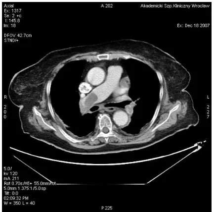
Fig. 2. Thrombo-embolic material located in the central portions of both pulmonary arteries and at the division of the main pulmonary artery – the acute phase of pulmonary embolism
Ryc. 2. Materiał zatorowo-zakrzepowy umiejsco-wiony centralnie w świetle obu tętnic płucnych oraz w podziale pnia płucnego – ostra faza zatorowości płucnej
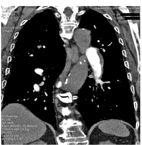
Fig. 3. Chronic phase of pulmonary embolism – the thrombo-embolic material with calcifications visible perimurally along the wall of the branch of the left pulmonary artery
of the MPA and PA were also present in most of the patients with pulmonary venous congestion.
In 44 of 102 patients (43.1%), non-embolic pa-thologies were demonstrated including evidence of pulmonary venous congestion (n = 21), pneumo-nia (n = 19) and malignancy (n = 4). In 26 patients (25.5%) massive uni- or bilateral hydrothorax was shown, which required an emergency puncture of the pleural cavities.
The authors did not notice any statistically sig-nificant inter-observer discrepancies. The evalua-tion of the lobar, segmental and proximal subseg-mental arterial branches showed no inter-observer differences. There was inter-observer discrepancy regarding the assessment of the peripheral subseg-mental branches of 1–2 pulmonary segments in 2 of 32 patients with CTPA evidence of PE. The in-ter-observer discrepancy neither proved statistical-ly significant nor influenced the radiological report and subsequent clinical management.
Discussion
Pulmonary embolism constitutes a common clinical problem particularly in older age group pa-tients. This is in agreement with the present results – the median age in the subset with recognized PE was 73.4. In authors’ material, PE was detected in nearly one third of the patients referred for CTPA with suspicion of either acute or chronic pulmo-nary embolism. However, in most of the patients who had undergone CTPA due to suspected PE, the result of the examination was negative. This is one of the reasons why conventional catheter dig-ital subtraction angiography (DSA) should not be the imaging modality of choice and should be re-served for selected clinically severe life-threating cases with strong clinical suspicion of PE to en-able emergent thrombolysis and/or fragmentation of the main clots with the intention to cause dis-tal migration of thrombo-embolic material [8–10].
Fig. 4. The acute phase of pulmonary embolism com-plicated by right lung segmental infarction
Ryc. 4. ostra faza zatorowości płucnej powikłana zawałem miąższu płuca prawego
Fig. 5. The acute phase of pulmonary embolism – mas-sive embolic material in the right pulmonary artery
Ryc. 5. ostra faza zatorowości płucnej – masywny materiał zatorowy w prawej tętnicy płucnej
Fig. 6. Ground-glass appearance in the course of chronic pulmonary embolism
Baile et al. regarded CTPA as comparable to tradi-tional pulmonary angiography more than 10 years ago [11]. In 2007, Wittram et al. presented discrep-ancies between DSA and CTPA [12].
Pulmonary CTA does not take a long time, therefore it can be performed even in patients with a severe clinical state. The initial evaluation of ob-tained scans is also quick, which is worth empha-sizing because prompt diagnosis leads to prompt proper treatment and improves clinical outcomes. Additionally, CTPA is a minimally invasive pro-cedure – the only potential risk of adverse events
is associated with intravenous contrast adminis-tration [13, 14]. However, there were no contrast-related complications in the present group. Gen-erally, the currently widespread use of iso-osmolal and low viscosity non-ionic contrast media re-sults in low risk of any CTA. Nevertheless, the au-thors always checked creatinine/GFR levels pri-or to CTPA [14]. Because of its high sensitivity in the detection of vascular anatomy and pathology, at present a sixty-four detector CTA is widely ap-plied in diagnostic imaging of other vascular ar-eas [15, 16].
Fig. 8. Dual energy CT – the iodine/ water ratio is significantly decreased, which reflects perfusion defects in the 10th segments of both lungs
Ryc. 8. Dwuenergetyczna tomografia komputerowa. Stosunek stężenia jod/ woda jest wyraźnie obniżony w seg-mentach 10. obu płuc, co odzwiercie-dla zaburzenie perfuzji
Fig. 7. Dual energy CT – Color Z map of pulmonary embolism. Red color – normal perfusion, blue color – focal lung perfusion defect predominantly in the X segments of both lungs
Exposure to radiation constitutes a disadvan-tage of any CT examination. Therefore there are some strategies that are meant to reduce the ra-diation-associated risks. In the case of CTPA, one of the techniques is associated with the reduction of the radiation dose to superficial radiosensitive organs (female breasts and thyroid gland) with-out a negative impact on the quality of obtained scans [17]. Another technique is an application of bismuth-layered radioprotective shields placed on the superficial organs, which reduces breast radia-tion exposure by 57% and thyroid gland dose by 60% [18].
Based on authors’ experience, evaluation of peripheral arterial branches should be carried out using thin slices to avoid missing small peripheral-ly located thrombo-embolic material [19]. Simul-taneous assessment of coronal and axial images makes it possible to avoid mistaking small pulmo-nary veins for distal arterial branches and to pre-cisely assess the placement and extent of clots.
If a patient’s condition is severe, a “first look” rough analysis should be performed directly fol-lowing the scanning, at the radiographer’s work-station, to make the further clinical decisions as prompt as possible. Afterwards, the images ob-tained should be analyzed in detail using first of all thin slices, followed by maximum intensity projec-tions (MIP) and possibly VR (volume rendering) reconstructions. It is worth stressing, the CTPA enables a detailed analysis of pulmonary paren-chyma, which makes it possible to detect peripher-al pulmonary infarction as a typicperipher-ally cone-shaped peripheral zone of consolidation in the areas of occluded vessels. Pulmonary CTA also enables the detection of other chest pathologies such as co-existing alveolar/interstitial edema, atelectatic/in-flammatory changes and enlargement of the right heart chambers as well as the main pulmonary ar-tery (MPA) as a sign of right heart overload. Ad-ditionally, CTA allows assessment of any hilar or mediastinal pathology e.g. enlarged lymph nodes, malignant tumor.
In authors’ opinion, evaluation of peripher-al pulmonary arteriperipher-al branches may be burdened with observer-dependent subjectivity [19]. It is mainly a consequence of the small lumen of pe-ripheral pulmonary arterial branches – according to present observations, small peripheral branch-es should only be evaluated using thin submilli-meter slices. Both thin and thick MIP reconstruc-tions should be reserved for evaluation of large and medium-sized pulmonary arterial branches. Visualization of peripheral branches using MIP and VR reconstruction may demonstrate proper-ly enhanced vessels whereas the same branches in submillimeter reconstructions show filling-defects
corresponding to the presence of thrombo-embol-ic material. Average volume artifact is the cause of misleading images when using MIP or VR re-construction. In spite of the limitations mentioned above, the authors regard CTPA as a highly sensi-tive and specific imaging diagnostic tool and thus a valuable imaging modality in the morphological presentation of the filling defects of pulmonary ar-terial branches.
The ventilation/perfusion (V/Q) scan is a po-tential alternative to CTPA [20, 21]. However, ac-cess to this diagnostic method is rather limited. Moreover, a relatively high rate of the V/Q scans is not conclusive, which decreases its cost-effective-ness. In contrast to this, CTPA is an easily acces-sible imaging diagnostic modality, allows rapid di-agnosis and following emergent endovascular or open-surgery intervention when necessary. That is why it is reasonable to include pulmonary CTPA in the PE diagnostic protocols as the first-line im-aging modality in this entity.
Additionally, when required, the area of exam-ination of CTPA can be extended to enable evalu-ation of venous iliac axes which are often difficult to be visualized in Doppler ultrasound (Doppler-US) [22]. From authors’ experience, the extend-ed range of scan can be usextend-ed when the implan-tation of a temporary or permanent IVC filter is considered.
When necessary, lower limb venous Doppler-US can be substituted by CT venography (CTV), which can be performed immediately after CTPA using one contrast medium administration (re-duced nephrotoxicity). The method makes it pos-sible to simultaneously visualize and assess both pulmonary circulation and the iliac and lower limb deep venous system. This seems to be the future of CTA in patients with suspicion of both PE and DVT [23, 24].
Advanced technology by means of dual-energy CT-scanners makes possible to perform CT-perfu-sion of the lungs, which is an additional advantage allowing for not only visualization but also quan-titative assessment of contrast-deficit (hypoper-fused) areas and thus detection of the regions of microembolism – which can be helpful in predict-ing potential respiratory complications [25, 26].
media) [28]. on the other hand, it is more difficult to obtain simultaneously technically adequate MR-PA and MRV compared to CTMR-PA/CTV [28].
Single photon emission computed tomogra-phy (SPECT) can constitute another alternative or supplementation to CTPA. According to Pilecki et al., SPECT in some cases may demonstrate a lack of perfusion or areas of hypoperfusion whereas the CTPA result was normal [29]. Therefore the au-thors think that it is crucial to implement a CT perfusion examination in the diagnostic protocol of PE [25, 26].
In conclusion, modern multidetector CT-scan-ners in association with the good quality of the ex-am and an experienced radiological teex-am result in a highly conclusive CTPA report which is of the
highest importance for a clinician and/or inter-ventional specialist. Pulmonary CTA seems to be the method of choice in the routine imaging di-agnostics of PE. Recent advances in modern CT-scanners enable quantitative analysis of lung pa-renchyma perfusion, which is an extremely useful tool in the detection and assessment of the extent of pulmonary microembolism. Additionally, the high speed of scanning makes it possible to scan the whole body following one contrast medium administration, which offers the possibility of si-multaneous performance of 2 protocols, i.e. CTPA followed by CTV. This method, although associat-ed with a higher dose of radiation, allows for both a detailed assessment of the deep lower leg venous system and pulmonary arterial tree.
References
[1] Torbicki A, Perrier A, Konstantinides S, Agnelli G, Galič N, Pruszczyk P, Bengel F, Brady AJ, Ferreira D, Janssens U, Klepetko W, Mayer E, Remy-Jardin M, Bassand JP; ESC Committee for Practice Guidelines (CPG):
Guidelines on the diagnosis and management of acute pulmonary embolism: the Task Force for the Diagnosis and Management of Acute Pulmonary Embolism of the European Society of Cardiology (ESC). Eur Heart J 2008, 29, 2276–2315.
[2] Stein PD, Fowler SE, Goodman LR, Gottschalk A, Hales CA, Hull RD, Leeper KV Jr, Popovich J Jr, Quinn DA, Sos TA, Sostman HD, Tapson VF, Wakefield TW, Weg JG, Woodard PK; PIOPED II Investigators: Multidetector computed tomography for acute pulmonary embolism. N Engl J Med 2006, 354, 2317–2327.
[3] Nikolaou K, Thieme S, Sommer W, Johnson T, Reiser MF: Diagnosing pulmonary embolism: new computed tomography applications. J Thorac Imaging 2010, 25, 151–160.
[4] Kelly AM, Patel S, Carlos RC, Cronin P, Kazerooni EA: Multidetector row CT pulmonary angiography and indi-rect venography for the diagnosis of venous thromboembolic disease in intensive care unit patients. Acad Radiol 2006, 13, 486–495.
[5] Wells PS, Anderson DR, Rodger M, Ginsberg JS, Kearon C, Gent M, Turpie AG, Bormanis J, Weitz J, Chamberlain M, Bowie D, Barnes D, Hirsh J: Derivation of a simple clinical model to categorize patients prob-ability of pulmonary embolism: increasing the models utility with the SimpliRED d-dimer. Thromb Haemost 2000, 83, 416–420.
[6] Le Gal G, Righini M, Roy PM, Sanchez O, Aujesky D, Bounameaux H, Perrier A: Prediction of pulmonary embolism in the emergency department: the revised Geneva score. Ann Intern Med 2006, 144, 165–171.
[7] Miniati M, Pistolesi M: Assessing the clinical probability of pulmonary embolism. Q J Nucl Med 2001, 45, 287–293.
[8] Remy-Jardin M, Pistolesi M, Goodman LR, Gefter WB, Gottschalk A, Mayo JR, Sostman HD: Management of suspected acute pulmonary embolism in the era of CT angiography: a statement from the Fleischner Society. Radiology 2007, 245, 315–329.
[9] Stein PD, Woodard PK, Weg JG, Wakefield TW, Tapson VF, Sostman HD, Sos TA, Quinn DA, Leeper KV Jr, Hull RD, Hales CA, Gottschalk A, Goodman LR, Fowler SE, Buckley JD; PIOPED II Investigators: Diagnostic pathways in acute pulmonary embolism: recommendations of the PIoPED II investigators. Radiology 2007, 242, 15–21.
[10] Stein PD, Sostman HD, Bounameaux H, Buller HR, Chenevert TL, Dalen JE, Goodman LR, Gottschalk A, Hull RD, Leeper KV Jr, Pistolesi M, Raskob GE, Wells PS, Woodard PK: Challenges in the diagnosis of acute pulmonary embolism. Am J Med 2008, 121, 565–571.
[11] Baile EM, King GG, Muller NL, D’Yachkova Y, Coche EE, Paré PD, Mayo JR: Spiral computed tomography is comparable to angiography for the diagnosis of pulmonary embolism. Am J Respir Crit Care Med 2000, 161, 1010–1015.
[12] Wittram C, Waltman AC, Shepard JA, Halpern E, Goodman LR: Discordance between CT and angiography in the PIoPED II study. Radiology 2007, 244, 883–889.
[13] Bierry G, Kellner F, Barnig C: Management of patients with history of adverse effects to contrast media when pulmonary artery CT angiography is required. Radiology 2007, 245, 919–921.
[14] Waybill MM, Waybill PN: Contrast media-induced nephrotoxicity: identification of patients at risk and algo-rithms for prevention. J Vasc Interv Radiol 2001, 12, 3–9.
[16] Waliszewska M, Jakubiak A, Guziński M, Sąsiadek M: Application of the 64-slice computed tomography as a diagnostic method in acute posttraumatic ischaemia of the upper limbs – 3 case reports. Pol J Radiol 2010, 75, 94–97.
[17] Hurwitz LM, Yoshizumi TT, Goodman PC Nelson RC, Toncheva G, Nguyen GB, Lowry C, Anderson-Evans C:
Radiation dose savings for adult pulmonary embolus 64-MDCT using bismuth breast shields, lower peak kilovolt-age, and automatic tube current modulation. AJR 2009, 192, 244–253.
[18] Parker MS, Hui FK, Camacho MA, Chung JK, Broga DW, Sethi NN: Female breast radiation exposure during CT pulmonary angiography. AJR 2005, 185, 1228–1233.
[19] Stein PD, Henry JW, Gottschalk A: Reassessment of pulmonary angiography for the diagnosis of pulmonary embolism: relation of interpreter agreement to the order of the involved pulmonary arterial branch. Radiology 1999, 210, 689–691.
[20] The PIOPED Investigators: Value of the ventilation/perfusion scan in acute pulmonary embolism. Results of the prospective investigation of pulmonary embolism diagnosis (PIoPED). JAMA 1990, 263, 2753–2759.
[21] Sostman HD, Miniati M, Gottschalk A, Matta F, Stein PD, Pistolesi M: Sensitivity and specificity of perfusion scintigraphy combined with chest radiography for acute pulmonary embolism in PIoPED II. J Nucl Med 2008, 49, 1741–1748.
[22] Goodman LR, Stein PD, Matta F, Sostman HD, Wakefield TW, Woodard PK, Hull R, Yankelevitz DF, Beemath A: CT venography and compression sonography are diagnostically equivalent: data from PIoPED II. AJR 2007, 189, 1071–1076.
[23] Kalva SP, Jagannathan JP, Hahn PF, Wicky ST: Venous thromboembolism: indirect CT venography during CT pulmonary angiography – should the pelvis be imaged? Radiology 2008, 246, 605–611.
[24] Goodman LR, Stein PD, Beemath A, Sostman HD, Wakefield TW, Woodard PK, Yankelevitz DF: CT venog-raphy for deep venous thrombosis: continuous images versus reformatted discontinuous images using PIoPED II data. AJR 2007, 189, 409–412.
[25] Hoey ET, Gopalan D, Ganesh V, Agrawal SK, Qureshi N, Tasker AD, Clements L, Screaton NJ: Dual-energy CT pulmonary angiography: a novel technique for assessing acute and chronic pulmonary thromboembolism. Clin Radiol 2009, 64, 414–419.
[26] Kraśnicki T, Podgórski P, Guziński M, Czarnecka A, Tupikowski K, Garcarek J, Sąsiadek M: Novel clinical applications of Dual Energy Computed Tomography. Adv Clin Exp Med 2012, 21, 6, 831–841.
[27] Bauman G, Eichinger M: Ventilation and perfusion magnetic resonance imaging of the lung. Pol J Radiol 2012, 77, 37–46.
[28] Stein PD, Chenevert TL, Fowler SE, Goodman LR, Gottschalk A, Hales CA, Hull RD, Jablonski KA, Leeper KV Jr, Naidich DP, Sak DJ, Sostman HD, Tapson VF, Weg JG, Woodard PK; PIOPED III Investigators: Gadolinium-enhanced magnetic resonance angiography for pulmonary embolism: a multicenter prospective study (PIoPED III). Ann Intern Med 2010, 152, 434–443.
[29] Pilecki S, Gierach M, Lasek W, Drobik P, Junik R: Single photon emission computed tomography in pulmonary embolism – estimation of selected, scintigraphic regions of interests. Pol J Radiol 2010, 75, 43–47.
Address for correspondence:
Jacek Kurcz
Department of General and Interventional Radiology and Neuroradiology Chair of Radiology
Wroclaw Medical University Borowska 213
50-556 Wrocław Poland
Tel.: +48 71 733 16 60 E-mail: [email protected]
Conflict of interest: None declared