Younes Shafigh
1, b, c, Akram beheshti
2, A, f, Malihe charkhchian
3, c–e,
fatemeh Samiee Rad
4, f, gSuccessful Treatment of Pilonidal Disease
by Intense Pulsed Light Device
1 Department of general Surgery, Qazvin University of Medical Sciences, Iran 2 Department of Dermatology, Qazvin University of Medical Sciences, Iran 3 Department of Internal Medicine, Qazvin University of Medical Sciences, Iran 4 Department of Pathology, Qazvin University of Medical Sciences, Iran
A – research concept and design; B – collection and/or assembly of data; C – data analysis and interpretation;
D – writing the article; E – critical revision of the article; F – final approval of article; G – other
Abstract
Background. Pilonidal disease is foreign body reactions accompanied by chronic inflammation that most com-monly arises in the hair follicles of the natal cleft or other hair-bearing areas. Today, surgical intervention remains the treatment of choice. but surgical treatment is an invasive method with a high failure rate and recurrence.
Objectives. The authors’ objective was to assess the efficacy of intense pulsed light (IPL) device on pilonidal disease.
Material and Methods. This case series study was carried out between 2008 and 2012 on patients with pilonidal sinus in Qazvin university of Iran.All patients received 6 session treatments with IPL hair removal with 4–6 weeks interval until most of the hair was removed. This was repeated 2.5 ± 0.3 years after treatment. In cases with acute phase pilonidal sinus histopathological examination was done.
Results. IPL hair removal procedure was performed on 30 patients with their ages ranging from 16 to 41 years, with a mean (SD) of 23.1 (6.2) years. In this study 13 patients were presented with acute and 6 patients were pre-sented with chronic phase. 11 patients had a positive history of one surgical treatment and prepre-sented recurrences. The overall recurrence rate after IPL treatment in this study was seen in 4 (13.3%) patients. The histopathological examination of our study showed that the hair fragments create a foreign body type granulomatous inflammatory reaction. This process could be triggering factor of the disease.
Conclusions. IPL hair removal in affected area could be an alternative treatment to surgery or a choice treatment post surgery to decrease recurrence rate (Adv Clin Exp Med 2014, 23, 2, 277–282).
Key words: intense pulsed light, pilonidal disease.
Adv clin exp Med 2014, 23, 2, 277–282 ISSN 1899–5276
ORIgINAL PAPeRS
© copyright by Wroclaw Medical University
Pilonidal disease is a foreign body reaction ac-companied by chronic inflammation that most com-monly arises in the hair follicles of the natal cleft or other hair bearing areas [1]. Usually in the last part of the second decade of life, after the onset of pu-berty, sex hormones affect the pilosebaceous glands, with the hair follicles becoming distended with kera-tin, and it is then that pilonidal sinus predominantly seen and is rare after the age of 40 years or before pu-berty [2]. Although the origin of the problem is un-known but the reports claim that genetic, hormonal stimulation of special sweet gland, obesity, personal hygiene and hirsutism are risk factors [3, 4]. The eti-ologic agent remains in question, as does the optimal
treatment and current accepted treatment options vary from traditional conservative treatments to ag-gressive surgery. The ideal therapy should be directed to treat the underlying etiological factor and would be a quick cure that allowed patients to return rapidly to normal activity, with minimal morbidity and a low risk of complications. Unfortunately, surgical treat-ment is an invasive method with a high failure rate and recurrence [5].
the treatment of pilonidal sinus has been limited. Therefore, the aim of this study is to investigate the efficacy of IPL device on pilonidal disease.
Methods
The research was performed on 30 patients with pilonidal sinus at the laser therapy clinic of Qazvin University of Medical Sciences, Iran, from May 2008 to May 2012.
The inclusion criteria were defined as patients with early acute phase, chronic course and recur-rent symptom after at least one surgical treatment. early acute phase involved patients with the onset of symptoms, usually pain and discharge. Occasionally a painless lump or swelling may be discovered by the patient while washing, or the characteristic midline pits may be found during a routine physical exami-nation. Patients were classified as chronic if present-ing chronic pain and discharge, often with a histo-ry of up to 2 years [1]. On examination, a single, or occasionally, multiple sinuses may be seen. Tufts of hair or other debris, such as clothing fibres, are often visibly found in the sinus. Localised oedema, swelling and inflammation may be present masking the underlying sinus. Recurrent pilonidal sinus was defined as reinfection in neighbouring hair follicles or chronic infection from entry of hair or debris in-to a posin-toperative wound. Patients who use phoin-to sensitizer drugs like Tetracycline and Doxycycline,
are pregnant, or have acute pilonidal abscess (loc-alised fluctuant swelling and painful) were exclud-ed from the study. All patients signexclud-ed an informexclud-ed consent form. The study protocol was approved by the ethics committee of the university before its ini-tiation and the protocols used conformed to the eth-ical guidelines of the 1975 Helsinki Declaration.
The affected area in 25 patients was the natal cleft and five female patients were sternal or intramamery area. All patients in this study had fitzpatrick skin types as II–IV and were treated with IPL device (Jei-sys, South Korea) at a wavelength with 590 to 1100 nm filters and the following parameters 30 to 34 J/cm2,
15 mms, and a 30 to 50 mms delay. The hair removal was done on affected area and extending 5 cm lateral-ly (fig. 1, 2). All patients received 6 session treatments with IPL epilation with 4–6 weeks interval until the re-moval of most (more than 80%) of the hair. In cases with acute phase pilonidal sinus histopathological ex-amination was performed and antibiotics therapy was carried out for 1 week and in case of remission symp-toms, the IPL epilation was done. The affected areas of all patients were evaluated after each treatment session of sign or symptom of recurrence and then once again this was done 2.5 ± 0.3 years after treatments.
Results
IPL hair removal procedure was performed on 30 patients with ages ranging from 16 to 41 years,
with a mean (SD) of 23.1 (6.2) years. 13 patients (43.3%) in this study presented with an early acute phase and 6 patients (20%) presented with chron-ic course. Also 11 patients (36.6%) of all patients (2 male and 9 female) had at least a history of one surgical treatment and presented with recurrences. 10 patients (33.3%) had a positive family history of pilonidal disease.
The overall recurrence rate was seen in 4 (13.3%) patients. Of these 4 patients 1 patient had a relapse of folliculitis after one treatment ses-sion. This problem was resolved with antibiot-ics and the patient is presently well, with ongoing treatments. Also 1 patient had a positive history of surgery and 3 patients did not have any surgery treatments. These patients have received 2 to 5 ad-ditional treatments session. All patients had expe-rienced partial to complete remission of symptoms in the treated area and none of them have required further surgical treatment and no side effects or complications were reported to date (Table 1 sum-marized the results of our study).
Our histopathological examination of ex-cised lesions showed that the hair fragments cre-ate a foreign body type granulomatous inflamma-tory reaction with plentiful multi–nucleated giant cells that are surrounded by fibrous tissue with
lymphoplasmacytic infiltration. Of course in some cases, in sinus wall aspect, epidermal appendages including hair follicles were absent. In these condi-tions hair shafts present free and deep granulation or scar tissue without epithelial lining. Therefore, when our histopathological results are considered, the role of acquired triggering factor was very im-portant (fig. 3, 4).
Discussion
Pilonidal sinus was described for the first time by Mayo in 1833 [9]. The origin of pilonidal sinus has been a subject of interest for many years. In the 1950s, it was thought to be of congenital origin, involving the remnant of the medullary canal and the infolding of the surface epithelium or a faulty coalescence of the cutaneous covering in the early embryonic stage [2, 10] but now most authors be-lieve that the condition results from a reaction to
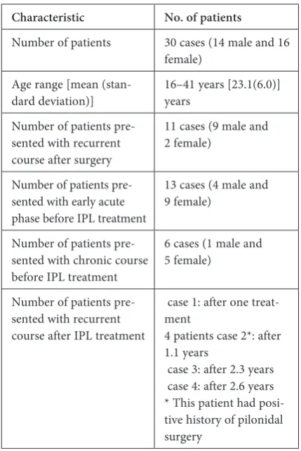
Table 1. baseline characteristics and outcome in patients with pilonidal sinus treated with the IPL epilation
Characteristic No. of patients
Number of patients 30 cases (14 male and 16 female)
Age range [mean
(stan-dard deviation)] 16–41 years [23.1(6.0)] years Number of patients
pre-sented with recurrent course after surgery
11 cases (9 male and 2 female)
Number of patients pre-sented with early acute phase before IPL treatment
13 cases (4 male and 9 female)
Number of patients pre-sented with chronic course before IPL treatment
6 cases (1 male and 5 female)
Number of patients pre-sented with recurrent course after IPL treatment
case 1: after one treat-ment
4 patients case 2*: after 1.1 years
case 3: after 2.3 years case 4: after 2.6 years * This patient had posi-tive history of pilonidal surgery
Fig. 3. Acute phase, Infiltration of inflammatory cells associated with the hair shaft Hematoxylin and eosin stain, X 100
hairs embedded in the skin and that this is the trig-gering factor of the disease [2, 3, 10]. In our study we showed that the hair fragments create a foreign body type granulomatous inflammatory reaction. This process, attention to another study, can be performed, triggering the factors of the disease.
The clinical presentation of this disease var-ies from a fever or small dimple to a large painful abscess. Of course, the most common manifesta-tion of acute pilonidal disease is a painful fluctuant mass in the sacrococcygeal region [11]. In this con-dition another differential diagnosis was consider-ably varied from an anal fistula, boils, carbuncles or infected sebaceous cysts to an ischiorectal ab-scess. Also, the sacrum osteomyelitis and tubercu-losis, as an important condition, may be mistaken and must be kept in mind even if ruled out by ra-diologic studies.
Unfortunately, even after the abscess was re-solved, either by itself or with antibiotics, many patients develop recurrent infections and an in-flammation of the sinus tracts. The recurrent dis-ease causes episodes of fistula formation, secre-tion, pain, drainage, terminal abscess [12, 13] and extremely rare complications with malignant transformations [14]. The main pathophysiolo-gy of these changes similar to Marjolin’s ulcer, are chronic inflammation and resulting ominous pro-cesses of tissue damage and repair [15]. Therefore, some of the authors believe that for an accurate di-agnosis and treatment in cases with recurrent pilo-nidal sinus, attention to bacteriological and patho-logical examination can help to exclude the special organism such as Actinomycosis or tumoral le-sions, epithelial inclusion dermoid cysts, chordo-mas and teratochordo-mas [16, 17].
Today, surgery remains the treatment of choice. Different surgical techniques vary from cision with primary closure or open packing to ex-cision with skin grafting and flap reconstruction [18–20]. Although the surgical treatment is associ-ated with unsatisfactory results, including the risk of 1–2 weeks of significant pain [21], 2–10 days of hospitalization [12], 2–8 weeks of healing [12] and a 2 to 3 week of recovery period [22, 23], the recur-rence rate is 30–40% after emergency surgery and 5– 20% after chronic pilonidal sinus [24].
IPL systems deliver a band of wavelengths that are directed at the skin and may be selectively ab-sorbed by melanin or water. Absorption of light by melanin may destroy the melanin directly and this method could be of use as an alternative treat-ment. Of course, IPL is to be viewed in contrast to monochromatic light emitted from lasers that are often considered as a cheaper alternative to lasers, although there are few studies that compare their treatment efficacy [25]. However, IPL systems are
less specific than lasers in their delivery of light en-ergy to the required melanin. but this system may offer more flexibility than some lasers in the selec-tion of pulse duraselec-tions or in the ability to deliv-er trains of pulses that may limit thdeliv-ermal damage to the surrounding cutaneous tissue. The fDA ap-proved this method for permanent hair reduction. In fact the IPL can create an optional thermolysis and is very effective in the hair of people with skin types I–IV fitzpatrick.
The use of the IPL hair removal in the treat-ment of pilonidal disease has not been well stud-ied. In a case report in 2006 [7] 5 patients with recurrent pilonidal sinus treated with either a di-ode laser or IPL. Results of this study showed 80% of patients do not have any recurrence period for 7 months to more than 36 months. One patient ex-perienced a recurrence after 36 months, which was longer than his previous remission. These results suggest laser and IPL are alternative techniques to surgical intervention, as they may provide longer period intervals between disease and decrease re-currences. Also, to evaluate the effect of alexan-drite laser and IPL hair removal in pilonidal dis-ease, 6 men were treated with laser epilation in 2005 [26]. In this study most patients had a his-tory of at least one surgical treatment in the area (natal cleft), and all patients had experienced re-current symptoms for many years. An alexandrite laser was mostly used, although, occasionally, an IPL device was used. In conclusion, in this study, all patients had experienced complete remission and none of them had demonstrated complica-tions or required surgical intervention.
In another novel study that was published in 2011 [27] to evaluate the efficacy of IPL hair re-moval after surgical intervention, 34 patients with recurrent pilonidal sinus were treated with IPL de-vice for hair removal. In this study patients required from 3 to 8 IPL therapy sessions with 5 ± 1 weeks intervals. The mean follow up was 3.8 months. No major morbidity was reported. Improvement was achieved in all cases with progressive loss of hair in the intergluteal cleft. All patients were satisfied with no recurrence.
infection and none of them have required any fur-ther surgical intervention until this moment and no side effects were reported to date.
These results suggest that the IPL technique is an alternative treatment to traditional surgi-cal intervention, in early acute, chronic and re-current symptoms. These results suggest that de-creasing hair in the area of pilonidal sinus reduces the rate of recurrence. Using an IPL for remov-al of surrounding hair with or after surgicremov-al exci-sion of a pilonidal cyst represents a favorable alter-native to surgical intervention alone. because the etiology of pilonidal cysts appears to involve in-grown hairs and a nidus of bacterial overgrowth, effective treatment using laser and light sources may help prevent the initial pathology of the cysts. The likely mechanism of action is secondary to de-creased inflammation in the setting of diminished
hair density. Ultimately, removal of hair by IPL in areas prone to cyst formation may obviate the need for surgical treatment and may make it pos-sible to secondarily prevent pilonidal cysts from appearing.
In conclusion, the advantages of the IPL hair removal includes: safe, easy, quick, nearly painless, performed on the outpatient basis, done almost always without local anesthesia, simple to teach, cheaper than a laser and no long lasting morbid-ity. On the other hand, the surgical method in ad-dition to cost, the side effects of anesthesia, long-term recovery and higher recurrence of disease will be formation deformed scar at the site and IPL epi-lation in affected area could be an alternative treat-ment to surgery or a choice post surgery treattreat-ment to decrease the recurrence rate in patients with suf-ficient compliance for multiple sessions.
Acknowledgements. This work was supported by a grant from Qazvin University of Medical Sciences, Iran. The authors are grateful for its financial support.
References
Sondenaa K, Andersen E, Nesvik I, Soreide JA:
[1] Patient characteristics and symptoms in chronic pilonidal sinus disease. Int J colorectal Dis 1995, 10, 39–42.
Hull TL, Wu J:
[2] Pilonidal disease. Surg clin North Am 2002, 82, 1169–1185.
Benedetto AV, Lewis AT:
[3] Pilonidal sinus disease treated by depilation using an 800 nm diode laser and review of the literature. Dermatol Surg 2005, 31, 587–591.
Odidi J, Gault D:
[4] laser depilation of the Natal cleft and aid to healing the pilonidal disease. Annals J 2002, 84, 29–32.
Shakoor siddiqui S, Ahmed Khan M:
[5] pilonidal sinus. Pak J Med Sci 2008, 24, 845–848.
Schulze SM, Patel N, Hertzog D, Fares LG:
[6] Treatment of pilonidal disease with laser epilation. Am. Surg 2006, 72, 534–537.
Sadick NS, Yee-Levin J:
[7] Laser and light treatments for pilonidal cysts. cutis 2006, 78, 125–128.
Downs AMR, Palmer J:
[8] Laser hair removal for recurrent pilonidal sinus disease. J cosmet Laser Ther 2002, 4, 91.
Mayo OH:
[9] Observations on injuries and disease of rectum. burgess and Hill, London, 1833, 45–46 Quoted from da Silva JH. Pilonidal cyst: cause and treatment. Dis colon Rectum 2000, 43, 1146–1156.
Nivatvongs S:
[10] Pilonidal disease. In: gordonPH, NivatvongsS, editors. Principles and Practice of Surgery for the colon, Rectum, and Anus. New York: Informa Health care USA Inc 2007, 235–246.
Oram Y, Kahraman F, Karincaoğlu Y, Koyuncu E:
[11] evaluation of 60 Patients with Pilonidal Sinus Treated with Laser epilation after Surgery. American Society for Dermatologic Surgery 2010, 36, 88–91.
Allen-Mersh TG:
[12] Pilonidal sinus: finding the right track for treatment. br J Surg 1990, 77, 123–132.
Chintapatla S, Safarani N, Kumar S, Habooubi N:
[13] Sacrococcygeal pilonidal sinus: historical review, pathological insight and surgical options. Techn coloproctol 2003, 7, 3–8.
Kulaylat, MN, Gong M, Doerr RJ:
[14] Multimodality treatment of squamous cell carcinoma complicating pilonidal disease. Am Surg 1996, 62, 922–929.
Bolandparvaz SH, Mohammadi AA, Riazi H, Monabbati A, Geramizadeh B:
[15] Unusual Presentation of Squamous
cell carcinoma on long-standing Sacrococcygeal Pilonidal Sinus. Iran J Med Sci 2009, 34, 149–151.
Anscombe AR
[16] , Hofmeyr J: Perianal actinomycosis complicating pilonidal sinus. br J Surg 1954, 41, 666.
Chikkamuniyappa S, Scott RS, Furman J:
[17] Pilonidal sinus of the glans penis associated with actinomyces case reports and review of literature. Scientific World Journal 2004, 22, 908–912.
Theodoropoulos GE, Vlahos K, Lazaris AC, Tahteris E, Panoussopoulos D:
[18] Modified bascom’s asymmetric
midgluteal cleft closure technique for recurrent pilonidal disease: early experience in a military hospital. Dis colon Rectum 2003, 46, 1286–1291.
Garrido A, Ali R, Ramakrishnan V, Spyrou G, Stanley PR:
[19] Reconstruction of the natal cleft with a perforator-based flap. br J Plast Surg 2002, 55, 671–674.
Thomas D:
[20] comparison of three methods in surgical treatment of pilonidal disease. Aust N ZJ Surg 2002, 72, 561–564.
Zieger K:
[21] Komplikationer efter operation for cystis pilonidalis. Ugeskr Laeger 1999, 161, 6056–6058.
Testini M, Piccinni G, Miniello S, Di Venere B, Lissidini G, Nicolardi V:
[22] Treatment of chronic pilonidal sinus with
Ertan T, Koc M, Gocmen E, Aslar AK, Keskek M, Kilic M:
[23] Does technique alter quality of life after pilonidal sinus surgery? Am J Surg 2005, 190, 388–392.
Poulsen IM, Bisgaard T:
[24] Treatment of pilonidal cysts. Ugeskr Laeger 2008, 170, 2963–2968.
Ross EV:
[25] Laser versus intense pulsed light: competing technologies in dermatology. Lasers Surg Med 2006, 38, 261–272.
Landa N, Aller O, Landa-Gudin N, Torrontegui J, Azpiazu JL:
[26] Successful Treatment of Recurrent Pilonidal Sinus with Laser epilation. Dermatol Surg 2005, 31, 726–728.
Khalil AAM, El-alfy KA:
[27] combined Surgical correction and Intense Pulse Light (IPL) Depilation for Recurrent Pilonidal Sinus; Mansoura experience. egypt, J Plast Reconstr Surg,2011, 35, 183–185.
Address for correspondence:
Akram beheshti
clinical Research Development Unit
Qazvin University of Medical Sciences, Qazvin, Iran Tel.: +98 281 334 74 96
e-mail: [email protected] conflict of interest: None declared Received: 20.05.2013