E
LŻBIETAB
EREŚ−P
AWLIK1, A
NDRZEJG
ROBELNY1, M
ARCINF
RĄCZEK2,
M
ARIAZ
ALESSKA−K
RĘCICKA, T
OMASZK
RĘCICKI2Electrical Impedance Measurements
in Assessing Laryngeal Squamous Cell Carcinoma
Pomiary impedancji elektrycznej
w diagnostyce raka płaskonabłonkowego krtani
1 Institute of Telecommunications and Acoustics, Wrocław University of Technology, Poland
2 Department of Otolaryngology, Silesian Piasts University of Medicine in Wrocław, Poland
Adv Clin Exp Med 2006, 15, 4, 619–624 ISSN 1230−025X
ORIGINAL PAPERS
© Copyright by Silesian Piasts University of Medicine in Wrocław
Abstract
Background. Tissue differentiation may plausibly be based on measurements of its dielectric properties. It has been noted that cancerous cells differ from healthy ones in their local dielectric properties.
Objectives. In this study, impedance analysis, involving the measurement of voltage in response to the application of an alternating current over a wide frequency range, was used to study laryngeal carcinoma.
Material and Methods. The measurements were done on 16 tissues obtained from patients treated surgically for laryngeal squamous cell carcinoma. The acquisition system consisted of an AutoLAB impedance analyzer and a set of electrodes. The real and imaginary parts of the impedance were measured over a frequency range of 100 Hz to 1 MHz in cancerous and adjacent healthy epithelium. The modulus of impedance in the frequency function was then calculated.
Results.The values of the modulus of impedance |Z|were essentially higher in healthy mucosa than in cancerous epithelium (p< 0.05). The ratio of |Z|between them varied from 2.1 to 15.0. The impedance of healthy tissue rose significantly with decreasing frequency of the applied current.
Conclusion.The results of the study indicate that the impedance spectrum is quantitatively related to the structure of the tissue. The differences in the impedance values between the investigated areas enable differentiating healthy from cancerous mucosa of the human larynx with great selectivity (Adv Clin Exp Med 2006, 15, 4, 619–624).
Key words:impedance analysis, laryngeal carcinoma, diagnostics.
Streszczenie
Wprowadzenie. Różnicowanie ludzkich tkanek może być przeprowadzone za pomocą pomiaru ich własności dielektrycznych. Dotyczy to w szczególności nowotworów złośliwych, których wskaźniki bioelektryczne różnią się zasadniczo od stwierdzanych w tkankach niezmienionych nowotworowo.
Cel pracy.Ocena wartości impedancji raków płaskonabłonkowych krtani na podstawie pomiarów napięcia uzys− kanego w odpowiedzi na przyłożony do tkanek prąd zmienny w szerokim zakresie częstotliwości.
Materiał i metody. Pomiary przeprowadzono in vitrona 16 krtaniach uzyskanych od pacjentów po zabiegu laryn− gektomii całkowitej wykonanym w Klinice Otolaryngologii AM we Wrocławiu z powodu potwierdzonego histo− patologicznie raka płaskonabłonkowego. System pomiarowy składał się z urządzenia AutoLab oraz zestawu spe− cjalnie opracowanych elektrod igłowych. We wszystkich przypadkach dokonano oceny składowej rzeczywistej oraz urojonej impedancji w szerokim zakresie częstotliwości (100–1000 Hz) zarówno w tkance guza, jak i przyle− głym niezmienionym nabłonku krtani. Na podstawie uzyskanych danych obliczono moduł impedancji w funkcji częstotliwości.
Wyniki.Wartości modułu impedancji |Z|były istotnie stystycznie większe w zdrowym nabłonku w porównaniu z rakami krtani (p< 0.05). Stosunek modułu impedancji |Z|guzów krtani do niezmienionego nabłonka wynosił 2,1–15. Wartości impedancji w tkankach zdrowych wzrastały istotnie wraz ze spadkiem częstotliwości przyłożo− nego prądu elektrycznego.
Wnioski.Wyniki badań potwierdzają, że wartość impedancji bioelektrycznej jest ściśle zależna od struktury bada− nej tkanki. Uzyskane wyniki sugerują również możliwość wykorzystania analizowanych wskaźników w różnico− waniu raka płaskonabłonkowego krtani od nowotworowo niezmienionego nabłonka dróg oddechowych (Adv Clin Exp Med 2006, 15, 4, 619–624).
Studies on the electrical properties of tissues have been conducted since the end of the nine− teenth century [1]. One of the methods, called electrical impedance spectroscopy (EIS), mea− sures the electrical properties of tissue, i.e. con− ductivity (σ) and relative permittivity (ε), as func− tions of the frequency (ν) of the applied alternating current (ac). The work by Cole and Cole and later researchers led to techniques enabling the determi− nation of the impedance properties of healthy, damaged, and pathologically changed tissue in a wide range of frequencies [2, 3]. Although elec− trical impedance measurements of malignant tis− sues have been investigated for years, it has only been in recent years that achievements in computer science and data processing have enabled progress in this field.
Head and neck squamous cell cancer compris− es almost 5% of all malignant tumors diagnosed in Europe, making it the seventh most common malignancy. The majority of primary tumors in the head and neck region are located in the larynx. Although the diagnosis of head and neck cancer is established on the basis of the clinical and histo− logical pictures, each attempt to reduce the time of diagnosis is worth undertaking. One of the most challenging problems of head and neck oncology, especially in developing countries, is the high local advancement of the tumors at the moment of diagnosis. This is why special attention should be directed to screening efforts and the diagnosis of suspicious changes. Electrical impedance mea− surement is a technique which seems to be an interesting basis for a clinically significant, fast, and cost−effective methodology for the detection of cancerous tissue. This study aimed to assess the electrical impedance spectra in cancerous and macroscopically healthy laryngeal mucosa.
Material and Methods
The material comprised larynxes obtained from patients with squamous cell carcinoma treat− ed surgically at the Department of Otolaryng− ology, Silesian Piasts University of Medicine in Wrocław. All of the 16 patients included in the study underwent total laryngectomy. The diagnosis was made by histological examination pre− and postoperatively. Most of the patients (15) were males and one was female. Patient age ranged from 47 to 75 years old (mean: 56 years). The stage of the tumors assessed according to the TNM classifi− cation showed high local advancement, i.e. pT2 in 3 cases, pT3 in 11 cases, and pT4 in 2 cases.
The acquisition system consisted of an AutoLAB Analyser (Eco Chemie B.V., The
Netherlands) serially coupled with a personal computer. Impedance data were obtained under the control of Frequency Response Analyzer (FRA) software for Windows (Eco Chemie). In each case, the larynx was positioned into a cus− tom−built plastic chamber through which elec− trodes could be inserted to touch the surface of the mucosa. The intra−electrode distance was fixed in the plastic cover. Impedance measurements were made with needle electrodes 0.88 mm in diameter and 25 mm in length. The probes used in the study have the advantages of small size and the possibil− ity of being placed in the structure to be studied. The probe was calibrated in saline of known elec− trical conductivity. The method involves applica− tion of a sinusoidally varying current and the mea− surement of the ensuing voltage. A current was passed between an adjacent pair of electrodes. Measurements of impedance were carried out over a wide range of frequencies from 100 Hz to 1 MHz. At least two measurements were made on each tissue: the cancerous and the adjacent, macro− scopically normal epithelium. Electrical imped− ance was gauged from the center of the tumor. The regions of the tissues included in the impedance measurements were finally assessed histopatho− logically. The study had the approval of the local ethics committee.
Statistica 6.0 (StatSoft, Inc.) was used for sta− tistical calculations and graphical presentations. The obtained data were shown to have a normal distribution with a statistical significance of p= 0.05. The association between the values of impedance |Z| in healthy and cancerous tissue was tested by the Wilcoxon test. Pairs of measurements conduct− ed under similar conditions were compared. Differences with p< 0.05 were considered signif− icant.
Results
n
f [kHz]
Healthy epithelium (Tkanka zdrowa)
Cancerous tissue (Tkanka zmieniona nowotworowo)
Z’ [ Ω ]Z ” [ Ω ]| Z
h| [
Ω ] Z’ [ Ω ]Z ” [ Ω ]| Z
h| [
Ω ] Z’ [ Ω ]Z ” [ Ω ]| Z
c| [
Ω ] Z’ [ Ω ] Z” [ Ω ]| Z
c| [
Ω ] 1 100 2212.4 1492.4 2668.70 2038.4 1288.7 241 1.60 723.47 181.18 745.81 1 915.07 267.70 953.42 10 5795.2 4049.7 7069.96 5372.5 3555.3 6442.35 1207.6 396.02 1270.87 1553.4 444.03 1615.61 1 13923 9636.4 16932.52 12372 6756.5 14096.69 1739.8 486.46 1806.52 2120.4 627.21 221 1.21 2 100 1785.6 874.3 1988.15 1091.7 480.1 1 1 192.60 745.85 75.565 749.66 584.12 46.66 585.98 10 3614.6 1292.2 3838.63 2257.8 1 102.1 2512.42 856.67 82.16 860.60 654.53 62.96 657.55 1 5430.8 1587.9 5658.18 4176.8 1682.6 4502.97 972.16 206.47 993.84 753.54 220.84 785.23 3 100 2337 2236.1 3234.45 1589.6 1516.5 2196.95 1249.9 410.75 1315.66 1212.1 420.40 1282.93 10 8084.5 10187 13005.16 5519.1 6438.7 8480.40 191 1.2 373.77 1947.40 1926 401.29 1967.36 1 35177 25129 43230.64 22596 15770 27554.89 2302.5 454.13 2346.85 2340.5 438.62 2381.24 4 100 1462.6 471.67 1536.77 1 168.8 31 1.49 1209.59 697.16 78.186 701.53 542.69 67.38 546.85 10 2356.2 541.31 2417.58 1777.3 387.6 1819.07 816.94 1 14.84 824.97 652.15 83.49 657.47 1 3074.5 809.93 3179.39 2376.6 661.75 2467.01 1020.8 359.54 1082.26 783.05 236.80 818.07 5 100 2212.4 1492.4 2668.70 2038.4 1288.7 241 1.60 915.07 267.7 953.42 488.08 149.46 510.45 10 5795.2 4049.7 7069.96 5372.5 3555.3 6442.35 1553.4 444.03 1615.61 898.21 492.04 1024.15 1 13923 9636.4 16932.52 12372 6756.5 14096.69 2120.4 627.21 221 1.21 1804.6 886.67 2010.66 6 100 927.07 144.89 938.32 727.82 201.47 755.19 323.45 22.4 324.22 314.18 21.87 314.94 10 1 144.9 196.1 1 161.57 1243.5 433.84 1317.00 355.23 32.975 356.75 347.1 31.87 348.56 1 1460.3 561.16 1564.40 1816.7 658.34 1932.30 393.07 121.6 41 1.44 385.52 1 14.64 402.20 T able 1.
The real (Z’) and imaginary (Z”) parts of impedance and the modulus of impedance
|
Z
|
in six cases of cancerous and healthy laryngeal mucosa as a function of the frequency (f) of the
applied current Tabela 1.
W
artości pomiarów części rzeczywistej (Z’), urojonej (Z”) i
modułu impedancji
|
Z
|
w
paśmie częstotliwości (f) uzyskane w
tkance zdrowej i
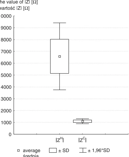
(average: 6581.59 Ω; SD: 8657.59 Ω), while |Zc| ranged from 314.94 Ω to 2381.24 Ω (average: 1099.79 Ω, SD643.43 Ω). Examples of the mag− nitudes of these parameters for the two different measurements for cancerous and healthy epitheli− um are shown in Table 1. Readings from the tis− sues showed that the values of |Z|differed signifi− cantly between the two groups. The values of |Zh| were statistically higher than those of |Zc| (p < 0.05) (Fig. 1). The ratio of |Zh|to |Zc|varied from 2.1 to 15.0. The impedance of healthy tissue clear− ly rose with decreasing frequency of the applied current. The same values did not increase signifi− cantly in cancerous epithelium (Fig. 2). This dif− ference enables one to differentiate healthy from cancerous areas of the human larynx with great selectivity. Despite the presence of some varia− tions in |Z| in both groups, the impedance in healthy epithelium was always higher.
Discussion
The body offers two types of resistance (R) to an electrical current: capacitative R (reactance), and resistive R (simply called resistance). Each human tissue contains elements that have either resistive and capacitative properties. The capaci− tance arises from cell membranes and the resis−
tance from extra− and intracellular fluid and the cell structure. Impedance is the term used to describe the combination of both. Measuring impedance in living tissues is difficult to achieve. Different inter− nal components determine the electrical properties of tissue at different frequency bands within an impedance spectrum [4]. Molecular structure is the main factor at frequencies over 1 GHz, whereas at low frequencies (< 100 Hz) it is charge accumula− tion at the membranes. At frequencies of a few kHz to 1 MHz, also called the βdispersion region, the electrical current passes mainly through the extra− cellular space, so the shape of the cells, their inter− cellular relationships, and their arrangement in the tissue are the crucial elements determining imped− ance. With increasing frequency, current can pene− trate the cell and flow through both the intracellu− lar and extracellular space. In such a case, the intra− cellular volume and the size of the nucleus can influence the current [5]. Consequently, electrical impedance spectral measurements open the way to assessing tissue structure. The relationship between capacitance, resistance, and thus impedance, reflects different electrical properties of tissues that are affected by disease and other factors, e.g. nutri− tion and dehydration. Electrical impedance mea− surement has recently been investigated for moni− toring tissue damage in vivo, e.g. the influence of ischemia on rat muscle [6], toxic injury of the liver [7], skin reaction after radiation [8], X−ray injury in rat muscle [9], and the detection of dental decay [10]. The altered electrical properties of cancerous tissue compared with healthy tissue are attributed to increased cellular water and salt content, altered
average średnia
± SD
H
0 1000 2000 3000 4000 5000 6000 7000 8000 9000 10000
± 1,96*SD
Z ZC
the value of |Z| [ ] wartość |Z| [ ]
Ω Ω
Fig. 1.Comparison of impedance in healthy (|Zh|) and
cancerous tissue (|Zc|) (p < 0.05)
Ryc. 1.Porównanie wartości modułów impedancji tkanki zdrowej (|Zh|) i zmienionej nowotworowo (|Zc|)
(p < 0,05)
normal mucous prawidłowa błona śluzowa normal mucous prawidłowa błona śluzowa cancerous mucous błona śluzowa zmieniona nowotworowo
|Z| [ ]Ω
f [Hz]
Fig. 2. Frequency (f) dependence of the modulus of the impedance |Z|in normal and cancerous laryngeal epithelium
membrane permeability, and altered packing den− sity and orientation of the cells [4]. In neoplastic tissues, the cell layering of normal squamous epithelium is destroyed, so current of low frequen− cies which does not penetrate the cell membrane does not have to track around the cell layers and the tissue impedance is expected to be lower. Due to enlargement of the cell nuclei in cancerous tis− sue, the conduction pathways through the intracel− lular space are smaller. Cancerous lesion is assumed to have higher conductivity than sur− rounding tissue. The dependence of lesion detectability on the conductivity ratio between lesion and the adjacent tissue is basic to EIS. Emtestam et al. [11] investigated the electrical impedance properties of nodular basal cell carci− noma (BCC). Among the set of the four indices they used (magnitude index, phase index, and the real and imaginary part index), only values of the modulus of magnitude and the imaginary part of the complex electrical impedance significantly decreased in BCC compare with normal skin, which is in accordance with presented results.
In presented study the reproducibility of the measurements in each case was high. In most of the tissues the shapes of the graph which represent measurements on the same tumor in different places were similar (Fig. 2). Disturbances in the form of the graphs could be caused by the elec− trode depth in the tissue, the presence of cartilage, and changes in mucous humidity [12, 13]. In− creased electrode depth in the investigated tissues led to a decrease in impedance.
Described results are in accordance with observations obtain by Jossinet et al., who worked with freshly excised breast tissue and observed large differences in dielectric parameters between neoplastic and normal areas [14]. The potential
application of this technique was also assessed in detecting neoplastic and precancerous changes in the cervix [5, 15] and bladder [16]. Current flow measurements can help in accurately diagnosing cervical neoplasia in patients with positive smear tests [7]. The clinical usefulness of these devices has not yet been fully reported.
Presented results indicate that the magnitude of impedance and its dependence on frequency are a function of the composition and structure of the tis− sue. The impedance method can be used to confirm the diagnosis of malignant tumors by endoscopy or directly before they are verified histologically. Such a procedure could facilitate taking a representative sample of mucous. Fortunately, some of the results of in vivomeasurements can be simply predicted. It was shown that to obtain corresponding values of the bioelectrical parameters at the same frequency for in vivoand ex vivomeasurements, a multiplying factor should be applied [13].
The future aim of authors’ research is to devel− op a diagnostic aid based on a multiple−electrode array enabling non−invasive real−time measure− ment of the local distribution of tissue electrical impedance at various frequencies from the surface of the skin. On the basis of the collected regional impedance data, further reconstruction into 2D could be done. Significant progress has been made in recent years in the technique known as electrical impedance imaging or tomography (EIT). The scope of the medical application of EIT has also greatly widened. EIT has been applied to the monitoring of lung ventilation [17], brain activity [18], gastric emptying [19], and diastolic function of heart [20]. Further investigation must be undertaken to specify the role of this technique in the diagnostics of head and neck tumors.
References
[1] Hermann L: Ueber eine Wirkung galvanischer Strome auf Muskein und Nerven [On the influence of galvanic currents on muscles and nerves]. Pflugers Arch gesamte Physiol 1871, 5, 223–275.
[2] Cole KS, Cole RH: Dispersion and absorption in dielectrics. Alternating current characteristics. J Chem Phys 1941, 9, 341–351.
[3] Ackmann JJ, Seitz MA:Methods of complex impedance measurements in biologic tissue. Crit Rev Biomed Eng 1984, 11, 281–311.
[4] Foster KR: Dielectrical properties of tissues and biological materials: a critical review. Crit Rev Biomed Eng 1989, 17, 25–104.
[5] Brown BH, Tidy JA: Relation between tissue structure and imposed electrical current flow in cervical neoplasia. Lancet 2000, 355, 892–895.
[6] Ristic B, Kun S, Peura RA: Muscle tissue ischaemia monitoring using impedance spectroscopy: quantitative results of animal studies. Proc 19thAnnu Int Conf IEEE 1997, 5, 2108–2111.
[7] Heroux P, Bourdages M: Monitoring living tissues by electrical impedance spectroscopy. Ann Biomed Eng 1994, 22, 328–337.
[8] Nuutinen J, Lahtinen T, Turunen M, Alanen E, Tenhunen M, Usenius T, Kolle R:A dielectric method for measuring early and late reactions in irradiated human skin. Radiother Oncol 1998, 47, 249–254.
[10] Longbottom C, Huysmans M−CDNJ, Pitts NB, Los P, Bruce PG: Detection of dental decay and its extent using a.c. impedance spectroscopy. Nature Medicine 1996, 2(2), 235–237.
[11] Emtestam L, Nicander I, Stenstrom M, Ollmar S: Electrical impedance of nodular basal cell carcinoma: a pilot study. Dermatology 1998, 197, 313–316.
[12] Molckovsky A, Wilson BC: Monitoring of cell and tissue responses to photodynamic therapy by electrical imped− ance spectroscopy. Phys Med Biol 2001, 46, 983–1002.
[13] Chauveau N, Dumont P, Aligne C, Rigaud B, Cros S, Morucci JP: In vivoand ex vivoimpedance spectrome− try of MCF−7 tumors on nude mice: measurement problems. Innov Tech Biol Med 1995, 16(6), 680–687.
[14] Jossinet J, Lobel A, Michoudet C, Schmitt M: Quantitative technique for bio−electrical spectroscopy. J Biomed Eng 1985, 7, 289–294.
[15] Comppleson M: An electronic approach to the detection of pre−cancer and cancer of the uterine cervix: a pre− liminary evaluation of Polarprobe Int J Gynaecol Cancer 1994, 4, 79–83.
[16] Walker DC, Smallwood RH, Keshtar A, Wilkinson BA, Hamdy FC, Lee JA:Modelling the electrical proper− ties of bladder tissue – quantifying impedance changes due to inflammation and oedema. Physiol Meas 2005, 26, 251–268.
[17] Mayer M, Brunner P, Merwa R, Scharfetter H: Monitoring of lung edema using focused impedance spec− troscopy: a feasibility study. Physiol Meas 2005, 26, 185–192.
[18] Tidswell A, Gibson A, Bayford RH, Holder DS: Electrical impedance tomography of human brain activity with a two−dimensional ring of scalp electrodes. Physiol Meas 2001, 22(1), 167–175.
[19] Vaisman N, Weintrop N, Blumental A, Yosefberg Z, Vardi P: Gastric emptying in patients with type 1 diabetes mellitus. Ann NY Acad Sci 1999, 873, 506–511.
[20] Noordegraaf AV:Noninvasive assessment of right ventricular diastolic function by electrical impedance tomog− raphy. Chest 1997, 111(5), 1222–1228.
Address for correspondence:
Tomasz Kręcicki
Silesian Piasts University of Medicine in Wrocław ul. Chałubińskiego 2
51−368 Wrocław Poland
tel.: +48 71 7842512 fax: +48 71 3270950
e−mail: [email protected]
Conflict of interest: None declared
Received: 8.09.2005 Revised: 13.05.2006 Accepted: 13.05.2006
Praca wpłynęła do Redakcji: 8.09.2005 r. Po recenzji: 13.05.2006 r.