O R I G I N A L A R T I C L E
Open Access
Ability of artificial intelligence to diagnose
coronary artery stenosis using hybrid
images of coronary computed tomography
angiography and myocardial perfusion
SPECT
Hiroto Yoneyama
1*, Kenichi Nakajima
2, Junichi Taki
2, Hiroshi Wakabayashi
2, Shinro Matsuo
2, Takahiro Konishi
1,
Koichi Okuda
3, Takayuki Shibutani
4, Masahisa Onoguchi
4and Seigo Kinuya
2* Correspondence:[email protected]. ne.jp
1Department of Radiological Technology, Kanazawa University Hospital, 13-1 Takara-machi, Kanazawa 920-8641, Japan Full list of author information is available at the end of the article
Abstract
Background:Detecting culprit coronary arteries in patients with ischemia using only myocardial perfusion single-photon emission computed tomography (SPECT) can be challenging. This study aimed to improve the detection of culprit regions using an artificial neural network (ANN) to analyze hybrid images of coronary computed tomography angiography (CCTA) and myocardial perfusion SPECT.
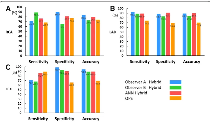
Methods:This study enrolled 59 patients with stable coronary artery disease (CAD) who had been assessed by coronary angiography within 60 days of myocardial perfusion SPECT. Two nuclear medicine physicians interpreted the myocardial perfusion SPECT and hybrid images with four grades of confidence, then drew regions on polar maps to identify culprit coronary arteries. The gold standard was determined by the consensus of two other nuclear cardiology specialist based on coronary angiography findings and clinical information. The ability to detect culprit coronary arteries was compared among experienced nuclear cardiologists and the ANN. Receiver operating characteristics (ROC) curves were analyzed and areas under the ROC curves (AUC) were determined. Results:Using hybrid images, observer A detected CAD in the right (RCA), left anterior descending (LAD), and left circumflex (LCX) coronary arteries with 83.6%, 89.3%, and 94.4% accuracy, respectively and observer B did so with 72.9%, 84.2%, and 89.3%, respectively. The ANN was 79.1%, 89.8%, and 89.3% accurate for each coronary artery. Diagnostic accuracy was comparable between the ANN and experienced nuclear medicine physicians. The AUC was significantly improved using hybrid images in the RCA region (observer A: from 0.715 to 0.835, p= 0.0031; observer B: from 0.771 to 0.843, p= 0.042). To detect culprit coronary arteries in perfusion defects of the inferior wall without using hybrid images was problematic because the perfused areas of the LCX and RCA varied among individuals.
Conclusions:Hybrid images of CCTA and myocardial perfusion SPECT are useful for detecting culprit coronary arteries. Diagnoses using artificial intelligence are comparable to that by nuclear medicine physicians.
Keywords:Cardiac evaluation, Nuclear cardiology, Ischemia, Computer-aided diagnosis, Neural network
Background
Myocardial perfusion imaging (MPI) is widely used to noninvasively assess rever-sible myocardial ischemia (Henzlova et al. 2016; Matsumoto and Hirayama 2017; Kiso et al. 2017; Matsuda and Takeishi 2016). Hachmovitch et al. reported that 10% ischemia of the left ventricle can be used as a guide to effective coronary revascularization, and a Japanese multicenter study also demonstrated that ≥5% of ischemic reduction led the improvement of patient outcome (Hachmovitch et al. 2003; Shaw et al. 2008; Nanasato et al. 2018). While importance of the assessment of ischemia is included in nuclear cardiology guidelines at present, all the insti-tutions do not necessarily have nuclear cardiology specialists. Artificial intelligence (AI) has also been applied to nuclear cardiology and might aid in the interpretation of single-photon emission computed tomography (SPECT) images (Arsanjani et al. 2013a; Motowani et al. 2016; Arsanjani et al. 2013b; Arsanjani et al. 2015; Guner et al. 2010; Nakajima et al. 2018; Nakajima et al. 2017; Nakajima et al. 2015; Johansson et al. 2014). Since the AI can identify possible abnormal regions with a degree of probability, it should be important to clarify how we can use AI for the clinical practice. On the other hand, although coronary angiography is regarded as the gold standard for a diagnosis of coronary artery disease (CAD), it is invasive and not the first choice for assessing patients. Thus, coronary computed tomo-graphy angiotomo-graphy (CCTA) is rapidly gaining clinical acceptance (Kiriyama et al. 2018), and it complements myocardial perfusion SPECT (MP-SPECT) in the assess-ment of CAD (Levine et al. 2011; Sato et al. 2015). Kirisli et al. reported that hy-brid images of MP-SPECT and CCTA offered the additional diagnostic benefit of allowing myocardial perfusion defect correlations with corresponding coronary arteries (Kirisli et al. 2014). Furthermore, Slomka et al. reported that CTA-guided MPI analysis correctly identified stenotic lesions that had been confirmed by in-vasive angiography, particularly in the right coronary (RCA) and left circumflex (LCX) arteries, whereas CTA alone and MPI unaided by CTA did not (Slomka et al. 2009). Several detailed studies have investigated the clinical usefulness of hybrid images. However, none have focused on interpreting hybrid images using cardio-REPO/EXINI Heart software (FUJIFILM Toyama Chemical Co. Ltd., Tokyo, Japan; EXINI Diagnostics AB, Lund, Sweden). The present study aimed to determine the diagnostic ability of this software with an AI component to detect culprit coronary arteries on hybrid images.
Methods
Patient population
MP-SPECT images
Acquisition
Patients were assessed by MP-SPECT using a Symbia T6 SPECT-CT system (Siemens, Erlangen, Germany; Siemens Japan, Tokyo, Japan). The equipment con-sisted of a dual-head gamma camera in 180° geometry equipped with a low-energy, high-resolution (LEHR) collimator. The SPECT data were acquired in a 64 × 64 matrix using the following parameters: zoom, × 1.45; pixel size, 6.6 mm, duration, and 35 s per projec-tion for a total acquisiprojec-tion period of 20 min. We obtained a total of 60 frames, at 6° per step over 360°. The cardiac cycle was divided into 16 frames for electrocardiography (ECG) gating. R-R intervals that averaged ± 25% on the ECG monitor were accepted for gating. The energy was centered at 140 keV, with a 15% window for SPECT/CT imaging.
Table 1Demographics of patients assessed by MP-SPECT and invasive coronary artery angiography
Mean ± SD or number (n)
Age (year) 70.2 ± 10.6
Gender (M vs. F) 43 vs. 14
Height (cm) 162.5 ± 10.4
Weight (kg) 61.9 ± 13.6
Body mass index (kg/m2) 23.2 ± 3.4
Hypertension 48 (81.4%)
Diabetes mellitus 42 (71.2%)
Dyslipidemia 34 (57.6%)
eGFR (mL/min/1.73 m2) 61.6 ± 27.0
No significant stenosis 20 (33.9%)
1-vessel disease 20 (33.9%)
2-vessel disease 11 (18.6%)
3-vessel disease 8 (13.6%)
Regions of vessels with≥50% stenosis
LAD 26 (44.1%)
LCX 15 (25.4%)
RCA 19 (32.2%)
Regions of vessels with≥75% stenosis
LAD 18 (30.5%)
LCX 11 (18.6%)
RCA 15 (25.4%)
SSS (LAD, LCX, RCA, total) 2.3 ± 2.8, 3.1 ± 3.4, 2.7 ± 3.1, 7.8 ± 6.8
SRS (LAD, LCX, RCA, total) 0.92 ± 1.7, 2.0 ± 2.8, 1.7 ± 2.8, 4.3 ± 5.4
SDS (LAD, LCX, RCA, total) 1.2 ± 1.7, 1.0 ± 1.3, 1.1 ± 1.5, 3.4 ± 3.1
EDV (mL) at rest 98.4 ± 60.3
ESV (mL) at rest 50.3 ± 54.0
LVEF (%) at rest 57.6 ± 17.2
Data are shown as means ± SD or numbers (n).EDVend-diastolic volume,eGFRestimated glomerular filtration rate,ESV
end-systolic volume,Ffemale,LADleft anterior descending coronary artery,LCXleft circumflex coronary artery,LVEFleft ventricular ejection fraction,Mmale,MPmyocardial perfusion,RCAright coronary artery,SDstandard deviation,SDS
Scatter and attenuation were not corrected. The detectors were placed close to the patient in circular mode at a radius of 24–25 cm.
Reconstruction
Data were reconstructed using a three-dimensional iterative method based on an ordered subset expectation maximization (3D-OSEM) algorithm with resolution recovery (RR; Flash 3D) and 120 updates (12 subsets × 10 iterations). This algorithm is available in the e.soft application package (Siemens). A Gaussian filter was used in the OSEM algorithm, and RC was used for smoothing. The full width at half maximum of the Gaussian filter was 13.2 mm.
Analysis and display
Gated SPECT images were automatically analyzed to calculate left ventricular (LV) volume and function, including end-diastolic volume (EDV), end-systolic volume (ESV), and left ventricular ejection fraction (LVEF), using quantitative gated SPECT (QGS) software (Cedars Sinai Medical Center, Los Angeles, CA, USA) and cardio-REPO/EXINI Heart software. Myocardial perfusion defects were semi-quantified using a 17-segment model with 5-point visual scoring. The %uptake by each segment was analyzed using quantitative perfusion SPECT (QPS) software (Cedars Sinai Medical Center). We used normal databases from the Japanese Society of Nuclear Medicine (JSNM) Working Group, which was created by the collaboration of multiple Japanese institutions (Nakajima et al. 2007). The databases have been used in Japanese clinical practice, and the population-specific JSNM database was superior to American data-base (Nakajima et al. 2009). Ischemia was defined as a summed difference score between stress and resting images of two or more.
Hybrid images of MP-SPECT and CCTA
Patients were assessed by CCTA using SOMATOM Definition Flash (Siemens Health-care, Erlangen, Germany; Siemens Japan, Tokyo, Japan), a 128-section, dual X-ray source CT scanner. Images were acquired using CCTA after ~ 60 mL of contrast medium (Iopamidol 370 mg/mL, Iopamiron 370 mg/mL; Bayer Yakuhin, Ltd., Osaka, Japan) was administered at a flow rate of 4 mL/s followed by a 40 mL saline flush at the same rate. The scanning parameters were as follows: collimation, 128 × 0.6 mm; gantry rotation, 280 ms; step-and-shoot mode; tube voltage, 120 kV; tube current of 340 mAs/rotation; and a 220-mm field of view. Axial images were reconstructed with a slice thickness of 0.75 mm and a reconstruction kernel of B35f (Heart View medium). All data were transferred to an Advantage Workstation 4.6 (GE Healthcare, Chicago, IL, USA; GE Healthcare Japan, Tokyo, Japan) and then manual registration of the CCTA and MP-SPECT images proceeded using pixel-shifting.
Artificial neural network
validated in 364 patients (Nakajima et al. 2017). At least two Japanese nuclear car-diology specialist determined abnormal stress defects and stress-induced ischemia by consensus. Areas of possible perfusion abnormalities in stress and rest images (stress and rest defects, respectively) were segmented, and the ANN judged candidate regions based on 16 features extracted from the shape, extent, location, count, perfusion homogeneity, regional motion, wall thickening, and sex to give a probability of abnor-mality (ANN probability) (Nakajima et al. 2018). The cutoff value was defined as the ANN probability of 50%.
Image interpretation
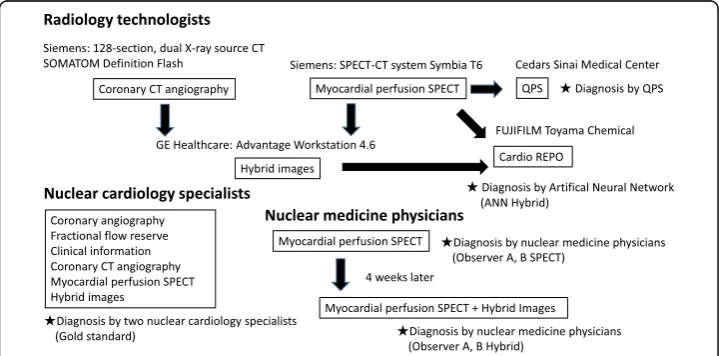
Figure 1 shows a flow chart of the image interpretation. A radiological technologist analyzed probabilities of perfusion abnormalities using the ANN and assigned the culprit coronary artery on hybrid images of CCTA and MP-SPECT. We defined the gold standard as the nuclear cardiology specialist interpretation with all available clinical information. Because patients with old myocardial infarction, percutaneous coronary intervention, and coronary artery bypass grafting were included, which is common in our clinical practice, the truth could not be simply defined by stenosis or fractional flow reserve (FFR). In addition, the AI was trained to learn specialist reading and not to identify possible stenosis or decreased flow reserve. The gold standard was, therefore, determined using coronary angiography and clinically available information including myocardial perfusion SPECT, CCTA, hybrid images, patient history, and sub-sequent clinical courses. Significant coronary artery disease was defined as stenosis in at least one coronary artery, as indicated by an FFR of 0.80 or less or stenosis diameter of > 70% using invasive coronary artery angiography or ischemia using hybrid images of CCTA and myocardial perfusion SPECT if FFR was not available. Ischemia using hybrid images of CCTA and myocardial perfusion SPECT was defined by inducible per-fusion abnormality judged by two nuclear cardiology specialists. Two other nuclear medicine physicians evaluated the SPECT images, then the hybrid images of CCTA
Fig. 1Basic flow chart of roles of radiology technologists, nuclear medicine physicians, and interpretation.
and MP-SPECT in the same way 4 weeks later, using four grades of confidence to evaluate their interpretation of regional abnormalities as follows: 1, definitely normal; 2, probably normal; 3, probably abnormal; and 4, definitely abnormal. This cutoff value was defined as 3. The same physicians then drew the regions on polar maps and eva-luated confidence in their interpretations of the culprit coronary artery as RCA, LCX, left anterior descending coronary artery (LAD), RCA or LCX (equivocal), LCX or LAD (equivocal), and LAD or RCA (equivocal). Diagnostic performance, including sensiti-vity, specificity, and accuracy in the detection of culprit coronary arteries, was com-pared among observer A, observer B, and the ANN.
Statistical analysis
Data were statistically analyzed using JMP version 12 (SAS Institute Inc., Cary, NC, USA). We evaluated CAD detectability based on areas under receiver operating characteristic (ROC) curves (AUC). All statistical tests were two-tailed, and values with p< 0.05 were considered statistically significant.
Results
Patient-based diagnosis
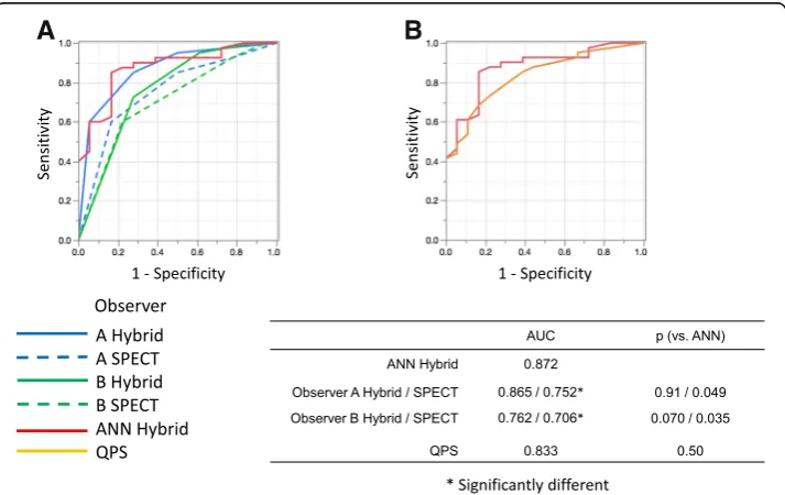
Table 2shows the likelihood ratios, Pearson statistics, andpvalues for observers A, B, and the ANN. The abilities of the ANN and nuclear medicine physicians to diagnose infarction and/or ischemia were equivalent, and the ANN outperformed conventional semi-quantitative defect scoring using QPS (Fig. 2). The sensitivity, specificity, and accuracy of patient-based diagnoses by observers A and B using hybrid images were 85.0%, 72.2%, and 81.0% and 73.1%, 73.2%, and 78.0%, respectively, and these values for the ANN and QPS were 87.8%, 72.2%, and 84.7%, and 73.2%, 77.8%, and 74.6%, respectively.
Coronary artery-based diagnosis
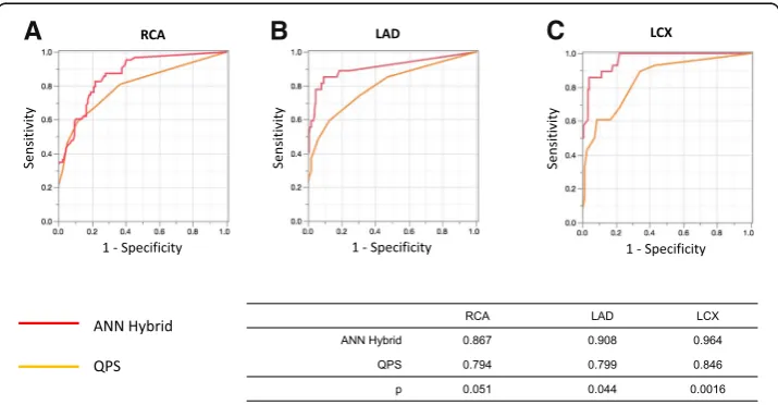
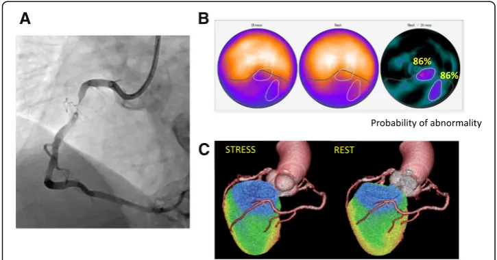
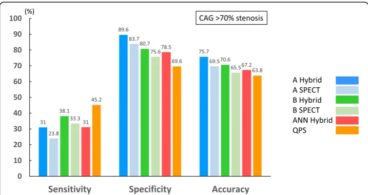
Figure3 shows the sensitivity, specificity, and accuracy of coronary-based diagnosis by observer A, observer B, the ANN, and QPS in identifying culprits among the RCA, LAD, and LCX. Figures 4 and 5 show the AUC of observer A, observer B, the ANN, and QPS. The AUC were significantly improved using hybrid images in the RCA region (observer A, p= 0.0031; observer B, p= 0.042). The culprit was most accurately diag-nosed in the LAD region. The AUC to detect RCA lesions were better for CTA-guided myocardial perfusion than MP-SPECT image analysis. Figure 6 shows a patient with single-vessel disease of the RCA in which the ANN system interpreted the images of a detected ischemic area.
Anatomical reference-based analysis
Table 2Contingency analysis of ANN and two observers True findings
Infarction Infarction + Ischemia Ischemia None Total
A. Observer A
Infarction 10 4 1 1 16
Infarction + Ischemia 2 4 6 0 12
Ischemia 0 1 6 4 11
None 3 2 2 13 20
Total 15 11 15 18 59
B. Observer B
Infarction 9 3 2 3 17
Infarction + Ischemia 5 6 7 5 23
Ischemia 1 1 5 3 10
None 0 1 1 7 9
Total 15 11 15 18 59
C. ANN
Infarction 10 0 1 2 13
Infarction + Ischemia 3 9 5 0 17
Ischemia 0 0 7 1 8
None 2 2 2 15 21
Total 15 11 15 18 59
D. Statistical significance
Likelihood ratio pvalue Pearson statistics pvalue
ANN 65.6 < 0.0001 68.1 < 0.0001
Observer A 44.8 < 0.0001 41.1 < 0.0001
Observer B 22.1 0.0087 22.4 0.0076
The likelihood ratios, Pearson statistics, andpvalues for observer A (A), observer B (B), and ANN (C)
Likelihood ratios, Pearson statistics, and probability (p) estimates for observers A, B, and artificial neural network (ANN)
A
B
Fig. 2Areas under ROC curves derived from patient-based analysis by ANN, two observers (a), and QPS (b).
AUC of ANN and QPS were 0.568 and 0.544, respectively. The AUC was not signifi-cantly improved using ANN.
Discussion
This study demonstrated that hybrid images of CCTA and MP-SPECT with neural net-work guidance could identify perfusion abnormalities effectively. When a radiology technologist analyzed hybrid images and MP-SPECT images using AI-based software to identify a culprit coronary artery, the diagnostic ability of the ANN was comparable to that of nuclear medicine physicians and outperformed conventional semi-quantitative defect scoring.
The need for applying AI software
MPI has been interpreted based on integrated understanding of myocardial perfusion distribution at stress and rest, and comparison between these two conditions using visual analysis and automatic scoring contributed to diagnosis of ischemia in clinical practice. However, while typical perfusion defects or ischemia could be unanimously judged as abnormal, nuclear physicians sometimes struggle to diagnose for equivocal findings; in such situation, a method to support objective and quantitative interpre-tation is considered useful. Nuclear medicine physicians tend to intentionally overlook or over-read MPI findings based on the experience of previous success or failure of interpretation. On the other hand, the ANN could emphasize possible abnormal regions with sufficient degrees of certainty and reproducibility. Since cardiologists and radiologists are not always specialists in nuclear cardiology, such ANN-based suggestions might enhance diagnostic confidence. The probability of abnormalities
A
B
C
provided by the ANN might be useful reference even for nuclear cardiology specialist. When a radiology technologist analyzed hybrid images and MP-SPECT images using AI-based software to identify a culprit coronary artery, the diagnostic ability of the ANN was comparable to that of nuclear medicine physicians and outperformed conventional semi-quantitative defect scoring. This finding was con-sistent with those of other studies (Nakajima et al. 2018; Nakajima et al. 2017; Nakajima et al. 2015; Johansson et al. 2014).
A
B
C
Fig. 4Areas under ROC curves derived from analysis of RCA (a), LAD (b), and LCX (c) by ANN and two observers using hybrid images and SPECT.ANNartificial neural network,LADleft anterior descending coronary artery,LCXleft circumflex coronary artery,RCAright coronary artery,ROCreceiver operating characteristic,SPECTsingle-photon emission computed tomography
A
B
C
Fig. 5Areas under ROC curves derived from analysis of RCA (a), LAD (b), and LCX (c) by ANN and QPS.
ANNartificial neural network,LADleft anterior descending coronary artery,LCXleft circumflex coronary artery,QPSquantitative perfusion single-photon emission computed tomography,RCAright coronary artery,
Identification of coronary artery lesions
Our analysis of diagnostic performance in specific vessels showed that CCTA-guided quantitation was useful in the RCA territory, and that the diagnostic accuracy was comparable in the LAD territory between nuclear physicians and software-guided tech-nologist interpretation. Clinicians must accurately match the anatomy of the coronary artery with the location of an abnormality in SPECT images to accurately assess myo-cardial ischemia. Generally, the LCX flows orthogonally to the LAD from the 11 o’clock position in polar maps. The obtuse marginal and posterolateral branches at 2–3 and 4–5 o’clock respectively emerge from the LCX in the left atrioventricular groove. The RCA runs in the right atrioventricular groove and sends out the posterior descending artery at 7 o’clock. The atrioventricular node artery branches from the poste-rior descending artery toward the 5–6 o’clock region (Nagano et al. 2016). However, using only SPECT to assign coronary artery territories is difficult because perfused regions of the LCX and RCA vary among individuals. Therefore, CCTA is needed to accurately identify culprit coronary arteries in patients with ischemia.
Accuracy of MP-SPECT to detect coronary artery disease
Nakajima et al. reported that the sensitivity, specificity, and accuracy of interpreting culprit coronary artery territories using MP-SPECT without hybrid images were of 77%, 74%, and 75%, respectively (Nakajima et al. 2010). Quantitative analysis based on summed QPS stress scores of ≥4 was used as threshold of abnormality; Johansson et al. reported that the specificity of computer-aided diagnostic software was higher than that of summed stress scores (86.8% vs. 73.6%; p< 0.001) at the same level of sensitivity. Danad et al. reported that quantitative analysis based on summed QPS
A
B
C
Fig. 6Highest stress defect score and ischemia determined by ANN and two observers in 79-year-old man with angina pectoris. Coronary angiography revealed 90% stenosis in RCA (a). Consensus interpretation diagnosed stress-induced ischemia and infarct in the inferior walls. Defect SSS, SRS, and SDS were 11, 6, and 5, respectively. Artificial neural network detected abnormality in inferior regions under stress-induced ischemia (black contour, ANN probability: 0.97) and ischemia (white contour, ANN probability: 0.86) images (b). Hybrid images of CCTA and myocardial perfusion SPECT identify culprit as RCA (c).ANNartificial neural network,
difference scores≥2 conferred an overall sensitivity, specificity, and accuracy of hybrid SPECT and CCTA for diagnosis of ischemia-causing coronary artery disease as indi-cated by FFR were 50%, 97%, and 76% (Danad et al. 2017). Although the diagnostic ability was dependent on the patient selection criteria, these results of diagnostic ability were comparable to our finding and support our conclusions.
Reduction of artifacts
Since the ANN does not consider body motion, body attenuation, or artifacts, these factors need to be reduced. Body motion tends to result in misdiagnosis, particularly when using the ANN. Although this can be addressed using motion correction software, reacquisition should proceed when large amounts of motion are detected by linography and/or sinography. Activity in sub-diaphragmatic organs can cause artifacts in images by simulating perfusion defects and concealing true defects. Iterative recon-struction suppresses streak artifacts caused by high extracardiac activity such as gallbladder activity. Reacquisition should also be considered when the extracardiac uptake of 99mTc is more intense than cardiac uptake, or when extracardiac uptake is not distinguishable from the inferior cardiac wall. Importantly, the ANN might not detect widespread, very mild ischemia in poor-quality images. Less noisy images are generally expected, even with limited radiation doses. Thus, dose and acquisition time should be balanced.
Disadvantages of hybrid images of CCTA and MP-SPECT
Although hybrid images of CCTA and MP-SPECT improves the detection of coronary artery disease and enhances diagnostic confidence, this procedure has not yet become routine in the clinical setting at many hospitals. The hybrid images of CCTA and MP-SPECT can require time-consuming manual intervention (Piccinelli et al. 2018). Radiation doses delivered to patients should also be considered. Coronary arteries can
be directly visualized by CCTA and coronary atherosclerosis can be detected and quan-tified. Individual coronary plaques can be identified, and information can be gathered regarding the total extent, severity, location, and composition of coronary artery disease (van Rosendael et al.2017). However, a phantom study found that the radiation dose of 128-section, dual-source CT coronary angiography was 195.7 mGy in the low-pitch spiral mode and 96.1 mGy in the step-and-shoot mode (Matsubara et al. 2012). Thus, the latter mode should initially be applied because less radiation is absorbed.
Study limitations
This single-center, retrospective, observational study has several limitations. We did not measure FFR in all patients and the cohort of patients was quite small. It was difficult to measure FFR in all cases and all stenotic coronary arteries. Therefore, data selection was biased by the clinical conditions that prompted the need for invasive angiography, SPECT, and CCTA, as only a minority of patients are evaluated for CAD using these modalities.
We did not apply fully automated quantitative analysis or image registration; the con-tour definitions and vascular territories were manually guided by co-registered CCTA anatomy.
Conclusions
Hybrid images of CCTA and MP-SPECT improved the ability to identify culprit coro-nary arteries in patients with stable ischemic heart disease. The diagnostic abilities of the ANN and nuclear medicine physicians were comparable. Because of interindividual differences in the perfused regions of the RCA and LCX, to identify a coronary artery responsible for perfusion defects of the inferior wall is difficult without using hybrid images.
Abbreviations
3D-OSEM:Three-dimensional iterative method based on ordered subset expectation maximization; AI: Artificial intelligence; ANN: Artificial neural network; AUC: Area under ROC curve; CAD: Coronary artery disease; CCTA: Coronary computed tomography angiography; ECG: Electrocardiography; EDV: End-diastolic volume; ESV: End-systolic volume; FFR: Fractional flow reserve; LAD: Left anterior descending coronary artery; LCX: Left circumflex coronary artery; LV: Left ventricular; LVEF: Left ventricular ejection fraction; MPI: Myocardial perfusion imaging; QGS: Quantitative gated SPECT; QPS: Quantitative perfusion SPECT; RCA: Right coronary artery; ROC: Receiver operating characteristics; RR: Resolution recovery; SPECT: Single-photon emission computed tomography
Acknowledgements
Authors thank Norma Foster for editorial assistance. We are grateful to K. Sakuda, H. Nunome, A. Mizutani, Y. Koshino, and all radiology technologists at Kanazawa University Hospital for creating hybrid images and providing technical support.
Funding
Kanazawa University Hospital provided funding for the design of the study, the collection, analysis, and interpretation of data and manuscript preparation.
Authors’contributions
Concept and design: HY and KN. Acquisition of data and ANN analysis: HY, TK, TS, and KO. Collection of clinical data: HY. Interpretation of data: KN, JT, HW, and SM. Data analysis and interpretation: KN. Manuscript preparation: MO and SK. All authors have read and approved the final manuscript.
Ethics approval and consent to participate
The Ethics Committee at Kanazawa University Hospital approved the protocol of this prospective study.
Consent for publication
Competing interests
K. Nakajima has collaborated with FUJIFILM Toyama Chemical, Tokyo, Japan to develop software.
Publisher’s Note
Springer Nature remains neutral with regard to jurisdictional claims in published maps and institutional affiliations.
Author details
1Department of Radiological Technology, Kanazawa University Hospital, 13-1 Takara-machi, Kanazawa 920-8641, Japan. 2
Department of Nuclear Medicine, Kanazawa University Hospital, Kanazawa, Japan.3Department of Physics, Kanazawa Medical University, Kahoku, Japan.4Department of Quantum Medical Technology, Institute of Medical, Pharmaceutical and Health Sciences, Kanazawa University, Kanazawa, Japan.
Received: 19 November 2018 Accepted: 19 February 2019
References
Arsanjani R, Dey D, Khachatryan T, Shalev A, Hayes SW, Fish M et al (2015) Prediction of revascularization after myocardial perfusion SPECT by machine learning in a large population. J Nucl Cardiol 22:877–884
Arsanjani R, Xu Y, Dey D, Fish M, Dorbala S, Hayes S et al (2013a) Improved accuracy of myocardial perfusion SPECT for the detection of coronary artery disease using a support vector machine algorithm. J Nucl Med 54:549–555
Arsanjani R, Xu Y, Dey D, Vahistha V, Shalev A, Nakanishi R et al (2013b) Improved accuracy of myocardial perfusion SPECT for detection of coronary artery disease by machine learning in a large population. J Nucl Cardiol 20:553–562
Danad I, Raijmakers PG, Driessen RS, Leipsic J, Raju R, Naoum C et al (2017) Comparison of coronary CT angiography, SPECT, PET, and hybrid imaging for diagnosis of ischemic heart disease determined by fractional flow reserve. JAMA Cardiol 10: 1100–1107
Guner LA, Karabacak NI, Akdemir OU, Karagoz PS, Kocaman SA, Cengel A et al (2010) An open-source framework of neural networks for diagnosis of coronary artery disease from myocardial perfusion SPECT. J Nucl Cardiol 17:405–413 Hachmovitch R, Hayes SW, Friedman JD, Cohen I, Berman DS et al (2003) Comparison of the short-term survival benefit
associated with revascularization compared with medical therapy in patients with no prior coronary artery disease undergoing stress myocardial perfusion single photon emission computed tomography. Circulation 107:2900–2906 Henzlova MJ, Duvall WL, Einstein AJ, Travin MI, Verberne HJ (2016) ASNM imaging guidelines for SPECT nuclear cardiology
procedures: stress, protocols, and tracers. J Nucl Cardiol 23:606–609
Johansson L, Edenbrandt L, Nakajima K, Lomsky M, Svensson SE, Tragardh E (2014) Computer-aided diagnosis system outperforms scoring analysis in myocardial perfusion imaging. J Nucl Cardiol 21:416–423
Kirisli HA, Gupta V, Shahzad R, Al Younis I, Dharampal A, Geuns RJ et al (2014) Additional diagnosis value of integrated analysis of cardiac CTA and SPECT MPI using the SMARTVis system in patients with suspected coronary artery disease. J Nucl Med 55:55–57
Kiriyama T, Fukushima Y, Hayashi H, Takano H, Kumita S (2018) Feasibility of combined risk stratification with coronary CT angiography and stress myocardial SPECT in patients with chronic coronary artery disease. Ann Nucl Med 32:22–33 Kiso K, Tateishi E, Imoto A, Terakawa Y, Murakawa K, Fukuda T (2017) The feasibility of quantitative assessment of myocardial
perfusion using single photon emission computed tomography imaging: current status and future challenges. Ann Nucl Cardiol 3:167–171
Levine GN, Bates ER, Blankenship JC, Bailey SR, Bittl JA, Cercek B et al (2011) 2011 ACCF/AHA/SCAI guideline for percutaneous coronary intervention. A report of the American College of Cardiology Foundation/American Heart Association Task Force on practice guidelines and the society for cardiovascular angiography and interventions. J Am Coll Cardiol 58:e44–e122
Matsubara K, Koshida H, Sakata K, Takata T, Horii J, Iida H et al (2012) Radiation dose and physical image quality in 128-section dual-source computed tomographic coronary angiography: a phantom study. J Appl Clin Med Phys 13:252–261 Matsuda A, Takeishi Y (2016) Choosing the appropriate examination for diagnosis of stable ischemic heart disease. Ann Nucl
Cardiol 2:167–173
Matsumoto N, Hirayama A (2017) Nuclear medicine is the best approach for detecting coronary artery disease: from JSNC 2016 evening seminar. Ann Nucl Cardiol 3:150–154
Motowani M, Dey D, Berman DS, Germano G, Achenbach S, Al-Mallah MH et al (2016) Machine learning for prediction of all-cause mortality in patients with suspected coronary artery disease: a 5-year multicentre prospective registry analysis. Eur Heart J 38:500–507
Nagano N, Hashimoto A, Miura T (2016) Basics of reading myocardial perfusion SPECT to identify coronary artery lesions. Ann Nucl Cardiol 2:158–161
Nakajima K, Kudo T, Nakata T, Kiso K, Kasai T, Taniguchi Y et al (2017) Diagnostic accuracy of an artificial neural network compared with statistical quantitation of myocardial perfusion images: a Japanese multicenter study. Eur J Nucl Med 44:2280–2283
Nakajima K, Kumita S, Ishida Y, Momose M, Hashimoto J, Morita K et al (2007) Creation and characterization of Japanese standards for myocardial perfusion SPECT: database from Japanese Society of Nuclear Medicine Working Group. Ann Nucl Med 21:505–511
Nakajima K, Matsuo S, Kawano M, Matsumoto N, Hashimoto J, Yoshinaga K et al (2010) The validity of multi-center common normal database for identifying myocardial ischemia: Japanese society of nuclear medicine working group database. Ann Nucl Med 24:99–105
Nakajima K, Okuda K, Kawano M, Matsuo S, Slomka P, Germano G et al (2009) The importance of population-specific normal database for quantification of myocardial ischemia: comparison between Japanese 360 and 180-degree databases and a US database. J Nucl Cardiol 16(3):422–430
Nakajima K, Okuda K, Watanabe S, Matsuo S, Kinuya S, Toth K et al (2018) Artificial neural network retrained to detect myocardial ischemia using a Japanese multicenter database. Ann Nucl Med 32:303–310
Nanasato M, Matsumoto N, Nakajima K, Chikamori T, Moroi M, Takehana K et al (2018) Prognostic impact of reducing myocardial ischemia identified using ECG-gated myocardial perfusion SPECT in Japanese patients with coronary artery disease: J-ACCESS 4 study. Int J Cardiol 267:202–207
Piccinelli M, Cooke DC, Garcia EV (2018) Multimodality image fusion for coronary artery disease detection: concepts and latest developments. Ann Nucl Cardiol 4:74–78
Sato A, Makiguchi N, Narita K, Brugada J, Boriani G, Bremerich J et al (2015) Cardiovascular imaging for nuclear cardiologists: first step of coronary computed tomography. Ann Nucl Cardiol 1:69–76
Shaw LJ, Berman DS, Marron DJ, Mancini GB, Hayes SW, Hartigan PM et al (2008) Optimal medical therapy with or without percutaneous coronary intervention to reduce ischemic burden: results from the clinical outcomes utilizing revascularization and aggressive drug evaluation (COURAGE) trial nuclear sub study. Circulation 117:1283–1291 Slomka PJ, Cheng VY, Dey D, Woo J, Ramesh A, Van Kriekinge S et al (2009) Quantitative analysis of myocardial perfusion
SPECT anatomically guided by coregistered 64-slice coronary CT angiography. J Nucl Med 50:1621–1630