R E V I E W A R T I C L E
Open Access
Functional connectivity studies in migraine:
what have we learned?
Kirill Skorobogatykh
1, Willem Sebastiaan van Hoogstraten
2, Diana Degan
3, Anastasia Prischepa
4,
Anastasya Savitskaya
4, Biondo Michela Ileen
5, Enrico Bentivegna
6, Iaroslav Skiba
7, Laura D
’
Acunto
8, Livia Ferri
6,9,
Simona Sacco
10, Jakob Møller Hansen
11, Faisal Mohammad Amin
11*and European Headache Federation School of
Advanced Studies (EHF-SAS)
Abstract
Background:
Resting-state functional connectivity (FC) MRI has widely been used to understand migraine
pathophysiology and to identify an imaging marker of the disorder. Here, we review what we have learned from FC
studies.
Methods:
We performed a literature search on the PubMed website for original articles reporting data obtained
from conventional resting-state FC recording in migraine patients compared with healthy controls or during and
outside of migraine attacks in the same patients.
Results:
We found 219 articles and included 28 in this review after screening for inclusion and exclusion criteria.
Twenty-five studies compared migraine patients with healthy controls, whereas three studies investigated migraine
patients during and outside of attacks. In the studies of interictal migraine more alterations of more than 20 FC
networks (including amygdala, caudate nucleus, central executive, cerebellum, cuneus, dorsal attention network,
default mode, executive control, fronto-parietal, hypothalamus, insula, neostriatum, nucleus accumbens, occipital
lobe, periaqueductal grey, prefrontal cortex, salience, somatosensory cortex I, thalamus and visual) were reported.
We found a poor level of reproducibility and no migraine specific pattern across these studies.
Conclusion:
Based on the findings in the present review, it seems very difficult to extract knowledge of migraine
pathophysiology or to identify a biomarker of migraine. There is an unmet need of guidelines for resting-state FC
studies in migraine, which promote the use of homogenous terminology, public availability of protocol and the a
priori hypothesis in line with for instance randomized clinical trial guidelines.
Keywords:
Resting-state fMRI, Functional connectivity, Neuroimaging, Migraine, Headache
Introduction
Pathophysiology of migraine is complex and, so far, no
biomarker for any of the phases of this cyclic disease
ex-ists. During the last decade, advanced neuroimaging
mo-dalities are increasingly used to understand migraine
pathophysiology and disease mechanisms in the search
for imaging markers of migraine. An often-used imaging
technique is the resting-state or the so-called functional
connectivity (FC) magnetic resonance imaging (fMRI),
which has been applied in increasing number of
mi-graine studies, since the first paper was published in
2011 [
1
]. Ideally, resting-state FC studies may be used to
unveil migraine mechanisms.
The migraine resting-state literature is often analyzed
and presented in several different ways, which makes it
hard to compare results across studies, and findings are
at times difficult to understand and are rarely
repro-duced. Thus, definitive imaging biomarkers for migraine
have still not been identified limiting the usefulness and
applicability of FC data.
Still, several well-performed resting-state FC studies
and reviews [
2
] are available but a systematic review of
the consistency of findings is missing. In the present
© The Author(s). 2019Open AccessThis article is distributed under the terms of the Creative Commons Attribution 4.0 International License (http://creativecommons.org/licenses/by/4.0/), which permits unrestricted use, distribution, and reproduction in any medium, provided you give appropriate credit to the original author(s) and the source, provide a link to the Creative Commons license, and indicate if changes were made.
* Correspondence:[email protected]
11Danish Headache Center, Department of Neurology, Rigshospitalet Glostrup, University of Copenhagen, Valdemar Hansens Vej 5, Glostrup, 2600 Copenhagen, Denmark
review, we wish to provide an overview of all published
conventional resting-state FC studies and discuss what
we have learned so far based on FC findings.
Methods
Literature search
Two authors (JMH and FMA) performed search on the
PubMed.com website to identify all original articles with
resting-state FC data in migraine patients. The literature
search was finalized on Pubmed.com September 20th,
2018. We used the following search terms: #1 resting state
fMRI and migraine, #2 functional connectivity and
mi-graine, and #3 functional connectivity fMRI and migraine.
The search was restricted to human studies published in
English language within 10 years, up to September 20th,
2018. Reviews, pediatric studies, case-reports, all other
headache diagnoses and letters were excluded. We also
assessed reference lists of the found articles for additional
relevant studies. Moreover, we excluded all studies that
did not use conventional resting-state analysis but other
modalities, e.g. functional connectivity density, Granger
causality, amplitude of low-frequency fluctuations, and
re-gional homogeneity. Articles, in which the method was
not properly described or if data on the comparison to a
non-headache control group was not available were also
excluded (expect if migraine attacks were compared to an
interictal phase). Finally, studies testing treatment effect
were also excluded. These exclusion criteria were chosen
to include comparable studies in this review.
Data extraction
To screen for inclusion and exclusion criteria, the senior
authors (JMH and FMA) assessed all abstracts found in
the initial search. The selected studies were then sent to
the co-authors (KS, WSvH, DD, AP, AS, BMI, EB, IS,
LDA, and LF) who then read the text and extracted
fur-ther information, i.e. origin of study, study population,
method and main findings.
Resting-state functional connectivity MRI
The imaging method is based on blood-oxygen-level
dependent (BOLD) recordings of the resting brain (i.e.
the person lying in the MRI scanner is relaxing with
closed eyes, but not sleeping). Every voxel in the
ob-tained image of the brain emits a signal with a specific
frequency. The higher the degree of synchronization of
signal frequency between two different voxels, the more
functional connected are these voxels, and vice versa.
Brain areas displaying a particular level of similarity
rep-resent a functional connectivity network. Thus, all areas
in the brain are more or less functionally connected to
each other. The use of this method depends on the
change in the functional connectivity between areas in a
network, when measured in two different conditions or
population samples.
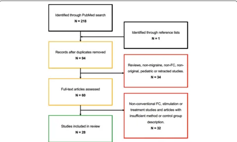
Results
Our search strategy was finalized September 20th, 2018
and resulted in a total of 219 results, including 94
unique results, from which following were excluded: 15
reviews, 12 stimulation studies, nine non-conventional
FC modalities, six examining effect of treatment
(acu-puncture), five non-migraine studies, five non-FC
stud-ies, four non-original articles, one pediatric study, and
one study was retracted. Further eight studies were
excluded because the method was not properly
de-scribed or lack of a non-headache control group. One
study was subsequently included from the reference lists.
We ended up with a total of 28 studies, including 25
during the interictal phase (Table
1
) and three during
the ictal phase (Table
2
) of migraine (Fig.
1
). The studies
were published between 2011 and 2017 and originated
from five different countries, including China = 11;
USA = 6; Italy = 6; Denmark = 4; Taiwan = 1.
Interictal migraine versus non-headache controls
Twenty-five published studies reported data comparing
interictal migraine with non-migraine non-headache
controls. In 12 studies a migraine without aura (MO)
population was examined, while pure migraine with aura
(MA) was only investigated in a single study. In four
studies, data for both MA and MO groups were reported
separately, whereas mixed results were reported in the
remaining eight studies.
When comparing migraine patient to controls, the
func-tional connectivity was changed within or with a number
of different networks or seed areas: periaqueductal gray
network [
1
,
23
], left [
3
,
7
] dorsal [
5
] and right [
3
,
25
]
anter-ior cingulate cortex, fronto-parietal-network [
4
], right
occipital lobe [
5
], left medial [
5
] and bilateral [
7
] prefrontal
cortex, right cerebellum [
5
], brainstem [
5
], bilateral central
executive network [
6
,
20
], left [
16
] salience network [
6
,
20
],
default mode network [
6
,
8
,
14
,
15
,
20
,
21
], right thalamus
[
7
], right [
7
] and anterior [
9
] insula, amygdala [
9
,
10
,
24
],
bilateral caudate [
11
], right nucleus accumbens [
11
],
hypo-thalamus [
12
], right executive control network [
13
], left
dorsal attention network [
16
], right cuneus [
16
], visual
net-work [
17
], marginal division of neostriatum [
18
], primary
visual cortex [
19
], primary auditory cortex [
19
] and
bilat-eral primary somatosensory cortex [
26
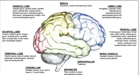
]. All areas with
abnormal connectivity to the above-mentioned networks
are shown in Table
1
and Additional file
1
and Fig.
2
.
Ictal migraine versus non-headache controls
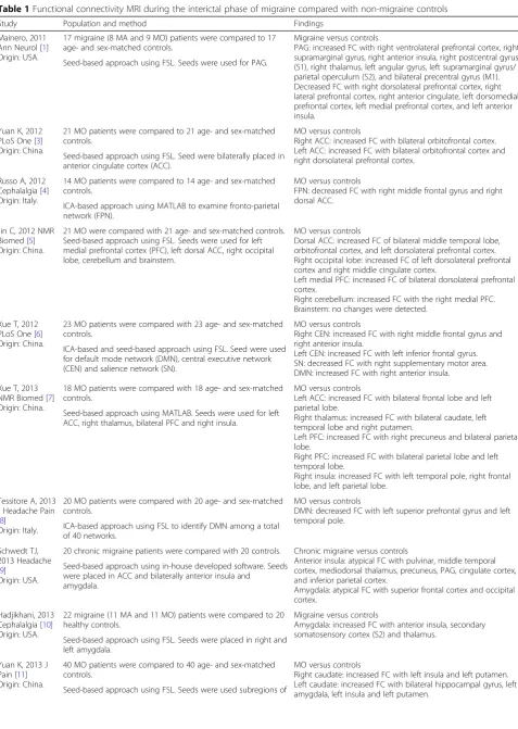
Table 1
Functional connectivity MRI during the interictal phase of migraine compared with non-migraine controls
Study Population and method Findings
Mainero, 2011 Ann Neurol [1] Origin: USA.
17 migraine (8 MA and 9 MO) patients were compared to 17 age- and sex-matched controls.
Migraine versus controls
PAG: increased FC with right ventrolateral prefrontal cortex, right supramarginal gyrus, right anterior insula, right postcentral gyrus (S1), right thalamus, left angular gyrus, left supramarginal gyrus/ parietal operculum (S2), and bilateral precentral gyrus (M1). Decreased FC with right dorsolateral prefrontal cortex, right lateral prefrontal cortex, right anterior cingulate, left dorsomedial prefrontal cortex, left medial prefrontal cortex, and left anterior insula.
Seed-based approach using FSL. Seeds were used for PAG.
Yuan K, 2012 PLoS One [3] Origin: China.
21 MO patients were compared to 21 age- and sex-matched controls.
MO versus controls
Right ACC: increased FC with bilateral orbitofrontal cortex. Left ACC: increased FC with bilateral orbitofrontal cortex and right dorsolateral prefrontal cortex.
Seed-based approach using FSL. Seed were bilaterally placed in anterior cingulate cortex (ACC).
Russo A, 2012 Cephalalgia [4] Origin: Italy.
14 MO patients were compared to 14 age- and sex-matched controls.
MO versus controls
FPN: decreased FC with right middle frontal gyrus and right dorsal ACC.
ICA-based approach using MATLAB to examine fronto-parietal network (FPN).
Jin C, 2012 NMR Biomed [5] Origin: China.
21 MO were compared with 21 age- and sex-matched controls. Seed-based approach using FSL. Seeds were used for left medial prefrontal cortex (PFC), left dorsal ACC, right occipital lobe, cerebellum and brainstem.
MO versus controls
Dorsal ACC: increased FC of bilateral middle temporal lobe, orbitofrontal cortex, and left dorsolateral prefrontal cortex. Right occipital lobe: increased FC of left dorsolateral prefrontal cortex and right middle cingulate cortex.
Left medial PFC: increased FC of bilateral dorsolateral prefrontal cortex.
Right cerebellum: increased FC with the right medial PFC. Brainstem: no changes were detected.
Xue T, 2012 PLoS One [6] Origin: China.
23 MO patients were compared with 23 age- and sex-matched controls.
MO versus controls
Right CEN: increased FC with right middle frontal gyrus and right anterior insula.
Left CEN: increased FC with left inferior frontal gyrus. SN: decreased FC with right supplementary motor area. DMN: increased FC with right anterior insula.
ICA-based and seed-based approach using FSL. Seed were used for default mode network (DMN), central executive network (CEN) and salience network (SN).
Xue T, 2013 NMR Biomed [7] Origin: China.
18 MO patients were compared with 18 age- and sex-matched controls.
MO versus controls
Left ACC: increased FC with bilateral frontal lobe and left parietal lobe.
Right thalamus: increased FC with bilateral caudate, left temporal lobe and right putamen.
Left PFC: increased FC with right precuneus and bilateral parietal lobe.
Right PFC: increased FC with bilateral parietal lobe and left temporal lobe.
Right insula: increased FC with left temporal pole, right frontal lobe, and left parietal lobe.
Seed-based approach using MATLAB. Seeds were used for left ACC, right thalamus, bilateral PFC and right insula.
Tessitore A, 2013 J Headache Pain [8]
Origin: Italy.
20 MO patients were compared with 20 age- and sex-matched controls.
MO versus controls
DMN: decreased FC with left superior prefrontal gyrus and left temporal pole.
ICA-based approach using FSL to identify DMN among a total of 40 networks.
Schwedt TJ, 2013 Headache [9]
Origin: USA.
20 chronic migraine patients were compared with 20 controls. Chronic migraine versus controls
Anterior insula: atypical FC with pulvinar, middle temporal cortex, mediodorsal thalamus, precuneus, PAG, cingulate cortex, and inferior parietal cortex.
Amygdala: atypical FC with superior frontal cortex and occipital cortex.
Seed-based approach using in-house developed software. Seeds were placed in ACC and bilaterally anterior insula and
amygdala.
Hadjikhani, 2013 Cephalalgia [10] Origin: USA.
22 migraine (11 MA and 11 MO) patients were compared to 20 healthy controls.
Migraine versus controls
Amygdala: increased FC with anterior insula, secondary somatosensory cortex (S2) and thalamus.
Seed-based approach using FSL. Seeds were placed in right and left amygdala.
Yuan K, 2013 J Pain [11] Origin: China.
40 MO patients were compared to 40 age- and sex-matched controls.
MO versus controls
Right caudate: increased FC with left insula and left putamen. Left caudate: increased FC with bilateral hippocampal gyrus, left amygdala, left insula and left putamen.
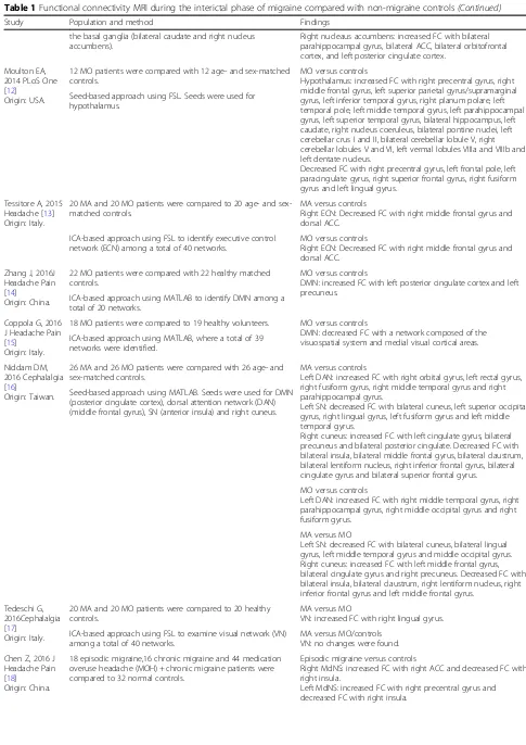
Table 1
Functional connectivity MRI during the interictal phase of migraine compared with non-migraine controls
(Continued)
Study Population and method Findings
Right nucleaus accumbens: increased FC with bilateral parahippocampal gyrus, bilateral ACC, bilateral orbitofrontal cortex, and left posterior cingulate cortex.
the basal ganglia (bilateral caudate and right nucleus accumbens).
Moulton EA, 2014 PLoS One [12]
Origin: USA.
12 MO patients were compared with 12 age- and sex-matched controls.
MO versus controls
Hypothalamus: increased FC with right precentral gyrus, right middle frontal gyrus, left superior parietal gyrus/supramarginal gyrus, left inferior temporal gyrus, right planum polare, left temporal pole, left middle temporal gyrus, left parahippocampal gyrus, left superior temporal gyrus, bilateral hippocampus, left caudate, right nucleus coeruleus, bilateral pontine nuclei, left cerebellar crus I and II, bilateral cerebellar lobule V, right cerebellar lobules V and VI, left vermal lobules VIIIa and VIIIb and left dentate nucleus.
Decreased FC with right precentral gyrus, left frontal pole, left paracingulate gyrus, right superior frontal gyrus, right fusiform gyrus and left lingual gyrus.
Seed-based approach using FSL. Seeds were used for hypothalamus.
Tessitore A, 2015 Headache [13] Origin: Italy.
20 MA and 20 MO patients were compared to 20 age- and sex-matched controls.
MA versus controls
Right ECN: Decreased FC with right middle frontal gyrus and dorsal ACC.
ICA-based approach using FSL to identify executive control network (ECN) among a total of 40 networks.
MO versus controls
Right ECN: Decreased FC with right middle frontal gyrus and dorsal ACC.
Zhang J, 2016J Headache Pain [14]
Origin: China.
22 MO patients were compared with 22 healthy matched controls.
MO versus controls
DMN: increased FC with left posterior cingulate cortex and left precuneus.
ICA-based approach using MATLAB to identify DMN among a total of 20 networks.
Coppola G, 2016 J Headache Pain [15]
Origin: Italy.
18 MO patients were compared to 19 healthy volunteers. MO versus controls
DMN: decreased FC with a network composed of the visuospatial system and medial visual cortical areas. ICA-based approach using MATLAB, where a total of 39
networks were identified.
Niddam DM, 2016 Cephalalgia [16]
Origin: Taiwan.
26 MA and 26 MO patients were compared with 26 age- and sex-matched controls.
MA versus controls
Left DAN: increased FC with right orbital gyrus, left rectal gyrus, right fusiform gyrus, right middle temporal gyrus and right parahippocampal gyrus.
Left SN: decreased FC with bilateral cuneus, left superior occipital gyrus, right lingual gyrus, left fusiform gyrus and left middle temporal gyrus.
Right cuneus: increased FC with left cingulate gyrus, bilateral precuneus and bilateral posterior cingulate. Decreased FC with bilateral insula, bilateral middle frontal gyrus, bilateral claustrum, bilateral lentiform nucleus, right inferior frontal gyrus, bilateral cingulate gyrus and bilateral superior frontal gyrus.
Seed-based approach using MATLAB. Seeds were used for DMN (posterior cingulate cortex), dorsal attention network (DAN) (middle frontal gyrus), SN (anterior insula) and right cuneus.
MO versus controls
Left DAN: increased FC with right middle temporal gyrus, right parahippocampal gyrus, right middle occipital gyrus and right fusiform gyrus.
MA versus MO
Left SN: decreased FC with bilateral cuneus, bilateral lingual gyrus, left middle temporal gyrus and middle occipital gyrus. Right cuneus: increased FC with left middle frontal gyrus, bilateral cingulate gyrus and right precuneus. Decreased FC with bilateral insula, bilateral claustrum, right lentiform nucleus, right inferior frontal gyrus and left middle frontal gyrus.
Tedeschi G, 2016Cephalalgia [17]
Origin: Italy.
20 MA and 20 MO patients were compared to 20 healthy controls.
MA versus MO
VN: increased FC with right lingual gyrus.
ICA-based approach using FSL to examine visual network (VN) among a total of 40 networks.
MA versus MO/controls VN: no changes were found.
Chen Z, 2016 J Headache Pain [18]
Origin: China.
18 episodic migraine,16 chronic migraine and 44 medication overuse headache (MOH) + chronic migraine patients were compared to 32 normal controls.
Episodic migraine versus controls
Right MdNS: increased FC with right ACC and decreased FC with right insula.
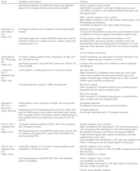
Table 1
Functional connectivity MRI during the interictal phase of migraine compared with non-migraine controls
(Continued)
Study Population and method Findings
Seed-based approach using MATLAB. Seeds were bilaterally placed in the marginal division of neostriatum (MdNS).
Chronic migraine versus controls
Right MdNS: increased FC with right middle temporal gyrus. Left MdNS: increased FC with bilateral middle frontal gyrus and left hippocampus.
MOH + chronic migraine versus controls
Right MdNS: increased FC with right interior temporal gyrus and left parahippocampal gyrus.
Left MdNS: increased FC with right middle frontal gyrus.
Hodkinson DJ, 2016 eNeuro [19] Origin: USA
40 migraine patients were compared to 40 matched healthy controls.
Migraine versus controls
V1: reduced anticorrelation to precuneus and decreased positive correlations to inferior occipital cortex/middle occipital cortex.
Seed-based approarch using M MATLAB. Seeds were used for networks of vision (V1), audition (primary auditory cortex) and somatosensation (S1).
Primary auditory cortex: reduced anticorrelation to PFC, dorsolateral PFC, precuneus, posterior cingulate cortex and lateral parietal cortex. Decreased positive correlations to insula, opercular cortex, posterior central sulcus and anterior temporal lobe.
S1: No changes were found.
Androulakis M, 2017 Neurology [20]
Origin: USA.
29 chronic migraine patients were compared to 29 age- and sex-matched controls.
Overall connectivity was decreased in all three networks in the chronic migraine group compared to controls.
Seed-based approach using MATLAB. Seeds were used for SN, CEN and DMN.
Changes were associated with moderate to severe headache and allodynia.
Lo Buono V, 2017 J Headache Pain [21]
Origin: Italy.
14 MA patients, 14 MO patients and 14 matched controls. MA versus MO
DMN: increased FC of bilateral central opercular cortex, right insular cortex, bilateral first and second Heschl’s gyrus, left superior temporal gyrus, bilateral lingual gyrus, right occpipital fusiform gyrus, and left occipital pole.
ICA-based approach using FSL. DMN was examined. MA versus controls
DMN: Increased FC of bilateral Heschl’s gyrus, bilateral planum temporale, and left superior temporal gyrus.
MO versus controls
DMN: Increased FC of bilateral lingual gyrus, occcipital fusiform gyrus, occipital pole, and cingulate gyrus.
Hougaard A, 2017 Eur J Neurol [22] Origin: Denmark.
40 MA patients were compared to 40 age- and sex-matched controls.
Seed-based approach
No difference was found in any examined network.
Seed-based and ICA-based approaches using FSL. Seeds were used for DMN, primary visual cortex, lateral geniculate nucleus, PAG, amygdala, inferior frontal gyrus, superior parietal lobule, in ferior parietal lobule, pars opercularis, visual area V2, V3A, V4 and V5.
ICA-based approach
No changes were detected in 30 analysed networks.
Chen Z, 2017 J Headache Pain [23]
Origin: China.
18 episodic migraine patients (15 MO, 3 MA) were compared with 18 healthy controls.
Episodic migraine versus controls
Right ventrolateral PAG: decreased FC with left precentral gyrus. Left ventrolateral PAG: decreased FC with left precentral gyrus, left middle frontal gyrus, left inferior frontal gyrus, bilateral middle temporal gyrus, right superior frontal gyrus and right supplementary motor area.
Left dorsolateral PAG: decreased FC with right pars triangularis of inferior frontal gyrus and the medial superior frontal gyrus. Seed-based approach using MATLAB. Seeds were used for PAG,
incl. Bilateral ventrolateral PAG, lateral PAG, dorsolateral PAG, and dorsomedial PAG.
Chen Z, 2017 J Headache Pain [24]
Origin: China.
18 episodic migraine and 16 chronic migraine patients were compared to 18 normal controls.
Episodic migraine versus controls
Left amygdala: increased FC with left middle cingulate gyrus and left precuneus.
Right amygdala: no change.
Seed-based approach using MATLAB. Seed were bilaterally placed in amygdala.
Chronic migraine versus controls Left amygdala: no change.
Right amygdala: decreased FC with right inferior occipital lobe and right middle occipital lobe.
Chronic versus episodic migraine
Left amygdala: inferior temporal gyrus, right orbital part of superior frontal gyrus, left fusiform, right postcentral gyrus, left rectus, right amygdala and left precentral gyrus.
Table 1
Functional connectivity MRI during the interictal phase of migraine compared with non-migraine controls
(Continued)
Study Population and method Findings
gyrus, left orbital part of medial frontal gyrus, left temporal pole, right orbital part of inferior frontal gyrus, right anterior cingulate gyrus and left orbital part of inferior frontal gyrus.
Yu D, 2017 Mol Pain [25] Origin: China.
31 MO patients were compared with 31 age- and education-matched controls.
MO versus controls
Right ACC: decreased FC with PFC and posterior cingulate cortex.
Left PFC: decreased FC with left insula and posterior parietal cortex.
Seed-based and ICA-based approaches using FSL. Seeds were used for DMN (medial PFC and posterior cingulate cortex), CEN (dorsloteral PFC and posterior parietal cortex) and SN (frontoin sular cortex and ACC).
No increased FC was found.
Zhang J, 2017 J Neurol [26] Origin: China.
30 MO patients were compared to 31 healthy controls. MO versus controls
Left S1: increased FC with left anterior parietal lobe, right superior parietal lobe, right S1, bilateral premotor cortex, right inferior frontal gyrus, right insula, right temporal lobe, left primary motor cortex and right middle occipital gyrus. Right S1: decreased FC with bilateral premotor cortex, bilateral superior frontal gyrus, bilateral ACC, pons, left insula, bilateral S1, bilateral paracentral lobule, right temporal lobe, right cerebellum lobule VIIIb and left inferior parietal lobule.
Seed-based approach using MATLAB. Seeds were bilaterally placed in primary somatosensory cortex (S1).
MAMigraine with aura,MOMigraine without aura,FSLFMRIB Software Library,FCFunctional connectivity,ACCAnterior cingulate cortex,ICAIndependent component analysis,CENCentral executive network,DANDorsal attention network,DMNDefault mode network,ECNExecutive control network,FPN Fronto-parietal network,PAGPeriaqueductal gray,PFCPrefrontal cortex,SNSalience network,VNVisual network,MdNSMarginal division of neostriatum
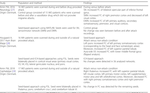
Table 2
Functional connectivity MRI during and outside of the ictal phase of migraine
Study Population and method Findings
Amin FM, 2016 Neurology [27] Origin: Denmark.
16 MO patients were scanned during and before drug provoked attack.
Control group consisted of 15 MO patients who were scanned before and after a vasodilator drug which did not provoke migraine attacks.
During versus before attack
SN: increased FC of bilateral opercular part of inferior frontal gyrus.
SMN: increased FC of right premotor cortex and decreased of left visual cortex.
DMN: increased FC of left primary auditory, secondary somatosensory, premotor, and visual cortices.
Seed-based approach using MATLAB. Seeds were used for SN, sensorimotor network (SMN) and DMN.
Control group
No change was seen between before and after attack recordings.
Hougaard A, 2017 Hum Brain Mapp [28] Origin: Denmark.
16 MA patients were scanned during and outside of a natural provoked attack.
Seed-based approach
Attack versus non-attack condition
Left pons: increased FC of left primary somatosensory cortex (corresponding to the head and face somatotopic areas). Moreover, increased FC of left superior parietal lobule. Aura-side V5: increased FC with lower middle frontal gyrus (flipped analysis).
Seed-based and ICA-based approaches using FSL. Seeds were bilaterally placed in cortical visual areas (primary visual cortex, V3, V4, V5), lateral geniculate nucleus, and pons.
ICA-based approach
No changes were detected in 56 analysed networks.
Amin FM, 2018 Cephalalgia [29] Origin: Denmark.
17 MO patients were scanned during and outside of a natural provoked attack.
Attack versus non-attack condition
Right thalamus: increased FC with left superior parietal lobule, left insular cortex, left primary motor cortex, left supplementary motor area and left orbitofrontal cortex. Moreover, decreased FC with right primary somatosensory cortex and right premotor cortex.
Seed-based approach using FSL. Seed were bilaterally placed in thalamus, pons, cerebellum crus I, and cerebellum lobule VI.
No change in FC was detected for the remaining seeds.
showed altered connectivity during the attack versus
outside of the attack: salience network [
27
],
somatosen-sory network [
27
], default mode network [
27
], left pons
[
28
] and right thalamus [
29
]. All areas with abnormal
connectivity to the above-mentioned networks and areas
are shown in Table
2
.
Discussion
Based on this first systematic review of isolated
conven-tional FC studies in migraine, we report that several
areas and networks throughout the brain, brainstem and
cerebellum showed altered connectivity in interictal and
ictal migraine studies.
The findings are very diverse, with change in FC in
many area thought to relevant for migraine as well as
several other areas. The fact that almost all published
studies report changes to some degree in all areas
stud-ied makes it difficult to gather the results into a coherent
model, of specific activation patterns of activation in
migraine.
All included studies (Tables
1
and
2
) shared many
characteristics; they used a 3 T MRI scanner, same type
of patients (either MA or MO according to the
Inter-national Classification of Headache Disorders criteria)
and controls and in addition analyzed data using almost
similar approaches (ICA or seed-based) in either the FSL
or
MATLAB-based
software
packages.
Seed-based
analysis can be affected by the chosen seed. Alterations
in the default mode network (DMN) is most frequently
reported. However, selection of different seed
coordi-nates for DMN could potentially be the reason why FC
changes in the DMN are different across studies. The
strength of ICA is that it is independent of seed selection
and more reproducible findings should be expected. The
ICA-approach has been used in 10 studies and even in
these studies different findings were reported.
Migraine is a heterogeneous disorder (with different
disease duration, attack frequency, co-morbidity, effect
of treatment, presence of aura), which might cause
vari-ation in results between studies. We did, however, only
include studies where headache was diagnosed according
to strict and uniform International Classification of
Headache Disorders criteria.
In recent resting-state fMRI studies supplementary
analyses like the Granger causality [
30–32
] have been
in-troduced to investigate if FC changes can be linked to
migraine phenotypes in the examined populations, but
even here findings cannot be reproduced. As it is clear
from Additional file
1
the findings are scattered and
show very little overlap (Additional file
1
). Moreover,
none of the reported FC changes may be specific for
mi-graine as other studies reported similar or exact same
network changes in several other conditions, including
fibromyalgia [
33
], Parkinsonian syndromes [
34
,
35
]
altered consciousness states [
36
], systemic lupus [
37
]
and chronic hepatitis C virus infection [
38
]. Thus, it can
be suspected that this FC method is at all not
reprodu-cible, which may be due to lack of sensitivity and
specifi-city. Furthermore, to the best of our knowledge no
sample size or power calculation guidelines are available
for resting-state FC, with the consequence that a
mean-ingful sample size for a resting-state FC study remains
unknown. To avoid spurious findings, it would be useful
to consider either sharing of data or joining patients in
multicenter studies to allow for better and more
repro-ducible studies.
As is already the norm for clinical trials, FC studies
should be based on publically available protocols. It is also
noteworthy that since very few studies report
“
negative
re-sults
”
or no changes in FC, primary endpoints should be
chosen before initiating studies, as is already the case for
randomized clinical trials (RCT). The fact that few (if any)
results are reproducible, strongly suggest that stricter
methodological guidelines for FC studies are warranted.
Almost half of the presented studies included only MO
patients which gives a total sum of 348 MO patients, where
120 MA patients can be calculated in our tables. The FC
method may be useful for the study of specific sub-types of
migraine if these are clearly selected beforehand, preferable
based on a calculation of the necessary number of patients,
and with a clear hypothesis to be tested.
The FC method is very versatile and may potentially help
improve
our
understanding
of
underlying
disease
mechanisms and even define biomarkers or migraine. Based
on this systematic review, we suggest that the current lack
of uniform study design, a priori hypothesis and diverse
analyses and terminology makes it difficult to apply the
available data for a coherent understanding of migraine.
Conclusions
Imaging, including FC studies could potentially help
im-prove our understanding of underlying disease
mecha-nisms, but so far no reproducible biomarkers of migraine
have been identified. Future FC studies should either pool
existing data to extract information about sub-phenotypes
of migraine patients or follow guidelines similar to RCT
guidelines in case of design of new FC studies.
Supplementary information
Supplementary informationaccompanies this paper athttps://doi.org/10. 1186/s10194-019-1047-3.
Additional file 1: Table S2.Schematic overview of regions with an altered functional connectivity to the examined networks throughout 25 studies of interictal migraine compared with healthy volunteers
Abbreviations
ACC:Anterior cingulate cortex; CEN: Central executive network; DAN: Dorsal
somatosensory cortex; SMN: Sensorimotor network; SN: Salience network; VN: Visual network
Acknowledgements
Authors thank Prof. Paolo Martelletti for organizing this EHF-SAS working group.
Authors’contributions
All authors contributed equally. KS, WSvH, DD, AP, AS, BMI, EB, IS, LDA, and LF are junior fellows, while SS, JMH and FMA are senior fellows of EHF-SAS. All authors contributed with data interpretation, drafting, revision of the manuscript and approved the final manuscript.
Funding
The article-processing charges for the article has been sponsored by the European Headache Federation.
Availability of data and materials
All included references in the present review article are available on the Internet.
Ethics approval and consent to participate
Not applicable.
Consent for publication
Not applicable.
Competing interests
KS has received personal fees, honoraria for lecturing or travel grants from TEVA, Novartis, Alder, Roche, and Allergan. SS has received personal fees, honoraria for lecturing or travel grants from Allergan, Eli Lilly, TEVA, and Novartis. FMA has participated in advisory boards and/or received personal fees, honoraria for lecturing or travel grants from Eli Lilly, TEVA, and Novartis. All authors (WSvH, DD, AP, AS, BMI, EB, IS, LDA, LF, and JMH) reports no competing interests.
Author details
1University Headache Clinic, Moscow, Russia.2Department of Neuroscience, Erasmus MC, Rotterdam, The Netherlands.3Department of Applied Clinical Sciences and Biotechnology, University of L’Aquila, L’Aquila, Italy. 4
Department of Neurology, Sechenov University, Moscow, Russia.5Sapienza University of Rome, Rome, Italy.6Internal Medicine Unit, Sant’Andrea Hospital, Sapienza University of Rome, Rome, Italy.7Neurology Department, Military Medical Academy, St. Petersburg, Russia.8Clinical Unit of Neurology, Department of Medical Sciences, University Hospital and Health Services of Trieste, University of Trieste, Trieste, Italy.9Department of Clinical and Molecular Medicine, Faculty of Medicine and Psychology, Rome, Italy. 10Clinical Neurology Section, Department of Applied Clinical Sciences and Biotechnology, University of L’Aquila, L’Aquila, Italy.11Danish Headache Center, Department of Neurology, Rigshospitalet Glostrup, University of Copenhagen, Valdemar Hansens Vej 5, Glostrup, 2600 Copenhagen, Denmark.
Received: 23 May 2019 Accepted: 12 September 2019
References
1. Mainero C, Boshyan J, Hadjikhani N (2011) Altered functional magnetic
resonance imaging resting-state connectivity in periaqueductal gray
networks in migraine. Ann Neurol 70:838–845
2. Schwedt TJ, Chiang CC, Chong CD, Dodick DW (2015) Functional MRI of
migraine. Lancet Neurol 14:81–91
3. Yuan K, Qin W, Liu P, Zhao L, Yu D, Zhao L, Dong M, Liu J, Yang X, von
Deneen KM, Liang F, Tian J (2012) Reduced fractional anisotropy of corpus callosum modulates inter-hemispheric resting state functional connectivity in migraine patients without aura. PLoS One 7:e45476
4. Russo A, Tessitore A, Giordano A, Corbo D, Marcuccio L, De Stefano M,
Salemi F, Conforti R, Esposito F, Tedeschi G (2012) Executive resting-state network connectivity in migraine without aura. Cephalalgia 32:
1041–1048
5. Jin C, Yuan K, Zhao L, Zhao L, Yu D, von Deneen KM, Zhang M, Qin W, Sun
W, Tian J (2013) Structural and functional abnormalities in migraine patients
without aura. NMR Biomed 26:58–64
6. Xue T, Yuan K, Zhao L, Yu D, Zhao L, Dong T, Cheng P, von Deneen
KM, Qin W, Tian J (2012) Intrinsic brain network abnormalities in migraines without aura revealed in resting-state fMRI. PLoS One 7: e52927
7. Xue T, Yuan K, Cheng P, Zhao L, Zhao L, Yu D, Dong T, von Deneen KM,
Gong Q, Qin W, Tian J (2013) Alterations of regional spontaneous neuronal activity and corresponding brain circuit changes during resting state in
migraine without aura. NMR Biomed 26:1051–1058
8. Tessitore A, Russo A, Giordano A, Conte F, Corbo D, De Stefano M, Cirillo S,
Cirillo M, Esposito F, Tedeschi G (2013) Disrupted default mode network connectivity in migraine without aura. J Headache Pain 14:89
9. Schwedt TJ, Schlaggar BL, Mar S, Nolan T, Coalson RS, Nardos B, Benzinger
T, Larson-Prior LJ (2013) Atypical resting-state functional connectivity of
affective pain regions in chronic migraine. Headache 53:737–751
10. Hadjikhani N, Ward N, Boshyan J, Napadow V, Maeda Y, Truini A, Caramia F,
Tinelli E, Mainero C (2013) The missing link: enhanced functional connectivity between amygdala and visceroceptive cortex in migraine.
Cephalalgia 33:1264–1268
11. Yuan K, Zhao L, Cheng P, Yu D, Zhao L, Dong T, Xing L, Bi Y, Yang X, von
Deneen KM, Liang F, Gong Q, Qin W, Tian J (2013) Altered structure and resting-state functional connectivity of the basal ganglia in migraine
patients without aura. J Pain 14:836–844
12. Moulton EA, Becerra L, Johnson A, Burstein R, Borsook D (2014) Altered
hypothalamic functional connectivity with autonomic circuits and the locus coeruleus in migraine. PLoS One 9:e95508
13. Tessitore A, Russo A, Conte F, Giordano A, De Stefano M, Lavorgna L,
Corbo D, Caiazzo G, Esposito F, Tedeschi G (2015) Abnormal connectivity within executive resting-state network in migraine with
aura. Headache 55:794–805
14. Zhang J, Su J, Wang M, Zhao Y, Yao Q, Zhang Q, Lu H, Zhang H, Wang S, Li
GF, Wu YL, Liu FD, Shi YH, Li J, Liu JR, Du X (2016) Increased default mode network connectivity and increased regional homogeneity in migraineurs without aura. J Headache Pain 17:98
15. Coppola G, Di Renzo A, Tinelli E, Lepre C, Di Lorenzo C, Di Lorenzo G,
Scapeccia M, Parisi V, Serrao M, Colonnese C, Schoenen J, Pierelli F (2016) Thalamo-cortical network activity between migraine attacks: insights from MRI-based microstructural and functional resting-state network correlation analysis. J Headache Pain 17:100
16. Niddam DM, Lai KL, Fuh JL, Chuang CY, Chen WT, Wang SJ (2016) Reduced
functional connectivity between salience and visual networks in migraine
with aura. Cephalalgia 36:53–66
17. Tedeschi G, Russo A, Conte F, Corbo D, Caiazzo G, Giordano A, Conforti R,
Esposito F, Tessitore A (2016) Increased interictal visual network connectivity
in patients with migraine with aura. Cephalalgia 36:139–147
18. Chen Z, Chen X, Liu M, Liu S, Shu S, Ma L, Yu S (2016) Altered functional
connectivity of the marginal division in migraine: a resting-state fMRI study. J Headache Pain 17(1):89 Epub 2016 Sep 26. PubMed PMID: 27670428; PubMed Central PMCID: PMC5037100
19. Hodkinson DJ, Veggeberg R, Kucyi A, van Dijk KR, Wilcox SL, Scrivani SJ,
Burstein R, Becerra L, Borsook D (2017) Cortico-cortical connections of primary sensory areas and associated symptoms in migraine. eNeuro 3. https://doi.org/10.1523/ENEURO.0163-16.2016
20. Androulakis XM, Krebs K, Peterlin BL, Zhang T, Maleki N, Sen S, Rorden C,
Herath P (2017) Modulation of intrinsic resting-state fMRI networks in
women with chronic migraine. Neurology 8(9):163–169
21. Lo Buono V, Bonanno L, Corallo F, Pisani LR, Lo Presti R, Grugno R, Di
Lorenzo G, Bramanti P, Marino S (2017) Functional connectivity and cognitive impairment in migraine with and without aura. J Headache Pain 18:72
22. Hougaard A, Amin FM, Magon S, Sprenger T, Rostrup E, Ashina M (2015) No
abnormalities of intrinsic brain connectivity in the interictal phase of
migraine with aura. Eur J Neurol 22:702–e46
23. Chen Z, Chen X, Liu M, Liu S, Ma L, Yu S (2017) Disrupted functional
connectivity of periaqueductal gray subregions in episodic migraine. J Headache Pain 18:36
24. Chen Z, Chen X, Liu M, Dong Z, Ma L, Yu S (2017) Altered functional
25. Yu D, Yuan K, Luo L, Zhai J, Bi Y, Xue T, Ren X, Zhang M, Ren G, Lu X (2017) Abnormal functional integration across core brain networks in migraine without aura. Mol Pain 13:1744806917737461
26. Zhang J, Su J, Wang M, Zhao Y, Zhang QT, Yao Q, Lu H, Zhang H, Li GF, Wu
YL, Liu YS, Liu FD, Zhuang MT, Shi YH, Hou TY, Zhao R, Qiao Y, Li J, Liu JR, Du X (2017) The sensorimotor network dysfunction in migraineurs without
aura: a resting-state fMRI study. J Neurol 264:654–663
27. Amin FM, Hougaard A, Magon S, Asghar MS, Ahmad NN, Rostrup E,
Sprenger T, Ashina M (2016) Change in brain network connectivity during PACAP38-induced migraine attacks: a resting-state functional MRI study.
Neurology 86:180–187
28. Hougaard A, Amin FM, Larsson HB, Rostrup E, Ashina M (2017) Increased
intrinsic brain connectivity between pons and somatosensory cortex during
attacks of migraine with aura. Hum Brain Mapp 3(8):2635–2642
29. Amin FM, Hougaard A, Magon S, Sprenger T, Wolfram F, Rostrup E, Ashina
M (2018) Altered thalamic connectivity during spontaneous attacks of
migraine without aura: a resting-state fMRI study. Cephalalgia 38:1237–1244
30. Wang T, Zhan W, Chen Q, Chen N, Zhang J, Liu Q, He L, Zhang J, Huang H,
Gong Q (2016) Altered resting-state ascending/descending pathways associated with the posterior thalamus in migraine without aura.
Neuroreport 27:257–263
31. Ning Y, Zheng R, Li K, Zhang Y, Lyu D, Jia H, Ren Y, Zou Y (2018) The
altered Granger causality connection among pain-related brain networks in migraine. Medicine (Baltimore) 97:e0102
32. Wang T, Chen N, Zhan W, Liu J, Zhang J, Liu Q, Huang H, He L, Zhang J,
Gong Q (2015) Altered effective connectivity of posterior thalamus in migraine with cutaneous allodynia: a resting-state fMRI study with granger causality analysis. J Headache Pain 17:17
33. Napadow V, Harris RE (2014) What has functional connectivity and chemical
neuroimaging in fibromyalgia taught us about the mechanisms and
management of‘centralized’pain? Arthritis Res Ther 16:425
34. Wolters AF, van de Weijer SCF, Leentjens AFG, Duits AA, Jacobs HIL, Kuijf ML
(2018) Resting-state fMRI in Parkinson’s disease patients with cognitive
impairment: a meta-analysis. Parkinsonism Relat Disord S1353-8020(18):
30550–30559
35. Filippi M, Sarasso E, Agosta F (2019) Resting-state functional MRI in
Parkinsonian syndromes. Mov Disord Clin Pract 6:104–117
36. Heine L, Soddu A, Gömez F, Vanhaudenhuyse A, Tshibanda L, Thonnard M,
Charland-Verville V, Kirsch M, Laureys S, Demertzi A (2012) Resting state networks and consciousness: alterations of multiple resting state networks connectivity in physiological, pharmacological, and pathological consciousness states. Front Psychol 3:295
37. Mikdashi JA (2016) Altered functional neuronal activity in neuropsychiatric
lupus: a systematic review of the fMRI investigations. Semin Arthritis Rheum
45:455–462
38. Kharabian Masouleh S, Herzig S, Klose L, Roggenhofer E, Tenckhoff H, Kaiser
T, Thöne-Otto A, Wiese M, Berg T, Schroeter ML, Margulies DS, Villringer A (2017) Functional connectivity alterations in patients with chronic hepatitis
C virus infection: a multimodal MRI study. J Viral Hepat 24:216–225