0095-1137/09/$08.00
⫹
0
doi:10.1128/JCM.01264-08
Copyright © 2009, American Society for Microbiology. All Rights Reserved.
Development and Assessment of a Multiplex Real-Time PCR Assay
for Quantification of Human Immunodeficiency Virus Type 1 DNA
䌤
A. Beloukas,† D. Paraskevis,† C. Haida, V. Sypsa, and A. Hatzakis*
Department of Hygiene, Epidemiology and Medical Statistics, Medical School, University of Athens, Athens, Greece
Received 4 July 2008/Returned for modification 17 January 2009/Accepted 27 April 2009
Previous studies showed that high levels of human immunodeficiency virus type 1 (HIV-1) DNA are
associated with a faster progression to AIDS, an increased risk of death, and a higher risk of HIV RNA rebound
in patients on highly active antiretroviral therapy. Our objective was to develop and assess a highly sensitive
real-time multiplex PCR assay for the quantification of HIV-1 DNA (RTMP-HIV) based on molecular beacons.
HIV-1 DNA quantification was carried out by RTMP in a LightCycler 2.0 apparatus. HIV-1 DNA was
quantified in parallel with CCR5 as a reference gene, and reported values are numbers of HIV-1 DNA
copies/10
6peripheral blood mononuclear cells (PBMCs). The clinical sensitivity of the assay was assessed for
115 newly diagnosed HIV-1-infected individuals. The analytical sensitivity was estimated to be 12.5 copies of
HIV-1 DNA per 10
6PBMCs, while the clinical sensitivity was 100%, with levels ranging from 1.23 to 4.25 log
10HIV-1 DNA copies/10
6PBMCs. In conclusion, we developed and assessed a new RTMP-HIV assay based on
molecular beacons, using a LightCycler 2.0 instrument. This multiplex assay has comparable sensitivity,
reproducibility, and accuracy to single real-time PCR assays.
The rate of human immunodeficiency virus type 1 (HIV-1)
disease progression varies greatly among HIV-1-infected
indi-viduals (29), depending on several factors, such as baseline
HIV RNA level, baseline CD4
⫹T-cell level, age at
serocon-version, gender, etc. (17, 23, 28, 32). Quantification of HIV
RNA in plasma has routinely been used for assessment of the
efficacy of highly active antiretroviral therapy (HAART), and it
provides a strong predictor of HIV-1 disease progression (27,
28). Treatment with HAART allows a drastic reduction of the
plasma HIV RNA load, to such an extent that circulating
HIV-1 in plasma becomes undetectable (
⬍
50 copies/ml) by the
most sensitive viral detection assays (27, 28). However, virus
eradication is not feasible because viral reservoirs established
in body and cell compartments are either not susceptible to the
action of antiretroviral drugs or characterized by a long
half-life and slow turnover (1, 6, 7, 16, 31).
Replication and immortalization of the HIV genome occur
by the reverse transcription of linear double-stranded DNA
(dsDNA) and subsequent integration of the proviral HIV-1
DNA genome into human chromosomes. To accomplish
suc-cessful reverse transcription, HIV-1 undergoes two template
switches by using the R and U5 long terminal repeat (LTR)
regions and the primer-binding site of its genome. Although
the linear dsDNA molecule is the precursor to the provirus,
HIV infection of target cells generates a number of
noninte-grated DNA species (2, 23, 31). These unintenoninte-grated HIV-1
DNA forms, which are linear or circularized, arise when viral
dsDNA fails to integrate into the host genome. For example,
(i) the ends of the linear DNA may be joined to form a 2-LTR
circle, (ii) homologous recombination between the two LTRs
in a 2-LTR circle may yield circles with a single LTR (1-LTR),
or (iii) linear unintegrated HIV-1 DNA may form.
There-fore, aside from proviral DNA (HIV-1 dsDNA integrated
into the host genome), several unintegrated forms also
per-sist intracellularly. It is worth mentioning that
cell-associ-ated HIV-1 DNA can be found in both actively and latently
infected cells (10).
As a result of this multiplicity of forms, HIV-1 DNA has
prognostic value as a marker for disease progression (18, 22,
23, 33), in addition to other associated parameters (i.e.,
plasma HIV RNA or CD4
⫹T-cell count) (2). In particular,
higher levels of HIV-1 DNA in patients not receiving
com-bination therapy are associated with an increased risk of
disease progression and a higher rate of death (2, 13, 14, 23,
33, 36). Moreover, HIV-1 DNA was found to be a predictor
of a sustained virological response to treatment; patients
with lower cellular viral loads showed a more prolonged
therapy response (2, 18, 20, 34). The HIV-1 DNA load can
also be used as an additional viral marker for treated
indi-viduals who have prolonged suppression of HIV-1
replica-tion with HAART. In this case, HIV-1 DNA is the only
marker indicative of the residual viral replication in
periph-eral blood mononuclear cells (PBMCs), lymphoid tissues,
and other compartments (10, 35).
Given the importance of HIV-1 DNA as a prognostic
marker for disease progression and response to therapy,
inde-pendent of other parameters, it is crucial to use reliable assays
for HIV-1 DNA quantification, as well as universal units of
quantification. In this study, we report the development and
assessment of a multiplex real-time PCR assay using molecular
beacons as a detection system to quantify all HIV-1 DNA
forms (single-stranded DNA and dsDNA forms and integrated
and unintegrated linear or circular forms) (RTMP-HIV) and
also the number of PBMCs, in parallel. The newly developed
RTMP-HIV assay quantifies all HIV-1 DNA forms in PBMCs,
which comprise the major intracellular viral reservoir.
* Corresponding author. Mailing address: Department of Hygiene,
Epidemiology and Medical Statistics, Medical School, University of
Athens, M. Asias 75, GR-115 27 Athens, Greece. Phone: (30210)
7462090. Fax: (30210) 7462190. E-mail: [email protected].
† The first two authors contributed equally to this work.
䌤Published ahead of print on 6 May 2009.
2194
on May 16, 2020 by guest
http://jcm.asm.org/
MATERIALS AND METHODS
Preparation of standard DNA.The external DNA standards used in the RTMP-HIV assay were HIV-1 DNA and CCR5 amplicons with known nucleo-tide lengths and base compositions. Amplicons were derived from a randomly selected hemophiliac’s sample. The standards were used as external standards after quantification in a LightCycler 2.0 instrument (Roche, Molecular Bio-chemicals, Mannheim, Germany), using a PicoGreen dsDNA quantification kit (Molecular Probes Inc., Invitrogen Detection Technologies). The concentrations of the HIV-1 and CCR5 amplicons were estimated according to standard curves created in the range of 250 to 6.25 ng after 1:2 serial dilutions of the genomic DNA standard. Tenfold serial dilutions of the standards (106
to 10 DNA copies) were used for the generation of standard curves in the multiplex amplification assays, using molecular beacon-based real-time PCR. To mimic the real condi-tions for multiplex PCR amplification, we included a steady copy number (5⫻ 105
copies) of CCR5 standard in addition to the HIV-1 DNA standards (23). Similarly, 103copies of HIV-1 DNA were added to each of the CCR5 standards. DNA extraction.Total DNA from PBMCs was isolated by manual extraction using a QIAamp DNA blood mini kit (Qiagen GmbH, Germany) according to the manufacturer’s recommendations; extracted DNA was eluted with 100l of DNase-free water prior to PCR. PBMCs were isolated from whole blood by Ficoll-Hypaque density gradient centrifugation (Histopaque-1077; Sigma). Ali-quots of approximately 106PBMCs per cryotube were stored at⫺80°C for 24 h
and then placed in a liquid nitrogen tank (about⫺196°C). PBMC counting was performed using a Neubauer counter, while thawing and washing were per-formed by standard methods.
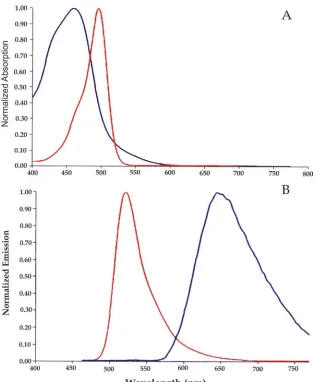
RTMP.PCR was optimized using two sets of primers and molecular beacon probes labeled with two different dyes for the amplification and detection of HIV-1 and CCR5 targets. More specifically, the primers and molecular beacon for HIV-1 amplification and detection were newly designed, targeting the con-served regions of the HIV-1 genome downstream of the LTR (HIV_sense) and gag(HIV_antisense) (in order to quantify all forms of HIV-1 DNA [single-stranded DNA and dsDNA forms and integrated and unintegrated linear or circulating forms, such as 1-LTR and 2-LTR]) (Table 1). Beacon and primer design was based on the alignment of all previously characterized HIV-1 sub-types (A to D, F to H, J, and K) and circulating recombinant forms (http://hiv -web.lanl.gov). In particular, the primers and molecular beacon target very-well-conserved genomic regions of all HIV-1 subtypes and circulating recombinant forms. Note that the target recognition sequence for the CCR5_beacon did not contain the⌬32deletion of the CCR5 gene and therefore can bind to both the wild-type CCR5 and mutant CCR5⌬32alleles, as described previously (23). The target recognition sequences for the primers used for HIV-1 DNA and CCR5 quantification are described in Table 1 (MWG-Biotech AG, Germany). The design of the primers and the molecular beacons was performed according to standard requirements, such as the following: (i) one must avoid primer-beacon annealing and primer dimers and (ii) the melting temperatures (Tms) of the primers must be similar and at least 5 to 10°C lower than theTms of the molecular beacons. The software tool used for the assessment of theTm calcu-lations was from the Virtual Genome Centre (http://frodo.wi.mit.edu/cgi-bin /primer3/primer3_www.cgi). For the simultaneous detection of the two PCR targets (in the same capillary tube), we used molecular beacons with two fluo-rescent dyes excited at overlapping wavelengths but showing no considerable overlap in their emission spectra (Fig. 1). The molecular beacons used in the RTMP-HIV assay were labeled with fluorescein (Biolegio BV, The Netherlands)
and Pulsar 650 (Biosearch Technologies, Inc., Novato, CA) fluorescent dyes for the detection of HIV-1 and CCR5 targets (37), respectively (Table 1). Prelimi-nary data showed that the RTMP-HIV assay could be used in both LightCycler 1.0 and LightCycler 2.0 instruments. The data reported in this study reflect the use of the 2.0 platform because of its enhanced testing characteristics.
The reaction mixture for real-time PCR (19) contained 2.5l 10⫻LightCycler FastStartTaqreaction mix (Roche, Molecular Biochemicals, Mannheim, Ger-many), 6 mM MgCl2, 1.1M HIV_sense primer, 1.1M HIV_anti primer, 0.08
M CCR5_sense primer, 0.08M CCR5_anti primer, 0.4M HIV_beacon, 1.2 M CCR5_beacon, 1.5 U FastStartTaqDNA polymerase, 1 U of uracil-DNA glycosylase, and 10l extracted DNA in a final reaction volume of 25l. The amplification conditions were optimized for LightCycler 1.0 and 2.0 as follows: 1 cycle of denaturation at 95°C for 10 min followed by 45 cycles of amplification at 95°C for 10 s, 60°C for 10 s, and 72°C for 10 s. Uracil-DNA glycosylase was used to eliminate PCR carryover contaminations from previous PCRs (24, 25). For each run, two standard curves were created in a 6-log range by 1:10 serial dilutions, one with HIV-1 DNA and one with CCR5 external standards. The slope and correlation coefficient of each standard curve were calculated based on the average cycle threshold (CT) value measured for each dilution point, ranging from 106to 101copies of standard DNA template. Quantification of HIV-1 DNA
and the CCR5 reporter gene in the unknown samples was performed by using these external standards for HIV-1 and CCR5. HIV-1 DNA was quantified by using the CCR5 gene as a reference gene, for which there is a steady number of copies per cell (two copies per cell), as described previously (23). Therefore, the numbers of HIV-1 DNA and CCR5 copies in a sample can easily be converted to HIV-1 DNA copies/106PBMCs by using the following formula: number of
HIV-1 DNA copies/number of CCR5 copies⫻2⫻106⫽
number of HIV-1 DNA copies per 106PBMCs, given that a single PBMC contains two copies of
the CCR5 gene (for example, 15 copies of HIV-1 DNA and 1,500 CCR5 copies in the same sample equals 2⫻104HIV-1 DNA copies per 106PBMCs).
Clinical specimens.HIV-1 DNA was quantified in total DNAs extracted from PBMCs from 115 randomly selected newly diagnosed HIV-1-infected individuals before the initiation of HAART. The clinical specimens (whole blood) were collected anonymously from September 2002 to September 2005. PBMCs were stored in a liquid nitrogen tank (about⫺196°C) until their use. Plasma HIV-1 RNA was measured with the Versant HIV-1 RNA 3.0 (bDNA) assay (Bayer, Tarrytown, NY), which has a dynamic range of 50 to 500,000 HIV-1 RNA copies/ml, according to the manufacturer’s recommendations. CD4⫹ T-cell counts were measured by flow cytometry by standard procedures.
Statistical analysis.The variability between replicate tests of HIV-1 DNA and CCR5 standards was described using the percent coefficient of variation (CV) for log10-transformed data. Pearson’s correlation coefficient calculation and
ordi-nary least-squares regression (OLR) on log10-transformed values for HIV-1
DNA and CCR5 standards were performed to compare experimentally observed to expected concentrations. Pearson’s correlation coefficient, OLR, and Deming regression were used to assess the association of HIV-1 DNA with HIV RNA and the CD4⫹T-cell count. The Deming regression is similar to the OLR but takes into account that bothyandxare measured with error (8).
RESULTS
The RTMP-HIV assay is the first to implement multiplex
methodology using molecular beacons as a detection system
with the LightCycler platform. Primary experiments were
car-ried out with a LightCycler 1.0 apparatus; therefore, the
ana-lytical sensitivity of the RTMP-HIV assay was primarily
esti-mated with the LightCycler 1.0 platform and thereafter with
the LightCycler 2.0 platform. These preliminary data showed
that the RTMP-HIV assay could be used on both platforms
(LightCycler 1.0 and LightCycler 2.0) and that the latter had
superior performance characteristics. The data reported in this
study with regard to the reproducibility of the RMTP-HIV
assay and testing of clinical samples reflect the use of the
LightCycler 2.0 platform because of its enhanced performance
characteristics.
[image:2.585.42.283.90.201.2]Analytical sensitivity of RTMP-HIV assay with LightCycler
1.0 and LightCycler 2.0 platforms.
The analytical sensitivity of
the RTMP-HIV assay was estimated by repetitively testing (20
TABLE 1. Real-time PCR amplicon sizes and locations of primers
and beacons within viral and human reference sequences
Primer or beacon Orientation or
fluorescent dye
Product
size (nt) Location (nt)
Primers
HIV_sense
Forward
221
686–708
aHIV_antisense
Reverse
884–907
aCCR5_sense
Forward
238
466–489
bCCR5_antisense
Reverse
681–704
bBeacons
HIV_beacon
6-Carboxyfluorescein
791–818
aCCR5_beacon
Pulsar 650
624–652
baRelative to GenBank accession no. K03455.
bRelative to GenBank accession no. AY463215.
on May 16, 2020 by guest
http://jcm.asm.org/
replicates) different concentrations of HIV-1 DNA standards,
ranging from 10
6to 3.125 copies/reaction (3.125, 6.25, 10, 12.5,
25, 10
2, 10
3, 10
4, 10
5, and 10
6) (Table 2). The seven highest
concentrations of the HIV-1 DNA proviral standards (12.5 to
10
6copies per reaction) were examined using the LightCycler
1.0 platform, while the three lowest concentrations (3.125 to 10
copies per reaction) were examined with the LightCycler 2.0
platform. The lowest concentration of HIV-1 DNA standard
that could be quantified at a frequency of 100% by
RTMP-HIV with the LightCycler 1.0 platform was 12.5 copies per
reaction (in a multiplex setting with 5
⫻
10
5copies of the
CCR5 standard), which equals 50 copies of HIV-1 DNA per
10
6PBMCs. The analytical sensitivity of the RTMP-HIV assay
was found to be higher with the LightCycler 2.0 platform
(3.125 copies per reaction, which equals 12.5 copies of HIV-1
DNA per 10
6PBMCs). The latter was set as the cutoff for the
quantification of HIV-1 DNA for the LightCycler 2.0 platform.
RTMP-HIV assay performance with LightCycler 2.0
plat-form.
To examine the reproducibility and linearity of the
RTMP-HIV assay, we quantified different concentrations of
[image:3.585.135.454.65.447.2]FIG. 1. (A) Absorption spectra for both dyes used as beacons. Red line, fluorescein-labeled beacon; blue line, Pulsar 650-labeled beacon.
(B) Emission spectra for these dyes. The two fluorescent dyes absorb in the same wavelength, but the emission spectra have very little overlap.
TABLE 2. Experimental results for repetitively quantified
(20 times) serial dilutions of HIV-1 DNA standards in
LightCycler 1.0 and LightCycler 2.0 platforms
aExptl platform
No. of HIV-1 DNA copies per reaction
No. of HIV-1 DNA copies/ 106PBMCs
Mean⫾SD of exptl valuesb
CV (%) of exptl valuesb
LightCycler 1.0
10
64
⫻
10
65.99
⫾
0.08
1.36
10
54
⫻
10
55.08
⫾
0.20
3.86
10
44
⫻
10
43.95
⫾
0.09
2.16
10
34
⫻
10
32.99
⫾
0.15
5.02
10
2400
2.21
⫾
0.27
12.37
25
100
1.44
⫾
0.27
18.87
12.5
50
0.79
⫾
0.21
25.93
LightCycler 2.0
10
40
1.03
⫾
0.13
12.99
6.25
25
0.87
⫾
0.31
35.32
3.125
12.5
0.17
⫾
0.40
232.26
a
Each reaction was performed in a multiplex setting with 5⫻105
copies of the CCR5 gene in a final reaction volume of 25l. The experimental results were 100% positive for all concentrations with both platforms.
b
Log10-transformed values.
on May 16, 2020 by guest
http://jcm.asm.org/
[image:3.585.299.541.564.692.2]HIV-1 DNA standards, ranging from 10 to 10
6copies per
reaction (10, 10
2, 10
3, 10
4, 10
5, and 10
6), in a multiplex setting
with CCR5 standards (5
⫻
10
5copies of CCR5 were present in
each sample) in 28 replicates and estimated the CV for each
concentration (Table 3). According to the estimated values,
the %CV was low (2.0% and 3.2%) for high concentrations of
HIV-1 DNA (for 10
6and 10
5copies per reaction, respectively),
while more variation (11.6% and 27.3%) was observed for
lower HIV-1 DNA values (10
2and 10 copies/reaction,
respec-tively) (Table 3). The standard deviation (SD) of experimental
values ranged from 0.12 (for 10
6copies per reaction) to 0.27
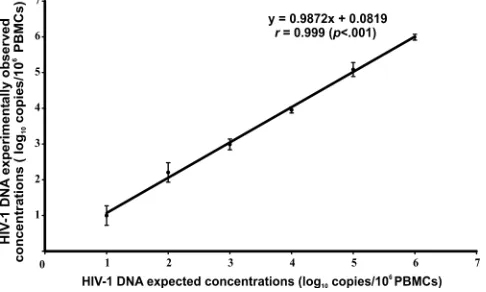
(for 10 copies per reaction). Furthermore, the theoretically
calculated values showed a very high correlation versus the
experimentally estimated concentrations (Pearson’s
r
⫽
0.999;
P
⬍
0.001) (Fig. 2).
CCR5 RTMP assay performance with LightCycler 2.0
plat-form.
The reproducibility and linearity of the newly developed
RTMP-HIV assay were estimated similarly for the CCR5
ex-ternal standard. In particular, the amount of CCR5 was
esti-mated repeatedly (12 replicates) for DNA concentrations
ranging between 10 and 10
6copies coamplified with HIV-1
DNA (10
3copies of HIV-1 DNA were included in each
reac-tion). The CV was calculated for each different serially diluted
concentration (Table 3). According to the estimated values,
the %CV was low (4.9% and 4.2%) for high concentrations
(for 10
6and 10
5copies of the CCR5 gene per reaction,
respec-tively) and was higher for 10
2and 10 copies per reaction (9.3%
and 27.6%, respectively) (Table 3). Interestingly, the SD of
experimental values ranged from 0.12 (for 10
4copies per
re-action) to 0.29 (for 10 and 10
6copies per reaction).
Further-more, the theoretically calculated values showed a very high
correlation versus experimentally estimated concentrations
(Pearson’s
r
⫽
0.999;
P
⬍
0.001) (Fig. 3).
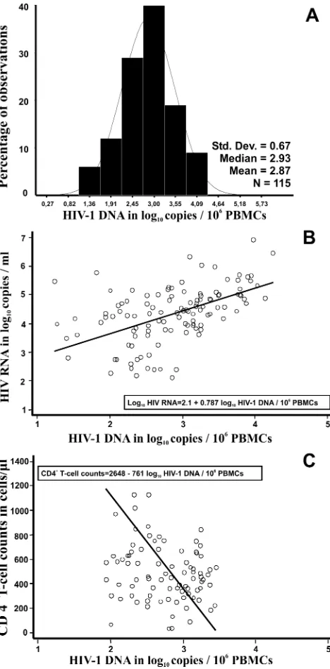
Assessment of RTMP-HIV assay with clinical samples.
The
RTMP-HIV assay was used for the quantification of HIV
DNA in 115 newly diagnosed HIV-1-infected individuals prior
to administration of therapy. HIV-1 DNA was detectable in
115 samples (100%). The log
10HIV-1 DNA copies/10
6PBMCs ranged from 1.23 to 4.25, with a mean
⫾
SD of 2.87
⫾
0.67 (Fig. 4A). Importantly, HIV DNA/10
6PBMC values fell
within a 3-log dynamic range. The log
10HIV-1 DNA copies/
10
6PBMCs and the number of CD4
⫹T cells per
l were
negatively correlated (Pearson’s
r
⫽ ⫺
0.461;
P
⬍
0.001), and
the strength of this linear relationship was similar to that of
HIV RNA levels and CD4
⫹T-cell counts (Pearson’s
r
⫽
⫺
0.353;
P
⬍
0.001) (data not shown). CD4
⫹T-cell counts
ranged from 32 to 1,328 cells/
l, with a median value (25th,
75th percentiles) of 444 cells/
l (279, 600 cells/
l) (Fig. 4C).
Finally, we found that the number of HIV-1 DNA copies/10
6PBMCs was positively correlated with the HIV-1 RNA level
(Pearson’s
r
⫽
0.488;
P
⬍
0. 001). Log
10HIV-1 RNA levels
ranged from 2.13 to 6.92, with a mean
⫾
SD of 4.35
⫾
0.98
(Fig. 4B).
DISCUSSION
[image:4.585.300.543.71.216.2]Our study aimed to develop an RTMP-HIV assay for the
quantification of total HIV-1 DNA per PBMC in a single-tube
capillary reaction by using a LightCycler instrument. For the
TABLE 3. Quantified values for serial dilutions of standards with
LightCycler 2.0 platform
aTarget DNA
No. of copies of DNA per
reaction
No. (%) of measurements above the limit of detection for 28 replicates
(HIV-1) or 12 replicates (CCR5)
Mean⫾SD of exptl valuesb
CV (%) of exptl valuesb
HIV-1
10
628 (100)
5.89
⫾
0.12
2.0
10
528 (100)
4.93
⫾
0.16
3.2
10
428 (100)
3.83
⫾
0.16
4.0
10
328 (100)
2.97
⫾
0.12
4.0
10
228 (100)
2.03
⫾
0.24
11.6
10
9 (32.1)
1.00
⫾
0.27
27.3
CCR5
10
612 (100)
5.92
⫾
0.29
4.9
10
512 (100)
5.03
⫾
0.21
4.2
10
412 (100)
3,94
⫾
0.12
2.9
10
312 (100)
3.06
⫾
0.20
6.6
10
212 (100)
2.05
⫾
0.19
9.3
10
12 (100)
1.05
⫾
0.29
27.6
a
Each reaction was performed in a multiplex setting, with each reaction mix including, in addition to the standards, 103
copies of HIV-1 DNA and 5⫻105
CCR5 copies for CCR5 and HIV-1 DNA quantifications, respectively, in a final volume of 25l.
b
[image:4.585.43.282.89.251.2]Log10-transformed data.
FIG. 2. Mean experimentally estimated concentrations (dots) for
six serial dilutions of HIV-1 DNA standards versus theoretically
cal-culated concentrations (log
10-transformed data). Error bars indicate
the SD for the six 10-fold serially diluted standards.
FIG. 3. Mean experimentally estimated concentrations (dots) for
six serial dilutions of CCR5 gene standards versus theoretically
calcu-lated concentrations (log
10-transformed data). Error bars indicate the
SD for the six 10-fold serially diluted standards.
on May 16, 2020 by guest
http://jcm.asm.org/
[image:4.585.43.286.542.688.2]accomplishment of this task, we quantified HIV-1 DNA by
using CCR5 as a reference gene, as there is a steady number of
copies of this gene per cell, as described previously (23).
Therefore, HIV-1 DNA quantifications can easily be converted
to copies of HIV-1 DNA/10
6PBMCs.
The analytical sensitivity of the RTMP-HIV assay was
higher with the LightCycler 2.0 platform than with the
Light-Cycler 1.0 platform because of the improved optical and
hard-ware settings installed in the latest version of LightCycler
in-struments. The analytical sensitivity of the RTMP-HIV assay
(12.5 HIV-1 DNA copies/10
6PBMCs) is very similar to those
of the most sensitive methods (5 to 10 copies of HIV-1 DNA
per reaction) based on single real-time PCRs (2), thus
suggest-ing that the RTMP-HIV assay can be used reliably for HIV-1
DNA quantification.
Prior to this study, several PCR-based methodologies had
been developed for the quantification of cell-associated HIV-1
DNA, based on end-point and real-time PCR methodology
(2). Implementation of real-time PCR methodology
consider-ably improved the quantification of HIV-1 DNA compared to
end-point PCR methodology. The former shows improved
ac-curacy and reproducibility and a wider range of linear
quanti-fication because the concentration of the target is estimated
during the linear phase rather than at the end point (plateau
phase) of the amplification step. Most home-brewed real-time
PCR methods have implemented TaqMan methodology (2),
while three others have used SYBR green for the detection
and quantification of HIV-1 DNA (3, 12, 21). Moreover,
Chris-topherson et al. reported a modification of the Amplicor
HIV-1 Monitor test (Roche Molecular Systems, Inc.,
Branch-burg, NJ) for total HIV-1 DNA quantification. This assay,
based on end-point PCR measurement, is almost identical to
the Amplicor HIV-1 Monitor assay for RNA (Roche
Molec-ular Systems, Inc., Branchburg, NJ) but differs in (i) the sample
preparation method, (ii) the use of plasmid DNA rather than
an RNA transcript as the quantitation standard, and (iii) the
normalization of DNA load to total cellular input (4).
In contrast with all previous methods, our assay combines
the advantages of real-time PCR (high dynamic range of
quan-tification, less intra- and interassay variability in
measure-ments, and improved sensitivity) with the direct quantification
of cell copy numbers by using the CCR5 gene as a reference
gene in a multiplex format. Despite the multiplex setting, our
assay performed very well with clinical samples, showing a
clinical sensitivity of 100% and a linear range of values within
3 log
10copies. Most in-house assays use two single PCRs for
the quantification of the amount of HIV-1 DNA per cell.
Importantly, the current method comprises the first assay with
a multiplex design using the LightCycler 2.0 platform for
quan-tification of the HIV-1 DNA concentration in PBMCs.
We should note that until now there has been no universal
way to report HIV-1 DNA values. In particular, some
investi-gators quantify the amount of HIV-1 DNA per 10
6PBMCs (2,
5, 9, 11, 23) by estimating the cell copy number directly or
indirectly or per number of resting CD4
⫹T cells (21). In the
latter method, the amount of detected HIV-1 DNA depends
on the subsets of PBMCs as well as on the methodology used
to estimate the number of cell equivalents.
In conclusion, we developed and assessed a novel
RTMP-HIV assay for RTMP-HIV-1 DNA and CCR5 quantification based on
molecular beacons and the use of a LightCycler instrument
(the assay applies to both LightCycler 1.0 and LightCycler 2.0
platforms). Although fluorescent molecular beacons have been
implemented in real-time PCR methodology for several years
(15, 26, 30), the RTMP-HIV assay is the first RTMP assay for
the LightCycler apparatus that uses molecular beacons as a
detection system. The assay with the LightCycler 2.0 platform
has comparable performance characteristics to those of several
previously described real-time PCR assays, as well as a
multi-FIG. 4. Distribution of log
10HIV-1 DNA copies/10
6
PBMCs
(A) and correlation of log
10HIV-1 DNA copies/10
6PBMCs with
plasma RNA levels (B) and with CD4
⫹T-cell counts (C) for 115 newly
diagnosed HIV-1-infected individuals. The solid lines in panels B and
C are fitted regression lines obtained by Deming regression.
on May 16, 2020 by guest
http://jcm.asm.org/
[image:5.585.46.280.68.542.2]plex design for high throughput and accurate detection of the
HIV-1 DNA concentration in PBMCs.
REFERENCES
1.Bailey, J. R., A. R. Sedaghat, T. Kieffer, T. Brennan, P. K. Lee, M. Wind-Rotolo, C. M. Haggerty, A. R. Kamireddi, Y. Liu, J. Lee, D. Persaud, J. E. Gallant, J. Cofrancesco, Jr., T. C. Quinn, C. O. Wilke, S. C. Ray, J. D. Siliciano, R. E. Nettles, and R. F. Siliciano.2006. Residual human immu-nodeficiency virus type 1 viremia in some patients on antiretroviral therapy is dominated by a small number of invariant clones rarely found in circulat-ing CD4⫹T cells. J. Virol.80:6441–6457.
2.Beloukas, A., D. Paraskevis, M. Psichogiou, and A. Hatzakis.2009. The role of HIV-1 DNA as an additional marker of HIV-1 infection. Curr. HIV Res.
7:255–265.
3.Casabianca, A., C. Gori, C. Orlandi, F. Forbici, C. F. Perno, and M. Mag-nani.2007. Fast and sensitive quantitative detection of HIV DNA in whole blood leucocytes by SYBR green I real-time PCR assay. Mol. Cell. Probes
21:368–378.
4.Christopherson, C., Y. Kidane, B. Conway, J. Krowka, H. Sheppard, and S. Kwok.2000. PCR-based assay to quantify human immunodeficiency virus type 1 DNA in peripheral blood mononuclear cells. J. Clin. Microbiol.
38:630–634.
5.Chun, T. W., L. Carruth, D. Finzi, X. Shen, J. A. DiGiuseppe, H. Taylor, M. Hermankova, K. Chadwick, J. Margolick, T. C. Quinn, Y. H. Kuo, R. Brook-meyer, M. A. Zeiger, P. Barditch-Crovo, and R. F. Siliciano.1997. Quanti-fication of latent tissue reservoirs and total body viral load in HIV-1 infec-tion. Nature387:183–188.
6.Chun, T. W., D. C. Nickle, J. S. Justement, D. Large, A. Semerjian, M. E. Curlin, M. A. O’Shea, C. W. Hallahan, M. Daucher, D. J. Ward, S. Moir, J. I. Mullins, C. Kovacs, and A. S. Fauci.2005. HIV-infected individuals receiv-ing effective antiviral therapy for extended periods of time continually replenish their viral reservoir. J. Clin. Investig.115:3250–3255.
7.Chun, T. W., L. Stuyver, S. B. Mizell, L. A. Ehler, J. A. Mican, M. Baseler, A. L. Lloyd, M. A. Nowak, and A. S. Fauci.1997. Presence of an inducible HIV-1 latent reservoir during highly active antiretroviral therapy. Proc. Natl. Acad. Sci. USA94:13193–13197.
8.Cornbleet, P. J., and N. Gochman.1979. Incorrect least-squares regression coefficients in method-comparison analysis. Clin. Chem.25:432–438. 9.Eriksson, L. E., T. Leitner, B. Wahren, A. C. Bostrom, and K. I. Falk.2003.
A multiplex real-time PCR for quantification of HIV-1 DNA and the human albumin gene in CD4⫹cells. APMIS111:625–633.
10.Furtado, M. R., D. S. Callaway, J. P. Phair, K. J. Kunstman, J. L. Stanton, C. A. Macken, A. S. Perelson, and S. M. Wolinsky.1999. Persistence of HIV-1 transcription in peripheral-blood mononuclear cells in patients re-ceiving potent antiretroviral therapy. N. Engl. J. Med.340:1614–1622. 11.Gibellini, D., F. Vitone, E. Gori, M. La Placa, and M. C. Re.2004.
Quanti-tative detection of human immunodeficiency virus type 1 (HIV-1) viral load by SYBR green real-time RT-PCR technique in HIV-1 seropositive patients. J. Virol. Methods115:183–189.
12.Gibellini, D., F. Vitone, P. Schiavone, C. Ponti, M. La Placa, and M. C. Re.
2004. Quantitative detection of human immunodeficiency virus type 1 (HIV-1) proviral DNA in peripheral blood mononuclear cells by SYBR green real-time PCR technique. J. Clin. Virol.29:282–289.
13.Goedert, J. J., T. R. O’Brien, A. Hatzakis, and L. G. Kostrikis.2001. T cell receptor excision circles and HIV-1 2-LTR episomal DNA to predict AIDS in patients not receiving effective therapy. AIDS15:2245–2250.
14.Goujard, C., M. Bonarek, L. Meyer, F. Bonnet, M. L. Chaix, C. Deveau, M. Sinet, J. Galimand, J. F. Delfraissy, A. Venet, C. Rouzioux, and P. Morlat.
2006. CD4 cell count and HIV DNA level are independent predictors of disease progression after primary HIV type 1 infection in untreated patients. Clin. Infect. Dis.42:709–715.
15.Gullsby, K., M. Storm, and K. Bondeson.2008. Simultaneous detection of Chlamydophila pneumoniaeandMycoplasma pneumoniaeby use of molecu-lar beacons in a duplex real-time PCR. J. Clin. Microbiol.46:727–731. 16.Hammer, S. M., M. S. Saag, M. Schechter, J. S. Montaner, R. T. Schooley,
D. M. Jacobsen, M. A. Thompson, C. C. Carpenter, M. A. Fischl, B. G. Gazzard, J. M. Gatell, M. S. Hirsch, D. A. Katzenstein, D. D. Richman, S. Vella, P. G. Yeni, and P. A. Volberding.2006. Treatment for adult HIV infection: 2006 recommendations of the International AIDS Society—USA panel. Top. HIV Med.14:827–843.
17.Hatzakis, A., G. Touloumi, R. Karanicolas, A. Karafoulidou, T. Mandalaki, C. Anastassopoulou, L. Zhang, J. J. Goedert, D. D. Ho, and L. G. Kostrikis.
2000. Effect of recent thymic emigrants on progression of HIV-1 disease. Lancet355:599–604.
18.Hatzakis, A. E., G. Touloumi, N. Pantazis, C. G. Anastassopoulou, O.
Kat-sarou, A. Karafoulidou, J. J. Goedert, and L. G. Kostrikis.2004. Cellular HIV-1 DNA load predicts HIV-RNA rebound and the outcome of highly active antiretroviral therapy. AIDS18:2261–2267.
19.Heid, C. A., J. Stevens, K. J. Livak, and P. M. Williams.1996. Real time quantitative PCR. Genome Res.6:986–994.
20.Hoen, B., D. A. Cooper, F. C. Lampe, L. Perrin, N. Clumeck, A. N. Phillips, L. E. Goh, S. Lindback, D. Sereni, B. Gazzard, J. Montaner, H. J. Stellbrink, A. Lazzarin, D. Ponscarme, S. Staszewski, L. Mathiesen, D. Smith, R. Finlayson, R. Weber, L. Wegmann, G. Janossy, and S. Kinloch-de Loes.2007. Predictors of virological outcome and safety in primary HIV type 1-infected patients initiating quadruple antiretroviral therapy: QUEST GW PROB3005. Clin. Infect. Dis.45:381–390. 21.Kabamba-Mukadi, B., P. Henrivaux, J. Ruelle, N. Delferriere, M. Bodeus,
and P. Goubau.2005. Human immunodeficiency virus type 1 (HIV-1) pro-viral DNA load in purified CD4⫹cells by LightCycler real-time PCR. BMC Infect. Dis.5:15.
22.Katzenstein, T. L., R. S. Oliveri, T. Benfield, J. Eugen-Olsen, C. Nielsen, and J. Gerstoft.2002. Cell-associated HIV DNA measured early during infection has prognostic value independent of serum HIV RNA measured concomi-tantly. Scand. J. Infect. Dis.34:529–533.
23.Kostrikis, L. G., G. Touloumi, R. Karanicolas, N. Pantazis, C. Anastasso-poulou, A. Karafoulidou, J. J. Goedert, and A. Hatzakis.2002. Quantitation of human immunodeficiency virus type 1 DNA forms with the second tem-plate switch in peripheral blood cells predicts disease progression indepen-dently of plasma RNA load. J. Virol.76:10099–10108.
24.Kwok, S., and R. Higuchi.1989. Avoiding false positives with PCR. Nature
339:237–238.
25.Longo, M. C., M. S. Berninger, and J. L. Hartley.1990. Use of uracil DNA glycosylase to control carry-over contamination in polymerase chain reac-tions. Gene93:125–128.
26.Martinez-Lopez, J., J. J. Lahuerta, P. Salama, R. Ayala, and J. M. Bautista.
2004. The use of fluorescent molecular beacons in real time PCR of IgH gene rearrangements for quantitative evaluation of multiple myeloma. Clin. Lab. Haematol.26:31–35.
27.Mellors, J. W., L. A. Kingsley, C. R. Rinaldo, Jr., J. A. Todd, B. S. Hoo, R. P. Kokka, and P. Gupta.1995. Quantitation of HIV-1 RNA in plasma predicts outcome after seroconversion. Ann. Intern. Med.122:573–579.
28.Mellors, J. W., C. R. Rinaldo, Jr., P. Gupta, R. M. White, J. A. Todd, and L. A. Kingsley.1996. Prognosis in HIV-1 infection predicted by the quantity of virus in plasma. Science272:1167–1170.
29.Pantaleo, G., C. Graziosi, and A. S. Fauci. 1993. New concepts in the immunopathogenesis of human immunodeficiency virus infection. N. Engl. J. Med.328:327–335.
30.Pattyn, F., F. Speleman, A. De Paepe, and J. Vandesompele. 2003. RTPrimerDB: the real-time PCR primer and probe database. Nucleic Acids Res.31:122–123.
31.Pierson, T., J. McArthur, and R. F. Siliciano.2000. Reservoirs for HIV-1: mechanisms for viral persistence in the presence of antiviral immune re-sponses and antiretroviral therapy. Annu. Rev. Immunol.18:665–708. 32.Powderly, W. G., M. S. Saag, S. Chapman, G. Yu, B. Quart, and N. J.
Clendeninn.1999. Predictors of optimal virological response to potent an-tiretroviral therapy. AIDS13:1873–1880.
33.Rouzioux, C., J. B. Hubert, M. Burgard, C. Deveau, C. Goujard, M. Bary, D. Sereni, J. P. Viard, J. F. Delfraissy, and L. Meyer.2005. Early levels of HIV-1 DNA in peripheral blood mononuclear cells are predictive of disease progression independently of HIV-1 RNA levels and CD4⫹T cell counts. J. Infect. Dis.192:46–55.
34.Sarmati, L., S. G. Parisi, E. Nicastri, G. d’Ettorre, C. Andreoni, L. Dori, F. Gatti, M. Montano, A. R. Buonomini, C. Boldrin, G. Palu, V. Vullo, and M. Andreoni.2007. Cellular HIV-1 DNA quantitation in patients during sim-plification therapy with protease inhibitor-sparing regimens. J. Med. Virol.
79:880–886.
35.Sharkey, M. E., I. Teo, T. Greenough, N. Sharova, K. Luzuriaga, J. L. Sullivan, R. P. Bucy, L. G. Kostrikis, A. Haase, C. Veryard, R. E. Davaro, S. H. Cheeseman, J. S. Daly, C. Bova, R. T. Ellison III, B. Mady, K. K. Lai, G. Moyle, M. Nelson, B. Gazzard, S. Shaunak, and M. Stevenson.2000. Persistence of episomal HIV-1 infection intermediates in patients on highly active anti-retroviral therapy. Nat. Med.6:76–81.
36.Tierney, C., J. L. Lathey, C. Christopherson, D. M. Bettendorf, R. T. D’Aquila, S. M. Hammer, and D. A. Katzenstein.2003. Prognostic value of baseline human immunodeficiency virus type 1 DNA measurement for dis-ease progression in patients receiving nucleoside therapy. J. Infect. Dis.
187:144–148.
37.Tyagi, S., and F. R. Kramer.1996. Molecular beacons: probes that fluoresce upon hybridization. Nat. Biotechnol.14:303–308.