0095-1137/11/$12.00 doi:10.1128/JCM.00108-11
Copyright © 2011, American Society for Microbiology. All Rights Reserved.
Rapid and Simultaneous Detection of
Mycobacterium tuberculosis
Complex
and Beijing/W Genotype in Sputum by an Optimized DNA Extraction
Protocol and a Novel Multiplex Real-Time PCR
䌤
Eric T. Y. Leung,
1* L. Zheng,
1Rity Y. K. Wong,
2Edward W. C. Chan,
1T. K. Au,
1Raphael C. Y. Chan,
1Grace Lui,
2Nelson Lee,
2and Margaret Ip
1Department of Microbiology1and Department of Medicine and Therapeutics,2The Chinese University of Hong Kong, Hong Kong, China
Received 19 January 2011/Returned for modification 22 February 2011/Accepted 2 May 2011
Rapid diagnosis and genotyping ofMycobacterium tuberculosisby molecular methods are often limited by the
amount and purity of DNA extracted from body fluids. In this study, we evaluated 12 DNA extraction methods and developed a highly sensitive protocol for mycobacterial DNA extraction directly from sputa using surface-coated magnetic particles. We have also developed a novel multiplex real-time PCR for simultaneous
identi-fication ofM. tuberculosiscomplex and the Beijing/W genotype (a hypervirulent sublineage ofM. tuberculosis)
by using multiple fluorogenic probes targeting both theM. tuberculosisIS6110and theRv0927c-pstS3intergenic
region. With reference strains and clinical isolates, our real-time PCR accurately identified 20 non-Beijing/W
and 20 Beijing/WM. tuberculosisstrains from 17 different species of nontuberculosisMycobacterium(NTM).
Further assessment of our DNA extraction protocol and real-time PCR with 335 nonduplicate sputum
spec-imens correctly identified all 74 M. tuberculosisculture-positive specimens. In addition, 15 culture-negative
specimens from patients with confirmed tuberculosis were also identified. No cross-reactivity was detected with
NTM specimens (n ⴝ 31). The detection limit of the assay is 10 M. tuberculosisbacilli, as determined by
endpoint dilution analysis. In conclusion, an optimized DNA extraction protocol coupled with a novel
multi-probe multiplex real-time PCR for the direct detection ofM. tuberculosis, including Beijing/WM. tuberculosis,
was found to confer high sensitivity and specificity. The combined procedure has the potential to compensate for the drawbacks of conventional mycobacterial culture in routine clinical laboratory setting, such as the lengthy incubation period and the limitation to viable organisms.
Fighting tuberculosis (TB) remains a global public health challenge. The World Health Organization has estimated that
over 2 billion people are infected withMycobacterium
tubercu-losis, the causative agent of TB (21, 22), and that 10% of them will eventually develop active disease. Despite the fact that numerous control programs have been attempted worldwide, there were still 9.4 million new cases and 1.7 million reported deaths in the year 2009 (22). The spread of the disease is also complicated by the emergence of hypervirulent strains from the Beijing/W lineage (9, 19). In contrast to non-Beijing strains, recent clinical and epidemiological studies have shown that Beijing/W strains have evolved unique properties, includ-ing the abilities to acquire drug resistance more frequently (9, 13, 15), to disseminate more efficiently (3, 6), and to evade host immune defense and the protective effect of BCG vac-cination (12, 14, 15, 17, 23). In some reports, Beijing/W strains have been closely associated with treatment relapse (5, 19).
Central to effective control of the disease is the development
of rapid and reliable methods for diagnosis ofM. tuberculosis
infection. Conventional identification requires a culture period from 6 to 8 weeks, while Beijing genotyping by traditional
IS6110fingerprinting method takes another 3 days after
suc-cessful culture has been obtained. The latter also requires technical skills and equipment that are often unavailable in routine clinical microbiology laboratories. Despite the avail-ability of a number of commercial diagnostic systems for rapid
detection of the M. tuberculosis complex, the use of these
systems in routine clinical laboratories is limited by their pro-hibitory costs and/or their requirement of specific equipments. Moreover, clinical detection and reporting of Beijing/W clones are worthwhile but not common due to the lack of convenient and reliable procedures. We therefore see the advantages of developing an in-house multiprobe multiplex real-time PCR protocol for simultaneous TB diagnosis and the detection of Beijing/W genotype.
Current understanding of the underlying genetics ofM.
tu-berculosishas facilitated development of such protocol. Recent studies by Jiang et al. compared the DNA sequences of
Bei-jing/W and non-BeiBei-jing/WM. tuberculosisisolates and found a
characteristic point mutation, G3A (bp⫺127) in theRv0927c
-pstS3intergenic region, that is specific to Beijing/W isolates (10). This mutation disrupts the promoter region and
down-regulates the expression ofRv0927c, which encodes a putative
short-chain dehydrogenase. Since the mutation is a molecular marker for Beijing/W, it could be incorporated into a multiplex real-time PCR for simultaneous detection.
To allow high sensitivity of detection ofM. tuberculosis
nu-cleic acids directly from sputum specimens, the choice of method for bacterial nucleic acid extraction takes the center stage. Two steps in the extraction procedure—cell lysis and
* Corresponding author. Mailing address: Department of Microbi-ology, The Chinese University of Hong Kong, Prince of Wales Hospi-tal, 30-32 Ngan Shing St., Shatin, New Territories, Hong Kong SAR, China. Phone: (852) 2632 2573. Fax: (852) 2647 3227. E-mail: ericleung @cuhk.edu.hk.
䌤Published ahead of print on 18 May 2011.
2509
on May 16, 2020 by guest
http://jcm.asm.org/
DNA purification—are determinants of detection sensitivity. It is particularly challenging for these two steps to be
accom-plished effectively in the situation ofM. tuberculosisin sputum,
considering the thick durable bacterial cell wall, the uneven distribution of organisms in the fluid due to the high tendency of clumping, the abundance of PCR inhibitors in sputum, and
the minute amount of M. tuberculosis DNA that is often
quenched by a rich human DNA background. Previous studies have made valuable attempts to overcome these obstacles.
UsingM. tuberculosis-spiked pleural fluids, Santos et al.
ana-lyzed the yield and the presence of PCR inhibitors in DNA extracted by different methods (16), including phenol-chloro-form based methods, freeze-thaw-boiling, detergent-based procedures, Chelex resin, a QIAmp DNA Minikit, Roche Co-bas TNAI-AMPLI-Prep, and finally the Promega DNA IQ Casework sample kit with Maxwell 16 robot. Among these commercial and in-house methods, it was found that, surpris-ingly, only three methods—the QIAmp DNA Minikit, Chelex 100, and Cobas TNAI-AMPLI-Prep—could efficiently remove
PCR inhibitors and yield PCR-detectable amount ofM.
tuber-culosisDNA at low-level spiking (⬍100 bacilli). It is noted that Cobas TNAI-AMPLI-Prep is intended to be used in combina-tion with specific machine which may not be accessible by some laboratories. Another report by Aldous et al. (1) compared the
sensitivities of DNA extraction methods for M. tuberculosis
-spiked sputum and found that IDI lysis tubes (Infectio Diag-nostics, Inc.), which function by actual mechanical disruption with glass beads, released the greatest amount of mycobacte-rial DNA. Detergent-based methods, however, could not com-pletely remove PCR inhibitors. Although these findings were generated by spike analysis only, they provide important hints for the search of the potentially most sensitive extraction pro-tocol tailored for routine clinical sputum specimens.
Among the limited options of reagents for body fluid spec-imens, a couple merit consideration. Dynabeads MyOne SIL-ANE magnetic beads (Invitrogen, Carlsbad, CA) are small
magnetic particles (1m in diameter) with a silica-like surface
to capture nucleic acids. The product is reported to have a high affinity for nucleic acids and is compatible with biological sam-ples. The beads are available alone as well as in kits that provide extraction buffers. It could be used manually or in conjunction with automated platforms for large throughput. The QIAmp MinElute virus spin kit (Qiagen) utilizes carrier RNA to enhance binding of nucleic acids to silica membrane, which may be particularly useful if there are few target mole-cules in the sample. It is also optimized for purification from human body fluids. Neither method has been evaluated for extraction from sputum. The present study aims to (i) develop
an extraction protocol with high sensitivity forM. tuberculosis
detection directly from sputum specimens; (ii) develop a mul-tiplex real-time PCR for TB diagnosis and simultaneous tection of the Beijing/W genotype; and (iii) evaluate the de-veloped assay, including both extraction and real-time PCR protocols, with routine clinical specimens.
MATERIALS AND METHODS
Bacterial strains.M. tuberculosisH37Rv (ATCC 27294) was used for the spike comparison of DNA extraction methods. For the evaluation of Beijing/W detec-tion by real-time PCR, 40 clinical isolates ofM. tuberculosis(20 Beijing/W and 20 non-Beijing/W) previously collected from Prince of Wales Hospital, Hong Kong,
during the period from 2004 to 2009 were used. The genotypes were confirmed by the deletion-targeted multiplex PCR (DTM-PCR) method as described by Chen et al. (7). In addition,M. bovisand 17 reference strains of nontuberculosis mycobacteria (NTM) were used for primer specificity testing (Table 1).
Specimens collection and processing.The use of clinical materials in the present study was approved by the Joint CUHK-NTEC Clinical Research Ethics Committee (CRE-2008.500). A total of 352 nonduplicate sputum specimens were collected during the period January 2009 to April 2010. Among the 352 specimens, 17 were used for the comparison of DNA extraction methods, and 335 were used for evaluation of the final developed assay, which consisted of both optimized extraction and real-time PCR protocol. These specimens were se-lected from over 3,000 clinical respiratory specimens based on two criteria: (i) they were acid-fast bacillus (AFB) smear positive, and (ii) they were obtained from patients with a strong clinical presentation of TB or with severe pulmonary infection. The two criteria represent situations in which rapid molecular diag-nosis of TB is most needed. Sputum specimens were initially decontaminated by sodium hydroxide digestion and subjected to conventional mycobacterial culture and real-time PCR in parallel. For conventional mycobacterial culture, 250l of each decontaminated sputum was inoculated in duplicate onto two Lowenstein-Jensen medium slants. The slants were incubated at 37°C and monitored weekly for eight consecutive weeks. Positive mycobacterial cultures were species differ-entiated by previously described 16S real-time PCR (11), hsp65 PCR-RFLP (20) and, if these tests were inconclusive, 16S DNA sequencing (8) to identifyM. tuberculosisand NTM species.
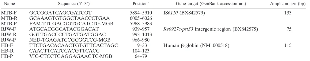
Design of primers and probes.Gene- specific primers and probes were de-signed by using Primer Express software version 3.0 (Applied Biosystems, Foster City, CA) (Table 2). Primers and TaqMan MGB probes targeting insertion sequence IS6110andRv0927c-pstS3intergenic region were based on theM. tuberculosisH37Rv genome sequence (GenBank accession numbers BX842579 and BX842575). The specificity of the primers was first verified by using the NCBI BLAST algorithm, followed by real-time PCR specificity testing with DNA extracted from the reference strains listed in Table 1.
Evaluation of 12 DNA extraction protocols with spiked specimens and clinical specimens.For the spiked specimens,M. tuberculosisH37Rv standard strain was grown at 37°C with orbital shaking (250 rpm) in Middlebrook 7H9 broth sup-plemented with oleic acid-albumin-dextrose-citric acid (OADC) to 1 McFarland standard. Then, 2-ml portions of 1:10 diluted culture were added to 50 ml of pooled culture-negative sputum specimens. Aliquots (2 ml) of spiked sputum were centrifuged at 5,000⫻g for 5 min. Each pellet was resuspended with 1 ml of TE buffer (pH 8.0) for subsequent DNA extraction. For the clinical specimens, 17M. tuberculosisculture-positive sputum specimens—9 AFB smear-positive and 8 AFB smear-negative specimens—were used. These specimens represented a range of different bacterial loads in a spectrum of different sputum viscosities.
TABLE 1. ATCC reference strains used in this study
Mycobacteriumstrain ATCC no.
MTCa
M. tuberculosisH37Rv...27294
M. bovis...35720
NTM M. asiaticum...25276
M. aurum...23366
M. avium...35718
M. chelonae...14472
M. chitae...19627
M. diernhoferi...19340
M. flavescens...14474
M. fortuitum... 6841
M. gastri...15754
M. gordonae...14470
M. intracellulare...13950
M. kansasii...12478
M. marinum... 927
M. neoaurum...25795
M. scrofulaceum...19981
M. smegmatis...19420
M. szulgai...35799
aMTC,M. tuberculosiscomplex.
on May 16, 2020 by guest
http://jcm.asm.org/
Aliquots (2 ml) of sputum were centrifuged and resuspended the same way as the spiked sputum above for subsequent DNA extraction.
DNA extraction.Three different types of beads used for mechanical disruption in the bacterial cell lysis step and four different downstream DNA purification methods were evaluated for a total of 12 protocols. The three different bead types included 0.1-mm zirconia beads (BioSpec Products, Inc., Bartlesville, OK), 0.2-mm glass beads (Sigma-Aldrich, St. Louis, MO), and 1-mm glass beads (Sigma Aldrich, St. Louis, MO). A portion (1 ml) of the TE-resuspended pellet was added to a 2-ml screw-cap microcentrifuge tube containing⬃200l of one of the three types of beads. The tubes were vortexed in a Disruptor Genie (Scientific Industries, Inc., Bohemia, NY) for 5 min and incubated at 95°C for 10 min for complete inactivation of the bacteria. After a brief centrifugation, 100-l aliquots of the lysate containing no debris were used for each DNA purification method.
Four different DNA purification methods were evaluated. Method C utilized Chelex 100 resins (Bio-Rad Laboratories, Hercules, CA). The lysate was added to an equal volume of 10% Chelex 100 resins, followed by incubation at room temperature for 5 min with occasional mixing. The mixture was centrifuged at 1,000⫻g for 2 min to collect the supernatant. The supernatants were directly used as DNA templates for real-time PCR. Method D utilized a QIAmp DNA minikit (Qiagen, Hilden, Germany). The lysate was processed according to the body fluid spin protocol supplied by the manufacturer. The purified DNA was eluted in 30l of buffer AE. Method V utilized a QIAmp MinElute virus spin kit (Qiagen). The lysate was processed according to the manufacturer’s recom-mended protocol (2010-Apr version). The purified DNA was eluted in 30l of Buffer AVE. Method HM: in-house buffers with Dynabeads MyOne SILANE magnetic beads (Invitrogen). The formulation of buffers is based on the buffer system previously described with slight modification (4). Briefly, binding buffer was prepared by dissolving 120 g of guanidium thiocyanate in 100 ml of 0.1 M Tris-HCl (pH 6.4). Subsequently, 22 ml of 0.2 M EDTA (pH 8.0) and 2.6 g of Triton X-100 were added to the solution. A wash buffer was prepared by com-bining 55 ml of ethanol and 45 ml of solution containing 3 M guanidium thio-cyanate, 10 mM Tris-HCl, and 10 mM NaCl (pH 8.0). To each 100-l portion of lysate, 200l of binding buffer was added, and the mixture was pulse vortexed. Then, 50l of Dynabeads and 200l of isopropanol were added, respectively, with brief vortexing in between. The tubes were incubated at room temperature for 3 min. Subsequently, the beads were immobilized by a magnet, and the supernatant was removed. The beads were washed twice with 800l of wash buffer and twice with 800l of 70% ethanol. The ethanol was removed by air drying the beads at room temperature for 10 min. DNA was eluted with 30l of TE buffer (pH 9.0). The extraction procedures for spiked specimens were re-peated nine times for each method for statistical analysis. For clinical specimens, each of the 17 specimens was extracted by all 12 evaluation methods.
Cloning of standards for real-time PCR.To monitor the efficiency of each run of real-time PCR, dilutions of standard with known quantities were included. For cloning, PCR was performed in a 50-l reaction by using an AmpliTaq Gold PCR reagent kit (Applied Biosystems), the primers MTB-F and MTB-R, and H37Rv genomic DNA. The reaction mixture was subjected to an initial poly-merase activation of 95°C for 10 min, followed by 30 thermal cycles of 94°C for 30 s, 60°C for 30 s, and 72°C for 30 s, with a final extension step at 72°C for 7 min in an Applied Biosystems 9700 thermal cycler. The amplicon appeared as a single band after electrophoresis. The amplicon was ligated to pCR2.1-TOPO vector by using a TOPO TA cloning kit (Invitrogen), and the recombinant vector was transformed into Top10Escherichia colicompetent cells according to the man-ufacturer’s recommendations. Transformants were isolated and subcultured for plasmid preparation by using a QIAprep spin miniprep kit (Qiagen). The DNA
sequence of the construct was confirmed by automatic DNA sequencing. Quan-tification of the plasmid was performed by measuring absorbance at 260 nm with a Nanodrop UV spectrophotometer (Invitrogen).
Multiprobe multiplex real-time PCR.Amplification was done in triplicates in 96-well optical reaction plates with an Applied Biosystems 7700 real-time PCR instrument. Amplification of human-globin gene by the primers HB-F and HB-R acts as internal amplification control to monitor PCR inhibition. The concentrations of the primers and probes were optimized one at a time by testing a range of concentrations for one component while fixing that of the others. The concentrations of the human-globin primers and probe were optimized to generate detectable signal when the other amplification is negative, so that the associated interference and resource competition is minimized. For comparison of the DNA extraction methods, each well contained 25l of reaction volume, including 12.5l of TaqMan Universal Master Mix, 500 nM MTB-F, 300 nM MTB-R, 50 nM MTB-P, and 2.5l of DNA extracts. For evaluation of the final developed assay with 335 clinical specimens, the 25-l reaction volume also contains an addition of 600 nM BJW-F, 900 nM BJW-R, 80 nM BJW-P, 50 nM HB-F, 30 nM HB-R, and 50 nM HB-P. The instrument was programmed to 95°C for 10 min, followed by 40 cycles of 95°C for 15 s and 60°C for 1 min. Quantified plasmid standards in 10-fold dilutions equivalent to a range of 20 to 2,000,000 IS6110copies were included in each run. Cycle threshold (CT) values of the
dilutions of standards were plotted against the number of copies of template to generate the standard curves. The values of slopes,yintercepts, and the corre-lation coefficients (r2
) of the linear regression lines were used as indicators of efficiency. Amplification that generated standard curves with slopes between 3.3 and 3.6,yintercepts between 39 and 40, andr2
values⬎0.995 was considered valid.
Statistical analysis.The efficiency of each DNA extraction method was eval-uated by comparing theCTvalues generated from real-time PCR, with the use of
the data analysis package included in Microsoft Excel software (Microsoft Corp., Redmond, WA). Statistical significance for different groups of spiked specimens extracted by different methods was analyzed by single-variance analysis of vari-ance (ANOVA). Detailed statistical significvari-ance findings for each method against each of any the other methods for spiked specimens and clinical speci-mens were tabulated by using the two-tailed Studentttest and pairedttest, respectively.Pvalues smaller than or equal to 0.05 were considered significant.
RESULTS
Primer specificities. Our real-time PCR correctly
identi-fied all 40M. tuberculosisisolates andM. bovis(members of
the M. tuberculosis complex). No cross-reaction with the NTM strains was observed. The 20 isolates with a Beijing/W genotype were also accurately differentiated from 20 non-Beijing/W strains, as verified by DTM-PCR results, provid-ing 100% specificity and sensitivity.
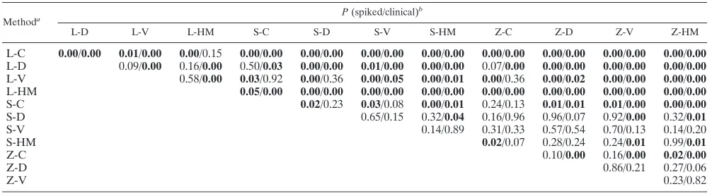
Comparison of DNA extraction methods.The efficiencies of
DNA extraction protocols were compared by theirCTvalues
generated from real-time PCR analysis of both spiked
speci-mens and clinical specispeci-mens (Table 3). The P value from
ANOVA analysis of the different protocols of extraction on
[image:3.585.41.547.83.184.2]spiked specimens was 2.57⫻10⫺17(Pⱕ0.05), which indicates
TABLE 2. Primers and probes used for real-time PCR
Name Sequence (5⬘–3⬘) Positiona
Gene target (GenBank accession no.) Amplicon size (bp)
MTB-F GCCGGATCAGCGATCGT 5894–5910 IS6110(BX842579) 133
MTB-R GCAAAGTGTGGCTAACCCTGAA 6005–6026
MTB-P FAM-TTCGACGGTGCATCTG-MGB 5968–5983
BJW-F ATGCACGGCATACGGACAT 939–957 Rv0927c-pstS3intergenic region (BX842575) 75
BJW-R GGTTGACCCCTGATGATGGAC 993–1013
BJW-P NED-TGAGATCCGCGGTCG-MGB 966–980
HB-F TTCTGACACAACTGTGTTCACTAGC 9–33 Human-globin (NM_000518) 115
HB-R CAACTTCATCCACGTTCACC 104–123
HB-P VIC-CTCCTGAGGAGAAGTC-MGB 64–79
aThat is, the base pair positions of the gene sequences in the corresponding GenBank accession.
on May 16, 2020 by guest
http://jcm.asm.org/
existence of significant difference among the efficiencies of different extraction protocols. Our results showed that me-chanical cell disruption with 0.2-mm glass beads (S) was more
efficient than with 1-mm glass beads (L). Contrasting theCT
values of protocols S-C versus L-C, S-D versus L-D, S-V versus L-V, and S-HM versus L-HM revealed cycle differences
(⌬CTs) from 2.1 to 3.7 with statistical significance (P⬍0.005)
(Table 4), suggesting 4.5- to 12.5-fold differences in the DNA yields. Zirconia beads (Z) have similar efficiencies as S (Z-C versus S-C, Z-D versus S-D, Z-V versus S-V, and Z-HM versus
S-HM;⌬CTsⱕ0.74;Pⱖ0.24). Comparison of the different
DNA purification methods using the same type of beads for cell lysis (i.e., L-C/L-D/L-V/L-HM, S-C/S-D/S-V/S-HM, and Z-C/Z-D/Z-V/Z-HM) showed that Chelex resins (C) had the
poorest efficiency among the four (0.96ⱕ⌬CTsⱕ2.49, 1.9- to
5.6-fold difference), while the other three purification methods
had similar efficiencies (⌬CTs ⱕ 0.81,P ⱖ 0.09). The three
most sensitive protocols with spiked specimens are S-HM,
Z-HM, and Z-D in increasing order ofCTvalues (decreasing
sensitivity).
Comparison using clinical specimens (including 9 AFB smear positive and 8 AFB smear negative sputum specimens) also suggested that Z and S were more efficient than L in
disruptingM. tuberculosiscells, which is in agreement with the
result from spike analysis. Zirconia beads in combination with magnetic beads (Z-HM) was found to be more efficient than
with 0.2-mm glass beads (⌬CT⫽0.97, P⫽ 0.01). The three
most sensitive protocols with clinical specimens are Z-HM,
Z-V, and Z-D in increasing order ofCTvalues.
To summarize the results from both spiked and clinical spec-imens, cell lysis by zirconia beads in combination with DNA purification by magnetic beads or spin columns consistently
produced the lowestCTvalues (highest yield of DNA) in
con-trast to the other protocols. Combinations involving large glass
beads or Chelex resin purification yielded significantly lessM.
tuberculosis DNA. Taking into consideration the sensitivity, technical simplicity of protocol, and potential for automation, the combination of zirconia beads and magnetic beads (Z-HM) was selected.
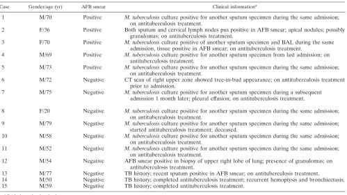
Evaluation of the developed extraction and real-time PCR
protocol.A total of 335 sputum specimens were extracted by
the Z-HM protocol, including 74M. tuberculosis
posi-tive, 31 NTM posiposi-tive, and 230 mycobacterial culture-negative specimens (Table 5). Of the 335 specimens, 89
(26.6%) were positive forM. tuberculosisby real-time PCR. All
74M. tuberculosis culture-positive specimens were positively identified by real-time PCR. Fifteen culture-negative speci-mens, including five that were AFB smear positive and ten that were AFB smear negative, were also determined to be positive by real-time PCR. Of these 15 specimens, 12 were obtained from patients diagnosed with TB (as determined either by histology/radiology or by conventional mycobacterial culture of other respiratory specimens from the same individual) and were undergoing TB chemotherapies (Table 6). The remaining three specimens originated from patients with records of re-cently treated TB. One of the three patients is currently on treatment for TB relapse. None of the NTM culture-positive
specimens was positive forM. tuberculosis by real-time PCR
[image:4.585.43.283.81.248.2](Table 5). Taking conventional mycobacterial culture as the
TABLE 3. MeanCTvalues of 12 different extraction methods
Methoda
CTb
Spiked specimens (n⫽9)
Clinical specimens (n⫽17)
Mean SD Mean SD
L-C 30.43 1.12 27.24 5.55
L-D 28.15 0.98 26.51 5.84
L-V 28.95 0.90 25.47 5.39
L-HM 28.96 1.11 27.77 6.03
S-C 27.79 1.19 25.51 5.15
S-D 26.02 1.55 25.16 5.42
S-V 26.31 1.26 24.60 5.45
S-HM 25.30 1.41 24.67 5.46
Z-C 27.05 1.41 25.14 5.38
Z-D 25.99 1.18 24.22 5.78
Z-V 26.09 1.33 23.79 5.27
Z-HM 25.31 1.36 23.70 5.57
a
L, large (1-mm) glass beads; S, small (0.2-mm) glass beads; Z, zirconia beads (0.1 mm); C, Chelex purification; D, DNA minikit (Qiagen); V, QIAmp Min-Elute virus spin kit; HM, in-house buffer with Invitrogen Dynabeads MyOne SILANE magnetic beads.
b
Comparisons ofCTvalues considered statistically significant (Pⱕ0.05) are
indicated in boldface.
TABLE 4. Pvalues for spiked and clinical specimens derived by statistical significance analysis between methods
Methoda P(spiked/clinical)
b
L-D L-V L-HM S-C S-D S-V S-HM Z-C Z-D Z-V Z-HM
L-C 0.00/0.00 0.01/0.00 0.00/0.15 0.00/0.00 0.00/0.00 0.00/0.00 0.00/0.00 0.00/0.00 0.00/0.00 0.00/0.00 0.00/0.00 L-D 0.09/0.00 0.16/0.00 0.50/0.03 0.00/0.00 0.01/0.00 0.00/0.00 0.07/0.00 0.00/0.00 0.00/0.00 0.00/0.00 L-V 0.58/0.00 0.03/0.92 0.00/0.36 0.00/0.05 0.00/0.01 0.00/0.36 0.00/0.02 0.00/0.00 0.00/0.00 L-HM 0.05/0.00 0.00/0.00 0.00/0.00 0.00/0.00 0.00/0.00 0.00/0.00 0.00/0.00 0.00/0.00 S-C 0.02/0.23 0.03/0.08 0.00/0.01 0.24/0.13 0.01/0.01 0.01/0.00 0.00/0.00
S-D 0.65/0.15 0.32/0.04 0.16/0.96 0.96/0.07 0.92/0.00 0.32/0.01
S-V 0.14/0.89 0.31/0.33 0.57/0.54 0.70/0.13 0.14/0.20
S-HM 0.02/0.07 0.28/0.24 0.24/0.01 0.99/0.01
Z-C 0.10/0.00 0.16/0.00 0.02/0.00
Z-D 0.86/0.21 0.27/0.06
Z-V 0.23/0.82
a
The methods are as defined in Table 3, footnotea.
b
Statistically significant results (Pⱕ0.05) are indicated in boldface.
on May 16, 2020 by guest
http://jcm.asm.org/
[image:4.585.41.544.572.711.2]gold standard, the overall sensitivity and specificity are 100% (74/74) and 94.3% (246/261), respectively. Taking into account the final clinical diagnosis of the patients, the specificity is 100% (246/246). Association of Beijing/W genotypes with AFB smear-positive and AFB smear-negative specimens were 59.6% (31/52) and 21.6% (8/37), respectively. The overall prev-alence of Beijing/W is 43.8% (39/89). The result was further confirmed by DTM-PCR (7) with complete concordance. The
detection limit of the assay is 10 bacilli, as determined by
endpoint dilution analysis. Aliquots of 100l of TE buffer were
included in each extraction and real-time PCR experiment as negative controls. Cross-contamination between specimens
was not detected. Internal controls remain positive for allM.
tuberculosis-negative specimens, indicating that inhibition of amplification was not encountered in this evaluation.
DISCUSSION
Sensitive detection of M. tuberculosis complex from body
fluids requires optimal cell lysis and efficient DNA purification to remove associated PCR inhibitors. The choice of reagents and methods is thus crucial. Chelex and the QIAmp DNA extraction kit are commonly used in DNA preparations, al-though the QIAmp DNA extraction kit is incompatible with mainstream automation for high throughput.
According to the results of the present study, the reagents and methods used in DNA extraction have a great impact on the DNA yield. Comparison using a spiked specimen pool suggests that S-HM is the most sensitive since it required the
fewest cycles to reach the threshold (CT) in real-time PCR
assay, followed by Z-HM, Z-D, Z-V, S-D, and S-V. In terms of sensitivity, these six protocols are better than the others tested. The evaluation with clinical specimens yielded similar but not identical order. Z-HM, Z-V, Z-D, and S-V were significantly more sensitive than other protocols, while the performance of S-HM and S-D were inferior. A possible explanation is that the efficiencies of mechanical disruption by beads could be affected by the viscosity and the actual bacterial load of sputum
speci-TABLE 5. Comparative sensitivities and specificities of real-time PCR with reference to conventional mycobacterial culture
Sputum specimens (n⫽335)a
No. of specimens detected by real-time PCRb
Non-Beijing/W Beijing/W
M. tuberculosisculture positive (n⫽74)
AFB smear positive (n⫽47) 47 28
AFB smear negative (n⫽27) 27 6
NTM culture positive (n⫽31)c
AFB smear positive (n⫽7) 0 0
AFB smear negative (n⫽24) 0 0
Culture negative for mycobacterium (n⫽230)
AFB smear positive (n⫽6) 5* 3
AFB smear negative (n⫽224) 10* 2
aAFB smear, acid-fast bacillus direct smear microscopy.
[image:5.585.42.282.91.252.2]b*, Clinical information regarding the corresponding patients is given in Table 6. cThis number includes 7M. avium, 6M. chelonae, 6 M. fortuitum, 1 M. gordonae, 4M. kansasii, 2M. neoaurum, 1M. simiae, 1M. terrae, and 3 Runyon group III mycobacterial specimens.
TABLE 6. Clinical information for 15 culture-negative cases determined to be positive by real-time PCR
Case Gender/age (yr) AFB smear Clinical informationa
1 M/70 Positive M. tuberculosisculture positive for another sputum specimen during the same admission; on antituberculosis treatment.
2 F/36 Positive Both sputum and cervical lymph nodes pus positive in AFB smear; apical nodules; possibly granulomas; on antituberculosis treatment.
3 F/70 Positive M. tuberculosisculture positive of another sputum specimen and BAL during the same admission, tissue positive in AFB smear; on antituberculosis treatment.
4 M/69 Positive M. tuberculosisculture positive for another sputum specimen from last admission; on antituberculosis treatment.
5 M/73 Positive M. tuberculosisculture positive for another sputum specimen during the same admission; on antituberculosis treatment.
6 M/72 Negative CT scan of right upper zone showed tree-in-bud appearance; on antituberculosis treatment prior to admission.
7 M/75 Negative M. tuberculosisculture positive for another sputum specimen during a subsequent admission 1 month later; pleural effusion; on antituberculosis treatment.
8 F/20 Negative M. tuberculosisculture positive for another sputum specimen during the same admission; on antituberculosis treatment.
9 M/79 Negative M. tuberculosisculture positive for another sputum specimen during the same admission; started antituberculosis treatment; deceased.
10 M/58 Negative M. tuberculosisculture positive for another sputum specimen during the same admission; on antituberculosis treatment.
11 M/52 Negative M. tuberculosisculture positive for another sputum specimen during the same admission; on antituberculosis treatment.
12 M/54 Negative AFB smear positive in biopsy of upper right lobe of lung; presence of granulomas; on antituberculosis treatment.
13 M/77 Negative TB history; recent sputum positive in AFB smear; on antituberculosis treatment.
14 M/50 Negative TB history; completed antituberculosis treatment; recurrent hemoptysis and bronchiectasis. 15 M/59 Negative TB history; completed antituberculosis treatment.
a
BAL, bronchoalveolar lavage.
on May 16, 2020 by guest
http://jcm.asm.org/
[image:5.585.44.543.440.721.2]mens. The results presented here suggest that zirconia beads are more compatible with the wide range of sputum viscosities. In both experiments using spiked specimens and clinical specimens, the performance of Chelex resins and large (1-mm)
glass beads was relatively poor. For instance, the CT of the
combination using large glass beads plus Chelex (L-C) with
spiked specimens was 30.43⫾1.12, which suggests the DNA
yield was⬃32-fold lower than that of S-HM. From our
obser-vation, 1-mm glass beads were relatively sluggish compared to the other two types of beads during vortex. The mild bombard-ment due to its heavy weight might lead to incomplete cell lysis and low DNA yield. On the other hand, Chelex purification suffers from a major disadvantage of dilution effect. While column and magnetic purification significantly concentrated the DNA by small-volume elution, Chelex purification inevita-bly caused a 2-fold dilution, which may account for its poor performance. Taking into consideration the results from the comparison analysis with both the spiked and the clinical spec-imens, the combination Z-HM was selected for its simplicity, high sensitivity, low cost relative to commercial spin columns, and potential for high-throughput automation.
Assays that detect insertion sequence IS6110for molecular
diagnosis of M. tuberculosis, such as the one described here,
benefits from the fact that IS6110often presents in high copy
numbers (⬎10 copies) in the genome of M. tuberculosis. A
common limitation of IS6110detections is the inability to
dis-criminate members of theM. tuberculosiscomplex such asM.
bovis and M. africanum, although the prevalence of these
agents in human is considerably less than that ofM.
tubercu-losis. In addition, a small portion (⬍2%) of M. tuberculosis
isolates does not contain IS6110 repeated segments in their
genome, which would give rise to false-negative results. Our novel multiprobe multiplex real-time PCR successfully detects
all 74 cultivableM. tuberculosisisolates with no cross-reaction
with NTM. Fifteen specimens determined to be positive by real-time PCR were culture negative. Since culture isolation is the gold standard for TB diagnosis, the possibility of false positivity among these specimens should not be excluded. However, when the clinical presentation of the corresponding patients and the results of repeated sampling are taken into consideration, the chance that these are real false positives is low (Table 6). Discrepancies between conventional mycobac-terial culture and PCR-based detection have been previously associated with TB patients who underwent antitubercular che-motherapies (2, 18). Since PCR could detect DNA from
non-viableM. tuberculosis as a result of antitubercular treatment
and also viableM. tuberculosisin insufficient quantity for
suc-cessful culture, the discrepancy found in the present study, together with the available clinical information, indicates an advantage in sensitivity of PCR-based detection over conven-tional mycobacterial culture.
The worldwide prevalence of Beijing genotype varies from 0
to⬎80% depending on geographic locations (19). Our finding
of 43.8% concurs with the characteristic high prevalence in the Asia-Pacific region. However, it should be noted that the spec-imens collected for this assay development were subjected to prior selection based on the two criteria stated; hence, the percentage is not necessarily a representative reflection of the regional status.
Contrasting conventional mycobacterial culture, the
molec-ular protocol described in the present study greatly reduces the turnaround time, which would be beneficial to both TB control and patient treatment outcome. It also avoids the hazards of maintaining a TB culture room. However, the protocol is more labor-intensive unless automation is available. Although the cost of reagents involved is significantly lower than that of commercially available rapid TB diagnosis systems, it is still higher than conventional mycobacterial culture, and this could be a barrier at the moment. As the cost of molecular reagents gradually drops, one may find this approach justifiable for its potential healthcare benefits in the foreseeable future. In con-clusion, the method used to extract DNA from sputum speci-mens greatly affects the outcome from molecular diagnosis. We describe here an optimized protocol for DNA extraction from sputum specimens and a multiprobe multiplex real-time
PCR for the simultaneous detection of M. tuberculosis and
Beijing/W genotype with high sensitivity and specificity.
ACKNOWLEDGMENTS
We sincerely thank Raymond Lai, Department of Microbiology, Prince of Wales Hospital, for his generous support and W. Y. Lau from the clinical microbiology laboratory for his assistance in specimen collection.
This study was solely supported by the Research Fund for the Con-trol of Infectious Diseases (RFCID 08070212), Food and Health Bu-reau, Hong Kong SAR government.
REFERENCES
1.Aldous, W. K., J. I. Pounder, J. L. Cloud, and G. L. Woods.2005. Compar-ison of six methods of extractingMycobacterium tuberculosisDNA from processed sputum for testing by quantitative real-time PCR. J. Clin. Micro-biol.43:2471–2473.
2.Bennedsen, J., et al.1996. Utility of PCR in diagnosing pulmonary tubercu-losis. J. Clin. Microbiol.34:1407–1411.
3.Bifani, P. J., B. Mathema, N. E. Kurepina, and B. N. Kreiswirth.2002. Global dissemination of theMycobacterium tuberculosisW-Beijing family strains. Trends Microbiol.10:45–52.
4.Boom, R., et al.2005. Rapid and simple method for purification of nucleic acids. J. Clin. Microbiol.28:495–503.
5.Burman, W. J., et al.2009. Relapse associated with active disease caused by Beijing strain ofMycobacterium tuberculosis.Emerg. Infect. Dis.15:1061– 1067.
6.Chang, J. R., et al.Genotypic analysis of genes associated with transmission and drug resistance in the Beijing lineage ofMycobacterium tuberculosis. Clin. Microbiol. Infect., in press.
7.Chen, J., et al.2007. Deletion-targeted multiplex PCR (DTM-PCR) for identification of Beijing/W genotypes ofMycobacterium tuberculosis. Tuber-culosis (Edinb.)87:446–449.
8.Edwards, U., T. Rogall, H. Blo¨cker, M. Emde, and E. C. Bo¨ttger.1989. Isolation and direct complete nucleotide determination of entire genes: characterization of a gene coding for 16S rRNA. Nucleic Acids Res.17:
7843–7853.
9.European Concerted Action on New Generation Genetic Markers and Tech-niques for the Epidemiology and Control of Tuberculosis.2006. Beijing/W genotypeMycobacterium tuberculosisand drug resistance. Emerg. Infect. Dis.
12:736–743.
10.Jiang, X., et al.2007. Identification of unique genetic markers in Rv0927c among Mycobacterium tuberculosis W-Beijing strains. Microbes Infect.
9:241–246.
11.Leung, K. L., et al.2009. Development of a simple and low-cost real-time PCR method for the identification of commonly encountered mycobacteria in a high throughput laboratory. J. Appl. Microbiol.107:1433–1439. 12.Manca, C., et al.2005. HypervirulentMycobacterium tuberculosisW/Beijing
strains upregulate type I IFNs and increase expression of negative regulators of the Jak-Stat pathway. J. Interferon Cytokine Res.25:694–701. 13.Rad, M. E., et al.2003. Mutations in putative mutator genes of
Mycobacte-rium tuberculosisstrains of the W-Beijing family. Emerg. Infect. Dis.9:838– 845.
14.Reed, M. B., et al.2004. A glycolipid of hypervirulent tuberculosis strains that inhibits the innate immune response. Nature431:84–87.
15.Reed, M. B., S. Gagneux, K. Deriemer, P. M. Small, and C. E. Barry 3rd.
2007. The W-Beijing lineage ofMycobacterium tuberculosisoverproduces triglycerides and has the DosR dormancy regulon constitutively upregulated. J. Bacteriol.189:2583–2589.
on May 16, 2020 by guest
http://jcm.asm.org/
16.Santos, A., et al.2010. Comparison of methods of DNA extraction for real-time PCR in a model of pleural tuberculosis. APMIS118:60–65. 17.Sun, Y. J., et al.2006. Tuberculosis associated withMycobacterium
tubercu-losisBeijing and non-Beijing genotypes: a clinical and immunological com-parison. BMC Infect. Dis.6:105.
18.Traore, H., A. van Deun, I. C. Shamputa, L. Rigouts, and F. Portaels.2006. Direct detection ofMycobacterium tuberculosiscomplex DNA and rifampin resistance in clinical specimens from tuberculosis patients by line probe assay. J. Clin. Microbiol.12:4384–4388.
19.van Soolingen, D., et al.1995. Predominance of a single genotype of Myco-bacterium tuberculosisin countries of east Asia. J. Clin. Microbiol.33:3234– 3238.
20.Wong, D. A., P. C. Yip, D. T. Cheung, and K. M. Kam.2001. Simple and
rational approach to the identification ofMycobacterium tuberculosis, Myco-bacterium aviumcomplex species, and other commonly isolated mycobacte-ria. J. Clin. Microbiol.39:3768–3771.
21.World Health Organization.2006. The stop TB strategy: building on and enhancing DOTS to meet the TB-related millennium development goals. Report WHO/HTM/TB/2006.368. World Health Organization, Geneva, Switzerland.
22.World Health Organization. 2010. Global tuberculosis control. Report WHO/HTM/TB/2010.7. World Health Organization, Geneva, Switzerland. 23.Wu, S., et al. 2009. Activation of theeis gene in a W-Beijing strain of
Mycobacterium tuberculosiscorrelates with increased SigA levels and en-hanced intracellular growth. Microbiology155:1272–1281.