for Determining the
In Vitro
Activity of Daptomycin versus
Staphylococcus aureus
and Enterococci
Thomas J. Kirn,a,bElizabeth Onyeaso,bMadiha Syed,bMelvin P. Weinsteina,b
Departments of Medicine (Infectious Disease)b
and Pathology and Laboratory Medicine,a
Rutgers Robert Wood Johnson Medical School, New Brunswick, New Jersey, USA
We systematically evaluated 5 methods for testing daptomycin versus 48Enterococcus faecalis, 51Enterococcus faecium, and 50
Staphylococcus aureusisolates using (i and ii) broth microdilution (BMD) with 50-mg/liter calcium medium supplementation
(reference method) and 30-mg/liter calcium medium supplementation (BMD30 method), (iii) Etest, and (iv and v) MicroScan panel 33 using 2 methods to prepare the bacterial inoculum (MicroScan turbidity and MicroScan Prompt). Isolates were catego-rized as susceptible (S) or nonsusceptible (NS) based on measured MICs. Essential (ⴞ1 dilution) agreement (EA) and categorical (S/NS) agreement (CA) for each method were compared to the reference method. ForE. faecium, categorical agreement was poor between the reference method and BMD30 as well as with the three commercial methods, with frequent false-NS results (30 for BMD30, 18 for Etest, 22 for MicroScan Prompt, and 25 for MicroScan turbidity). AllE. faecalisisolates were judged to be S by the reference method; two of these isolates were categorized as NS using the BMD30 method, and one was categorized as NS by all three commercial methods. AllS. aureusisolates were judged to be S using all five methods. MIC values determined by the comparator methods tended to be higher than those for the reference method, especially forE. faeciumisolates. EAs between the reference BMD and BMD30, Etest, MicroScan Prompt, and MicroScan turbidity were 63%, 63%, 63%, and 56%, respectively, for
E. faecium, 87%, 83%, 98%, and 80%, respectively, forE. faecalis, and all 100% forS. aureus.
D
aptomycin is a cyclic lipopeptide with activity against Gram-positive organisms. It was approved by the Food and Drug Administration initially in 2003 for the treatment of complicated skin and skin structure infections caused byStaphylococcus aureus, vancomycin-susceptibleEnterococcus faecalis, and some strepto-coccal species. Subsequently, in 2006, it was approved for the treatment ofS. aureusbacteremia and right-sided endocarditis. It is one of the few antibiotics that exhibitsin vitrobactericidal ac-tivity against enterococci (1).An evaluation of daptomycin activity trends against methicil-lin-resistantStaphylococcus aureus(MRSA) and vancomycin-re-sistant enterococci (VRE) during a 6-year period (2005 to 2010) in 32 U.S. medical centers showed that daptomycin exhibited sus-tained activity against an extensive collection of clinical isolates of MRSA and VRE from numerous U.S. medical centers over the last 6 monitored years (2). However, daptomycin-nonsusceptible (NS) enterococcal isolates have been reported and multiple mech-anisms of resistance have been described (3–7).
Currently, daptomycin is frequently used to treat infections caused by MRSA and VRE. In 2009, the clinical microbiology laboratory at Robert Wood Johnson University Hospital (RWJUH) (New Brunswick, NJ) began routinely testing Staphy-lococcus aureusand enterococci for daptomycin susceptibility us-ing the MicroScan Pos Combo panel type 33 (PC33) MIC plate (Siemens Healthcare Diagnostics, Tarrytown, NY) instead of the epsilometer test (Etest) (bioMérieux, Durham, NC). Following this change in methodology, we noted an increase in enterococci NS to daptomycin when tested using the MicroScan system. We suspected that the MicroScan method itself, rather than a change in prevalence of truly NS isolates, was responsible for this antibi-ogram trend. To address our concerns, we tested 11Enterococcus
spp. isolates in parallel using the MicroScan Pos combo type 33
panel microtiter plate method, the reference broth microdilution (BMD) method, and the Etest. The results of this pilot investiga-tion revealed numerous discrepancies between the reference method, the MicroScan susceptibility method, and the Etest. Al-though the MICs determined for the discrepant isolates were typ-ically within one dilution of each other, the MicroScan method resulted in NS interpretations in 5 isolates, whereas the reference method results were interpreted as susceptible (S).
Others have reported that isolates judged to be NS to dapto-mycin using MicroScan methods were frequently characterized as susceptible (S) when tested by Etest or BMD (8–10). Palavecino and Burnell compared daptomycin MIC results obtained by MicroScan and by Etest forS. aureusand enterococci and found that the MicroScan method demonstrated a rate of false nonsus-ceptible results as high as 88% forEnterococcus faecium, 90% forE. faecalis, and 87% forS. aureus(10). Bryant et al. evaluated 150 enterococcal isolates judged to be NS to daptomycin using an automated commercial method (MicroScan) by repeat testing with a variety of methods and demonstrated that only 20% were confirmed as NS (9). In both of these studies, enterococcal isolates were initially selected because they were judged to be NS to dap-tomycin using the routine testing methodology in the laboratory.
Received11 December 2013Returned for modification28 January 2014
Accepted12 March 2014
Published ahead of print19 March 2014
Editor:P. Bourbeau
Address correspondence to Thomas J. Kirn, [email protected].
Copyright © 2014, American Society for Microbiology. All Rights Reserved.
doi:10.1128/JCM.03439-13
on May 16, 2020 by guest
http://jcm.asm.org/
centrations of calcium.
MATERIALS AND METHODS
Isolate selection.FiftyS. aureus, 48E. faecalis, and 51E. faeciumblood culture isolates collected between January 2009 and April 2011 were se-lected from our frozen stock collection. Our collection consists of bacte-rial isolates from all positive blood cultures (except coagulase-negative staphylococci) identified at Robert Wood Johnson University Hospital and is limited to one isolate per episode of bacteremia. To minimize clon-ality, the first two isolates from each month of the study period were selected, with the exception of January and March of each calendar year, during which only the first isolate was selected.
Media and antimicrobial susceptibility testing. All isolates were tested for daptomycin susceptibility in triplicate using the MicroScan Pos Combo panel type 33 (PC33) MIC microtiter panel on the MicroScan instrument, the Etest, and the manual broth microdilution methods. In each case, the replicates were performed from a single inoculum. Clinical and Laboratory Standards Institute (CLSI) guidelines were used to cate-gorize isolates as susceptible (S) or NS (14). ForS. aureus, the S category includes isolates with MICs ofⱕ1g/ml and for enterococci, it includes isolates with MICs ofⱕ4g/ml. Organisms with MICs greater than these breakpoints are considered NS. For the MicroScan method, the turbidity and proprietary inoculation (Prompt) methods were performed for all isolates.
The reference broth microdilution method was performed in-house using 2-fold dilutions of daptomycin between 0.25g/ml and 64g/ml in BBL cation-adjusted Mueller-Hinton broth II (CA-MHB) (BD, Sparks, MD) supplemented to either 30 or 50 mg/liter Ca2⫹. CA-MHB mixtures with final Ca2⫹concentrations of 30 mg/liter and 50 mg/liter were pre-pared by adding 0.1 M CaCl2-2H2O. The amounts of 0.1 M CaCl2-2H2O added to the CA-MHB mixtures to achieve the desired final Ca2⫹ concen-trations were empirically determined (Ca2⫹measurements performed at Laboratory Specialists, Inc., Westlake, OH) for the specific lot of CA-MHB used in the study. Cubist Pharmaceuticals (Lexington, MA) pro-vided the daptomycin used in this study, and a single vial was used to prepare dilutions for all BMD assays. Aliquots of daptomycin diluted to a concentration of 1,280g/ml were stored at⫺70°C. The CLSI-specified control organism (E. faecalisATCC 29212) was included on each BMD plate.
The PC33 microtiter panels were inoculated according to the manu-facturer’s protocol using either the Prompt method or using a standard-ized turbidity-based method. The Prompt method employs the use of a collared inoculating wand that is touched to three isolated colonies. Re-traction of the wand through the collar results in the suspension of the bacteria in 30 ml of stabilized 0.1% pluronic D solution. This solution is then transferred to the microtiter plate. Inoculation of the microtiter plate using the turbidity method entailed transferring 100l of Tryptic soy broth adjusted to a 0.5 McFarland standard to a tube containing 0.1% pluronic D. The mixture was then transferred to an inoculating tray and a
broth supplemented with 50 mg/liter Ca⫹as the reference method and calculated essential (⫾1 dilution) agreement (EA) and categorical (S/NS) agreement (CA) for each commercial method compared to the reference method.
Patient privacy.The study protocol was reviewed by the Institutional Review Board (IRB) at Rutgers-Robert Wood Johnson Medical School, which determined that this study did not meet the regulatory definition of human subjects research provided in 45 CFR 46.102 and thus did not require IRB approval.
RESULTS
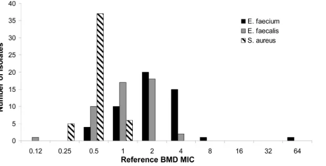
MIC distributions.Figure 1depicts the range of distributions of MICs obtained for each species tested using the reference BMD method. It is evident that theE. faeciumisolates tended to exhibit higher daptomycin MICs than did theE. faecalisandS. aureus
isolates. Notably, a significant number of theE. faeciumisolates (15/51) exhibited an MIC that was at the S/NS breakpoint (4
g/ml).
Reproducibility. Overall, excellent intrarun reproducibility was observed for all methods and all species. In most cases, the triplicate measurements were identical (data not shown). When rare deviations from identical triplicate measurements of MICs were observed, the differences were never⬎2-fold (1 dilution).
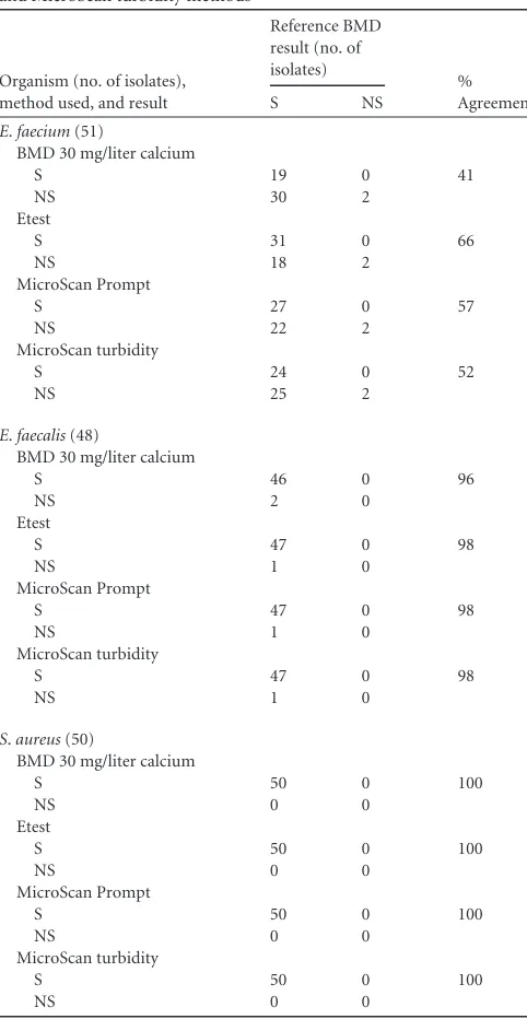
Categorical agreement.Categorical agreement between the reference BMD method, the BMD method using suboptimal cal-cium supplementation, and each of the commercial methods is depicted inTable 1. Categorical agreement between the reference method and the comparator methods forE. faeciumwas poor; 30 isolates were miscategorized using the BMD method with 30 mg/ liter calcium supplementation, 18 by Etest, 22 by MicroScan Prompt, and 23 by MicroScan turbidity. Categorical agreement levels forE. faeciumusing each of these four comparator methods were 41%, 66%, 57%, and 52%, respectively. Conversely, agree-ment was excellent forE. faecalis, for which only 2 isolates were miscategorized as NS using the suboptimally calcium-supple-mented BMD method and 1 was miscategorized as NS using the commercial methods. Similarly, categorical agreement forS. au-reuswas 100% for all comparator methods relative to the reference BMD method. Method-specific quality control results were within acceptable limits for each isolate tested by all five methods.
Figure 2demonstrates that, with the exception of 1 plate, BMD control tests, as specified by CLSI guidelines (E. faecalisATCC 29212), were routinely within the specified range (MICs, 1 to 4
g/ml) regardless of whether or not the Ca2⫹concentration was optimal (50 mg/liter) or suboptimal (30 mg/liter) (14).
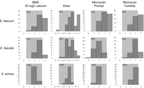
Essential agreement.Essential agreement (EA) between the
on May 16, 2020 by guest
reference BMD method, the suboptimally supplemented BMD method, and the three commercial methods varied widely among species (Fig. 3). Consistent with the observations for CA, EA be-tween the BMD reference method, the suboptimally calcium-sup-plemented BMD method, and all three commercial methods was poor forE. faeciumbut substantially better forE. faecalisandS. aureus. A trend toward higher MIC determinations, most pro-nounced forE. faeciumbut also present forE. faecalis, was evident when MICs were determined using the BMD method with subop-timal calcium supplementation, the Etest, and the MicroScan tur-bidity methods. ForE. faecium, EA was 63% for BMD with sub-optimal calcium supplementation, Etest, and MicroScan Prompt and 56% for MicroScan turbidity. ForE. faecalis, EA was 87% for BMD with suboptimal calcium supplementation, 83% for Etest, 98% for MicroScan Prompt, and 80% for MicroScan turbidity compared to the reference method. Finally, forS. aureusisolates, EA was 100% for all four comparator methods.
DISCUSSION
The selection of appropriate antimicrobial therapy for serious bacterial infections is predicated on accurate and timelyin vitro
antimicrobial susceptibility testing performed in the clinical mi-crobiology laboratory. Daptomycin is increasingly being used to treatS. aureusand enterococcal infections, especially when other antibiotic choices are limited due to resistance to other classes of antibiotics. In the case of VRE infection, daptomycin often repre-sents one of the few therapeutic options available for patients with life-threatening infections. The miscategorization of enterococcal isolates, especially VRE isolates, as NS to daptomycin when they are in fact S may impede timely implementation of effective anti-biotic therapy and lead to poor outcomes for some patients.
The effects of media formulations and especially calcium con-centrations on the accurate determination ofin vitrosusceptibility of Gram-positive organisms to daptomycin have been recognized for several decades (15). Consequently, CLSI protocols specify supplementation of CA-MHB with calcium to a final concentra-tion of 50 mg/liter for BMD susceptibility testing (14). However, very few clinical microbiology laboratories perform BMD testing at all and even fewer do so routinely. Instead, most laboratories rely on MIC determinations performed by Etest or automated methods. Etest is typically performed on MHA plates, which may have variable calcium concentrations depending on lot and/or
manufacturer (16). Likewise, automated methods may not con-sistently contain optimal calcium concentrations for daptomycin susceptibility testing and/or may contain other matrix compo-nents that could lead to inaccurate daptomycin susceptibility re-sults. Such variability between methods has been demonstrated by numerous investigators (9,10,16).
Our study design differs from those of previously reported in-vestigations of false daptomycin NS categorization for enterococci andS. aureusin that we selected isolates from patients with blood-stream infection irrespective of the daptomycin susceptibility de-termined at the time of isolation. Thus, our analysis is not biased toward isolates that may demonstrate daptomycin MICs close to the S/NS breakpoint and allows us to evaluate the daptomycin MIC distributions for enterococci andS. aureusisolated in our institution. When isolates were tested using the reference broth dilution method as specified by CLSI (50 mg/liter Ca2⫹ supple-mentation), very few daptomycin NS isolates were observed, consistent with nationwide and worldwide trends observed previ-ously (17–18). However, when Ca2⫹supplementation of CA-MHB in broth microdilution tests was suboptimal (30 mg/liter Ca2⫹), a significant number ofE. faeciumisolates with daptomy-cin MICs that would be interpreted as NS were observed. This is a direct consequence of the fact that a large number of theE. faecium
isolates included in our study demonstrated reference BMD MICs that were 4g/ml, which is the enterococcal interpretive cutoff for daptomycin. A small change in calcium concentration in the BMD method led to MICs that were just one dilution higher for many isolates but resulted in a significant number of isolates miscatego-rized as NS. These observations clearly illustrate the care that must be taken to ensure that accurate Ca2⫹supplementation is achieved when performing BMD for daptomycin in the clinical laboratory. This is sometimes difficult, however, since preparations of MHB and CA-MHB may vary considerably in Ca2⫹concentrations, re-quiring manufacturer-specific and even lot-specific changes in the amounts of supplementation required to achieve the target final Ca2⫹concentrations. In some cases, empirical determination of Ca2⫹concentrations using specific reagents may be the only way to ensure accurate final Ca2⫹concentrations and thus accurate daptomycin MICs. Obviously, the analytical measurement of cal-cium in supplemented media is beyond the capacity of most clin-ical microbiology laboratories, so arrangements with a reference FIG 1Daptomycin MIC distributions determined for all isolates using the reference broth microdilution technique.
on May 16, 2020 by guest
http://jcm.asm.org/
[image:3.585.137.455.66.228.2]laboratory must be sought. It is also important to note that sub-optimal Ca2⫹supplementation (at least at levels ofⱖ30 mg/liter) are not reliably revealed by testing the recommendedE. faecalis
control strain since, in our study, daptomycin MICs determined for the control strain using BMD with 30 mg/liter Ca2⫹ supple-mentation were, with one exception, within the acceptable range specified by CLSI (14).
To our knowledge, our study is the first to compare daptomy-cin susceptibility testing using Etest and two MicroScan methods to the reference BMD method using randomly selected enterococ-cal andS. aureusisolates. The results are consistent, however, with those of other studies that have used isolates selected based on daptomycin NS determinations (9–10). Palavecino and Burnell
As also reported by Bryant et al., all of theE. faeciumisolates and the singleE. faecalisisolate miscategorized as daptomycin NS using the commercial methods had daptomycin MICs of 4g/ml as judged by the reference BM method (9). Since a large number of
E. faeciumisolates exhibited MICs of 4g/ml while E. faecalis
isolates tended to have lower MICs, it is not surprising that CA for
E. faecaliswas better than forE. faecium. Interestingly, however, it appears that the fact that the daptomycin population MIC values approach the breakpoint for susceptibility forE. faeciumdoes not completely explain our findings. The bias toward higher MIC de-terminations using all three commercial methods was greater for
E. faeciumthanE. faecalis. This suggests that whatever influences the method-dependent variability in daptomycin susceptibility testing does not equally impact all species tested. This conclusion is further supported by the fact that the essential agreement be-tween all three commercial methods with reference BMD was 100% forS. aureus.
In summary, we have confirmed the previously reported over-estimation of daptomycin NSE. faeciumusing MicroScan and Etest methods with an unbiased panel of clinical isolates. Addi-tionally, we have systematically studied a large number ofE. faeca-lisandS. aureusisolates and confirmed that this phenomenon is primarily limited toE. faeciumisolates. Clearly, daptomycin NS results obtained using the MicroScan should be confirmed. How-ever, the best method for confirmation is unequivocally BMD, which most laboratories do not routinely perform. Furthermore, given the technical complexities associated with Ca2⫹
supplemen-FIG 2Daptomycin MICs observed for the CLSI-recommended enterococcal (ATCC 29212) control organism in each BMD run with either suboptimal (30 mg/liter) Ca2⫹supplementation or reference (50 mg/liter) Ca2⫹
supplemen-tation.
S 27 0 57
NS 22 2
MicroScan turbidity
S 24 0 52
NS 25 2
E.faecalis(48)
BMD 30 mg/liter calcium
S 46 0 96
NS 2 0
Etest
S 47 0 98
NS 1 0
MicroScan Prompt
S 47 0 98
NS 1 0
MicroScan turbidity
S 47 0 98
NS 1 0
S.aureus(50)
BMD 30 mg/liter calcium
S 50 0 100
NS 0 0
Etest
S 50 0 100
NS 0 0
MicroScan Prompt
S 50 0 100
NS 0 0
MicroScan turbidity
S 50 0 100
NS 0 0
on May 16, 2020 by guest
[image:4.585.41.282.92.558.2] [image:4.585.300.540.554.684.2]tation of media used in BMD for daptomycin, many laboratories might find it difficult to employ such testing, especially on a spo-radic basis. If laboratories do choose to implement BMD for con-firmation of daptomycin NS organisms, calcium supplementation must be very carefully controlled, as suboptimal levels will have significant impacts on MIC determinations and lead to further miscategorization. In the absence of the ability to refer isolates judged to be daptomycin NS by MicroScan for BMD testing or perform it in-house, retesting using the same method or a differ-ent inoculation technique is not useful, but Etest may offer a slight increase in accuracy.
ACKNOWLEDGMENTS
This work was supported by an investigator-initiated study grant from Cubist Pharmaceuticals.
We thank Giovanni Divinagracia for expert technical assistance.
REFERENCES
1.Eliopoulos GM, Willey S, Reiszner E, Spitzer PG, Caputo G, Moellering RC, Jr.1986.In vitroandin vivoactivity of LY 146032, a new cyclic lipopeptide antibiotic. Antimicrob. Agents Chemother.30:532–535.http: //dx.doi.org/10.1128/AAC.30.4.532.
2.Sader HS, Moet GJ, Farrell DJ, Jones RN.2011. Antimicrobial suscep-tibility of daptomycin and comparator agents tested against methicillin-resistantStaphylococcus aureusand vancomycin-resistant enterococci: trend analysis of a 6-year period in US medical centers (2005–2010). Di-agn. Microbiol. Infect. Dis. 70:412– 416. http://dx.doi.org/10.1016/j .diagmicrobio.2011.02.008.
3.Arias CA, Panesso D, McGrath DM, Qin X, Mojica MF, Miller C, Diaz L, Tran TT, Rincon S, Barbu EM, Reyes J, Roh JH, Lobos E, Sodergren E, Pasqualini R, Arap W, Quinn JP, Shamoo Y, Murray BE, Weinstock GM.
2011. Genetic basis forin vivodaptomycin resistance in enterococci. N. Engl. J. Med.365:892–900.http://dx.doi.org/10.1056/NEJMoa1011138. 4.Davlieva M, Zhang W, Arias CA, Shamoo Y.2013. Biochemical
char-acterization of cardiolipin synthase mutations associated with daptomy-cin resistance in enterococci. Antimicrob. Agents Chemother.57:289 – 296.http://dx.doi.org/10.1128/AAC.01743-12.
5.Kelesidis T.2013. Transport of daptomycin resistance genes between animals and humans as a possible mechanism for development of de novo daptomycin resistance in enterococci. Epidemiol. Infect.141:2185–2186. http://dx.doi.org/10.1017/S0950268812002865.
6.Mishra NN, Bayer AS, Tran TT, Shamoo Y, Mileykovskaya E, Dowhan W, Guan Z, Arias CA.2012. Daptomycin resistance in enterococci is associated with distinct alterations of cell membrane phospholipid content. PLoS One 7:e43958. http://dx.doi.org/10.1371/journal.pone .0043958.
7.Palmer KL, Daniel A, Hardy C, Silverman J, Gilmore MS.2011. Genetic basis for daptomycin resistance in enterococci. Antimicrob. Agents Che-mother.55:3345–3356.http://dx.doi.org/10.1128/AAC.00207-11. 8.Gomez-Garces JL, Lopez-Fabal F, Burillo A, Gil Y.2010. Comparative
study of the susceptibility to daptomycin and other antimicrobials against
Staphylococcusspp. resistant to methicillin andEnterococcusspp. using Wider, E-test, and microdilution methods. Rev. Esp Quimioter23:87–92. (In Spanish.)
9.Bryant KA, Roberts AL, Rupp ME, Anderson JR, Lyden ER, Fey PD, Van Schooneveld TC.2013. Susceptibility of enterococci to daptomycin is dependent upon testing methodology. Diagn. Microbiol. Infect. Dis.
76:497–501.http://dx.doi.org/10.1016/j.diagmicrobio.2013.04.019. 10. Palavecino EL, Burnell JM.2013. False daptomycin-nonsusceptible MIC
FIG 3Differences (number of dilutions) in daptomycin MIC determinations between the reference method (BMD with 50 mg/liter Ca2⫹supplementation),
BMD with 30 mg/liter calcium supplementation, and three commercial methods. Theyaxes represent the number of organisms, and thexaxes represent the number of dilutions of difference between the reference method and the comparator methods. Positive differences indicate that the reference method established a lower MIC than the comparator. Shaded areas represent acceptable levels of disagreement (⫾1 dilution). The percentages of isolates falling within the acceptable range (percent essential agreement) are noted in the upper left corners of the shaded areas.