of Rapid Multiplex Ligation-Dependent Probe Amplification
Duy Pham Thanh,aNga Tran Vu Thieu,aChau Tran Thuy,aMartin Lodén,cKiki Tuin,cJames I. Campbell,a,eNguyen Van Minh Hoang,a Phat Voong Vinh,aJeremy J. Farrar,a,eKathryn E. Holt,bGordon Dougan,dStephen Bakera,e,f
The Hospital for Tropical Diseases, Wellcome Trust Major Overseas Programme, Oxford University Clinical Research Unit, Ho Chi Minh City, Vietnama
; Department of Microbiology and Immunology, The University of Melbourne, Melbourne, Australiab
; MRC-Holland, Amsterdam, The Netherlandsc
; The Wellcome Trust Sanger Institute, Hinxton, Cambridge, United Kingdomd
; Centre for Tropical Diseases, University of Oxford, Oxford, United Kingdome
; The London School of Hygiene and Tropical Medicine, London, United Kingdomf
Salmonella entericaserovar Typhi, the causative agent of typhoid fever, is highly clonal and genetically conserved, making iso-late subtyping difficult. We describe a standardized multiplex ligation-dependent probe amplification (MLPA) genotyping scheme targeting 11 key phylogenetic markers of theS. Typhi genome. The MLPA method demonstrated 90% concordance with single nucleotide polymorphism (SNP) typing, the gold standard forS. Typhi genotyping, and had the ability to identify isolates of the H58 haplotype, which is associated with resistance to multiple antimicrobials. Additionally, the assay permitted the detec-tion of fluoroquinolone resistance-associated mutadetec-tions in the DNA gyrase-encoding genegyrAand the topoisomerase gene parCwith a sensitivity of 100%. The MLPA methodology is simple and reliable, providing phylogenetically and phenotypically relevant genotyping information. This MLPA scheme offers a more-sensitive and interpretable alternative to the nonphyloge-netic subgrouping methodologies that are currently used in reference and research laboratories in areas where typhoid is endemic.
T
yphoid fever, caused bySalmonella entericaserovar Typhi (S.Typhi), remains a significant cause of mortality and morbidity
in many regions of the world (1,2).S. Typhi is a pathogen that is
exclusive to humans, and this exquisite level of host restriction is reflected in the genome and population structure of the organism
(3).S. Typhi isolates share an exceptionally high level of genetic
conservation, making informative subtyping for local and
inter-national epidemiology challenging (3). Many methods involving
both phenotypic and genotypic techniques have been developed
to differentiate betweenS. Typhi isolates. Phage typing was long
considered the standard method for strain subtyping; however, this technique provides little or no phylogenetic information, is technically intensive, and is only performed in specialized
refer-ence laboratories (4,5). Multilocus sequence typing (MLST)
can-not productively be applied toS. Typhi because of its
monophy-letic nature (6). Other molecular techniques, including random
amplified polymorphic DNA PCR (RAPD-PCR), pulsed-field gel electrophoresis (PFGE), and amplified fragment length polymor-phisms (AFLP), have some discriminatory power but provide a limited ability to define absolute phylogenetic and
epidemiologi-cal relationships between isolates (7–9). More recently, genome
sequencing and the identification of single nucleotide polymor-phisms (SNPs) have provided the first well-defined phylogenetic
framework within which to assignS. Typhi isolates to irrefutable
genotypes that can describe the population (10–13). These studies
revealed thatS. Typhi genotypes are globally dispersed but are
increasingly dominated by a single genotype (H58), which com-monly displays resistance to multiple antimicrobials. H58 isolates frequently harbor multidrug-resistant incompatibility group HI1
(IncHI1) plasmids, as well as mutations in thegyrAgene, which
confer reduced susceptibility to fluoroquinolones (7,13).
While SNP detection and genome sequencing are the gold
standards forS. Typhi subtyping (7,10,11), these methodologies
remain in the domain of research institutions, which have the
facilities, expertise, and infrastructure to support these activities. Therefore, there is a translation gap that stands in the way of
utilizing genomic information for subtypingS. Typhi in endemic
locations. To address this, we have developed a simple
transport-able method for genotypingS. Typhi that can be performed in a
laboratory with only modest molecular biology capabilities.
S. Typhi genomes harbor several genomic insertions and
dele-tions (indels) that we hypothesize to be phylogenetically informa-tive and may be used as markers for inferring genotypes within the
S. Typhi population (12). Therefore, we predicted that by assaying
for selected indels in theS. Typhi genome, we can infer genotype,
rather than having to perform genome-wide SNP analysis or ge-nome sequencing. Multiplex ligation-dependent probe amplifica-tion (MLPA) is a PCR method that permits multiple DNA
se-quence targets to be amplified with a single primer pair (14). The
ability of the method to detect DNA signatures, as well as individ-ual SNPs, increases its potential utility and provides a system that can be globally standardized. We have designed, validated, and
used an MLPA assay to genotypeS. Typhi isolates and investigate
the molecular epidemiology of typhoid in Asia. Our data show
thatS. Typhi MLPA is simple, robust, and comparable to the gold
standard of SNP typing, permitting the identification of the major
Received16 April 2013Returned for modification15 May 2013 Accepted24 June 2013
Published ahead of print3 July 2013
Address correspondence to Stephen Baker, [email protected].
Supplemental material for this article may be found athttp://dx.doi.org/10.1128 /JCM.01010-13.
Copyright © 2013 Pham Thanh et al. This is an open-access article distributed under the terms of theCreative Commons Attribution 3.0 Unported license. doi:10.1128/JCM.01010-13
on May 16, 2020 by guest
http://jcm.asm.org/
genotypes, as well as mutations that are associated with reduced susceptibility to fluoroquinolones. The method presented here can be applied to other organisms and represents an alternative to MLST, PFGE, genome sequencing, and serotyping when the abil-ity to perform these methods or the methods themselves are lim-ited.
MATERIALS AND METHODS
Bacterial isolates.A total of 227Salmonellaisolates (217S. Typhi and 10
S.entericaserovar Paratyphi A) were used in this study (see Table S1 in the supplemental material). All bacterial isolates originated from previous investigations conducted in southern Vietnam between 1993 and 2005 or across seven other Asian countries between 2002 and 2004, as previously described (15). NineteenS. Typhi isolates that were previously whole-genome sequenced were used as controls for SNP typing and validation of the MLPA method; these isolates were provided by the Wellcome Trust Sanger Institute (United Kingdom) collection and are additionally de-scribed in Table S1 in the supplemental material (12). The 10S. Paratyphi A isolates originated from India and were included as an out-group for SNP genotyping and MLPA analysis.
Microbiological methods.All bacterial isolates were identified by standard biochemical tests and agglutination withSalmonella-specific an-tiserum (Murex Diagnostics, Dartford, United Kingdom). Antimicrobial susceptibilities were tested at the time of isolation by the modified Bauer-Kirby disk diffusion method, as recommended by the CLSI guidelines (16). The antimicrobial disks tested were chloramphenicol (CHL) (30
g), ampicillin (AMP) (10g), trimethoprim-sulfamethoxazole (SXT) (1.25/23.75g), ofloxacin (OFX) (5g), ciprofloxacin (CIP) (5g), and nalidixic acid (NAL) (30g). MICs were determined by Etest against CIP, NAL, and OFX, according to the manufacturer’s recommendations (AB Biodisk, Sweden). Mueller-Hinton agar and antimicrobial disks were pur-chased from UniPath, Basingstoke, United Kingdom. Escherichia coli
strain ATCC 25922 andStaphylococcus aureusstrain ATCC 25923 were
used as control strains for these assays. The results were interpreted according to current CLSI guidelines (16). An isolate was defined as multidrug resistant (MDR) if it was resistant to chloramphenicol, tri-methoprim-sulfamethoxazole, and ampicillin.
PCR amplification and DNA sequencing.DNA was extracted from all
S. Typhi isolates using the Wizard genomic DNA extraction kit (Promega, Fitchburg, WI). Oligonucleotide primers for the amplification of the quinolone resistance-determining regions (QRDRs) in thegyrAandparC
genes inS. Typhi were as described inTable 1. PCRs were performed under the following conditions: 92°C for 45 s, 55°C or 62°C (depending on the primer pair) for 45 s, and 74°C for 1 min, for 30 cycles, followed by a final extension step at 74°C for 2 min. DNA sequencing reactions were performed using the CEQ Dye Terminator cycle sequencing (DTCS) quick start kit (Beckman Coulter) and were sequenced using a CEQ 8000 capillary sequencer (Beckman Coulter). All resulting DNA sequences were analyzed using the CEQuence investigator CEQ2000XL (Beckman Coulter) before verification, alignment, and editing in BioEdit software (http://www.mbio.ncsu.edu/BioEdit/bioedit. html). BLASTn at the NCBI was used to compare all resultinggyrAandparCsequences to those of globalS. Typhi isolates. PCR amplification was used to confirm the inser-tion or deleinser-tion of the target sequence across allS. Typhi isolates prior to probe design and MLPA analysis; the primers used are shown inTable 1. PCRs were performed using the following conditions: 92°C for 45 s, 55 to 62°C for 45 s, and 74°C for 45 s, for 30 cycles, followed by a final extension step at 74°C for 2 min (Table 1).
[image:2.585.44.547.78.355.2]MLPA.All MLPA reagents were manufactured and supplied by MRC-Holland (Amsterdam, The Netherlands). SixteenS. Typhi-specific MLPA probes were designed according to the manufacturer’s recommendations. The probes are described inTable 2and target 11S. Typhi indels (B, Q, A, K, E, F, S, C, H, N, and D) (Fig. 1). MLPA was performed according to a standard protocol developed by MRC-Holland, as previously described (14). Briefly,S. Typhi genomic DNA was diluted with Tris-EDTA (TE) buffer to final volume of 5l (50 to 200 ng) and heated at 98°C for 10 min
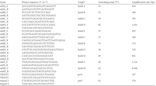
TABLE 1Conventional PCR primers targetingS. Typhi indels and SNPs
Primer Primer sequence (5=to 3=) Targeta Annealing temp (°C) Amplification size (bp)
indelA_F AGCGATGTGATGATCAGGATT Indel A 55 418
indelA_R AATGGCGTGTTCAGTGGATT
indelB_F TCCGTCTCTTTCTCCAGC Indel B 55 368
indelB_R AATTGATGCTGCTGCTGGACG
indelC_F ACGGGTGAAATACTCGAACG Indel C 58 783
indelC_R CACCAAGCAGATTGTTCAGG
indelD_F CGCTATTTTTTCCGCCCATGC Indel D 60 1,451
indelD_R TAATAACATCGGCGTGCCG
indelE_F CCGTCGCCAAAGTGACGC Indel E 55 993
indelE_R CCGTTGAATCGGAAGTAATAATCG
indelF_F AAGCAAATGCTTAGCACCAC Indel F 55 180
indelF_R CAATGCATAAAGTTAATTTAATCAGGA
indelK_F ATGGGTGAGCGCCTCTTTGG Indel K 55 155
indelK_R GACTGGCTGGACATTTTGTG
indelN_F ATGTTTCATGTGTGGGTAGGGTTGCC Indel N 58 720
indelN_R AAGAATGCCCATTGAGCGG
indelQ_F CAACACCCGTGCGGACGAT Indel Q 60 841
indelQ_R AGCTTACTTCCGGCTCCGAC
indelS_F TTGGTGATAAAATTGGCTCGGG Indel S 60 1,116
indelS_R AAAGAATGGAAACCAGAGTTTCC
indelH_F GTGGCAAAACAACGCATCG Indel H 62 6,560
indelH_R CGGTGGAGTTAGTGATGCTGA
GYRA/P1 TGTCCGAGATGGCCTGAAGC gyrA 55 347
GYRA/P2 TACCGTCATAAGTTATCCACG
StmparC1 CTATGCGATGTCAGAGCTGG parC 62 270
StmparC2 TAACAGCAGCTCGGCGTATT
aIndel sites correspond to those shown inFig. 1.
on May 16, 2020 by guest
http://jcm.asm.org/
in a thermocycler (Bio-Rad). After denaturation, the DNA was hybridized with MLPA reagents at 60°C. After overnight incubation, ligation was performed at 54°C for 15 min, followed by 98°C for 5 min. Ten microliters of the ligation mixture was added to 30l of PCR buffer. After equilibra-tion at 60°C, 10l of SALSA PCR master mix, containing the labeled forward primer 5=-FAM-GGGTTCCCTAAGGGTTGGA-3= (FAM, 6-carboxylfluorescein) and the unlabeled reverse primer 5=-GTGCCAGC AAGATCCAATCTAGA-3=, was added. PCR amplification was per-formed using 35 cycles of 95°C for 30 s, 60°C for 30 s, and 72°C for 45 s, followed by 72°C for 20 min. The resulting MLPA amplicons were exam-ined by agarose gel (3%) electrophoresis and by fragment analysis using an ABI 3130xl capillary electrophoresis system (Applied Biosystems). For fragment analysis, 0.5l of PCR amplicon was mixed with 9.25l of Hi-Di formamide and 0.25l 500 LIZ size standard (GeneScan; Applied Biosystems). The mixture was incubated for 3 min at 95°C, chilled for 10 min, and analyzed. The resulting fragment analysis data were analyzed using GeneMapper v4.0 (Applied Biosystems). Data were analyzed using BioNumerics (Applied Maths, Belgium), and phylogenetic trees were drawn using Dendroscope v2.3.
Single nucleotide polymorphism genotyping.A cross-sectional sub-set of 73S. Typhi isolates was arbitrarily selected to encompass a range of locations, antimicrobial resistance patterns, studies in Vietnam, and dates of isolation for gold-standard SNP genotyping using the Illumina GoldenGate platform (see Table S1 in the supplemental material). DNA was extracted from allS. Typhi isolates as described above, and concen-trations were assessed using the Quant-iT kit (Invitrogen, Carlsbad, CA)
prior to SNP typing. The chromosomal haplotype ofS. Typhi isolates was determined based on the SNPs present at 1,485 chromosomal loci identi-fied previously from genome-wide surveys (12,17). All loci were interro-gated using a GoldenGate (Illumina) custom assay according to the man-ufacturer’s standard protocols, as described previously (10,11,18,19). SNP calls were generated from raw fluorescence signal data by clustering with a modified version of Illuminus, as described previously (10,11,18,
19). SNP alleles were concatenated to generate a multiple-sequence ment; maximum likelihood phylogenetic trees were fitted to each align-ment using RAxML with a general time reversible plus gamma (GTR⫹␥) model and 1,000 bootstraps as previously described (11,20).
RESULTS
Development of MLPA to detect phylogenetically informative indels in theS. Typhi genome.Data gleaned from genome se-quencing predicted that the presence or absence of certain indels
within theS. Typhi chromosome might be phylogenetically
infor-mative markers for assigningS. Typhi isolates to the genotype
groups originally defined by SNP analysis (12). Consequently,
conventional PCR assays were designed to independently detect
11 of these previously described indels (12). Each target (A, B, C,
D, E, F, H, K, N, Q, and S) was selected to represent one of the
main branches of theS. Typhi phylogenetic tree, and where two
indels were present on one branch, one was selected to avoid
[image:3.585.40.546.77.416.2]re-dundancy (Table 1,Fig. 1).
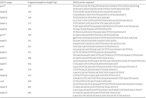
TABLE 2MLPA probe targets and sequences
MLPA target Fragment/amplicon length (bp) MLPA probe sequencea
parCb 130 TCAATACGACTCTAGATTAGACACCATGGCTTCATAGCAGGCGA
TGTCGCCATGCGGGTGATACTTACCTATTATGCTGAGC
Indel B 139 TTCCGGTCAGACTTGGACGCAAAATACAGGTA
CGAAAGGCCAGCTCGTGAAAATCCATAGAGGGGCT
Indel Q 158 TTATGGACGCTGGTGCAGCAGGAG
CCCTCCCATCCGTTAATTGTTAGTACCACGCTGAACACCG
Indel A 175 TTTTAGTCCATCACGCGCTTCAACATCCGTTC
TATCAGGATGTTGAAAAAGTGGTGCGCATTAATCCACTGAACAC
Indel K 204 TCGACTGGCTGGACATTTTGTGGTTC
TCTGCCGATGCGCTGGAGCAGCTTTGTGATGCGCT
gyrAb,c 238 CCAGCGAGAATGGCTGCGCCATACGAACGA
AATCGCCGTGGGGATGGTATTTACCGATTACGTCACCAACGAC
Indel E 252 GGAACATGGCGATATCAAATCCATCAATG
AAAGCGTCCAGTCCCTGTTTGATGCTCAGTCCCTG
aroC 267 GACAACAACGATAACGGAGCCGTGATGGCA
GGAAACACAATTGGACAACTCTTTCGCGTAACCACTTTCG
Indel S 275 GCTCTCTGGCTTTTTGAACACAGAAATGG
TCGAGTGAAATCAGCTTTGACTTAGCCCTTTTATAAAGCCTTGCGGC
Indel F 308 GCAGTCAGATTTGAACGTCCGGCATAA
AAATAAGTGCTATTAAGCTCTTCAACTGTATCCATACTCTAATTTCCTG
Indel C 362 CTAAAGCGTCGTTGATAATGGTGTAACGGT
GAACTGTCACAGGTCGTGGCGTTATCTTCGATAAACTTCACCT
fliC 374 CTGTTGACCCAGAATAACCTGAACAAATCC
CAGTCCGCACTGGGCACTGCTATCGAGCGTTTGT
Indel H 414 CTTGCTTTACCGAGCAGCATATTCTTTCCGCT
ATGACCGTTCAGTATTTGCAGAAGAAAGGCTTTCAGGTTCAGCC
ssaV 429 CCTGATTCCCTCACCTAACCATGAAC
GCATTGCGACTCCAGAAATTTTATTTGTCGATGATGTAATCGTAACC
Indel N 465 CCAGCACAGGCGTATTATCGCTAACACGCT
CACCAACATAGTCGATTTCATGCCAGTAAACTATTGGTAGCCTGGT
Indel D 494 CCTGGTCAAGCGTTAACTTTATTACTGCCCAT
GACGGCTTCACGCTGAACGATCTGGTGTCTTATAACGA
aProbes comprise left and right hybridization sequences.
b
Probe contains a nucleotide to detect a specific SNP in the QRDR region (boldface and underlined).
cgyrAprobe requires a spanning oligonucleotide to detect mutations at codons 83 and 87 (TGGTGTCATACACTGCGA).
on May 16, 2020 by guest
http://jcm.asm.org/
Conventional PCR amplification, detecting indels at the 11
tar-get loci, was performed on DNA extracted from the 19S. Typhi
isolates that had been whole-genome sequenced (12) (Table 3).
The PCR amplification results (PCR⫹/indel⫹and PCR⫺/indel⫺)
corresponded precisely with the genome sequencing data, con-firming the phylogenetic potential of these 11 indel targets in the selected isolates.
An MLPA probe set was designed to detect the same 11 indels,
as well as SNPs in thegyrAandparCgenes and sequences within
the conservedS. Typhi genesaroC,ssaV, andfliCto serve as
con-trols for hybridization, amplification, and detection. The probe sequences and the predicted amplicon length for each target are
shown inTable 2. ThegyrAandparCprobes were designed to
detect SNPs associated with reduced susceptibility to fluoro-quinolones within the QRDRs. Although several mutations have
been identified atgyrAcodon 83, the most common mutation
associated with fluoroquinolone resistance (TCC to TTC) (15,21)
changes serine to phenylalanine (S83F); thus, thegyrAprobe was
designed to target this mutation. Additionally, a spanning probe was added to differentiate mutations at codons 83 and 87 within
gyrA; it was designed so that a wild-type allele would not yield a
detectable DNA fragment and so other (non-S83F) mutations at codons 83 and 87 would generate a product of decreased intensity in comparison to any product yielded from the S83F mutation.
TheparCprobe was designed to detect the most common
fluoro-quinolone resistance mutation (AGC to ATC), which changes ser-ine to isoleucser-ine at codon 80, and to yield a product only when an
S. Typhi isolate contained the S80I mutation (15,21). The indel
type was inferred by the absence of amplification or of peaks
fol-lowing fragment analysis (Fig. 2).
Validation of MLPA forS. Typhi genotyping and detection of gyrAandparCmutations.MLPA was performed using DNA
pre-pared from 19 globalS. Typhi isolates that were previously
whole-genome sequenced (Table 3) (12). Analysis of the DNA fragments
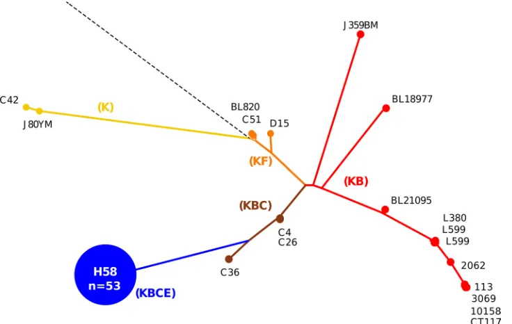
[image:4.585.47.536.63.330.2]FIG 1Indels in theSalmonellaTyphi genome. (a) Phylogenetic tree ofS. Typhi derived from SNP data, adapted from Holt et al. (12). The broken line with a black circle designates the ancestral root; all major haplotypes are highlighted (e.g., H58). Capitalized colored letters represent 17 different indels, the position of the indels on the tree indicates the branch with which they are associated, and the asterisks highlight the 11 indels selected for MLPA. (b) Genomic map ofS. Typhi strain CT18 from 1 to 4,800,000 bp (coordinates shown). Highlighted are the genomic positions and the strand of the 17 indels and thearoC,ssaV,fliC,gyrA, andparCgenes. In the center of the map is a description of the 17 indels (A to S), showing the genomic coordinates (with respect toS. Typhi CT18) and the affected coding sequence(s).
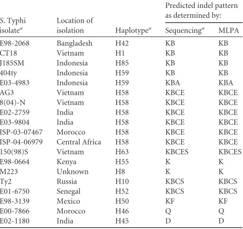
TABLE 3Comparison ofS. Typhi MLPA indel genotyping and genome sequencing
S. Typhi isolatea
Location of
isolation Haplotypea
Predicted indel pattern as determined by:
Sequencinga MLPA
E98-2068 Bangladesh H42 KB KB
CT18 Vietnam H1 KB KB
J185SM Indonesia H85 KB KB
404ty Indonesia H59 KB KB
E03-4983 Indonesia H59 KBA KBA
AG3 Vietnam H58 KBCE KBCE
8(04)-N Vietnam H58 KBCE KBCE
E02-2759 India H58 KBCE KBCE
E03-9804 India H58 KBCE KBCE
ISP-03-07467 Morocco H58 KBCE KBCE
ISP-04-06979 Central Africa H58 KBCE KBCE
150(98)S Vietnam H63 KBCES KBCES
E98-0664 Kenya H55 K K
M223 Unknown H8 K K
Ty2 Russia H10 KBCS KBCS
E01-6750 Senegal H52 KBCS KBCS
E98-3139 Mexico H50 KF KF
E00-7866 Morocco H46 Q Q
E02-1180 India H45 D D
aSee Holt et al. (12).
on May 16, 2020 by guest
http://jcm.asm.org/
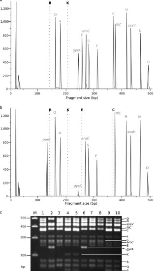
[image:4.585.41.287.484.716.2]FIG 2SalmonellaTyphi MLPA indel typing. (a) Fragment analysis generated by MLPA ofS. Typhi isolate 113 from Vietnam, demonstrating a KB (H1) indel pattern
S. Typhi by MLPA (KB peaks are missing, indicated by broken lines). Each peak corresponds with the fluorescence intensity of amplicons targeting specific loci (labeled at the top of each peak), including a large peak at 238 bp associated with a singlegyrAmutation at codon 83. (b) Fragment analysis generated by MLPA ofS. Typhi isolate 9019 from Vietnam, demonstrating a KBCE (H58) indel pattern by MLPA (KBCE peaks are missing, indicated by broken lines). This isolate has twogyrAmutations at codons 83 and 87 and aparCmutation, as determined by a diminished peak at 238 bp and large peak at 130 bp, respectively. (c) Representative fragment analysis ofS. Typhi MLPA amplicons by agarose gel electrophoresis. Fourteen bands are visible and correspond with the predicted MLPA amplicon lengths shown inTable 2. Lane M, 100-bp marker; lane 1,S. Typhi 113/Vietnam (panel a) KB (gyrAmutation⫺); lane 2,S. Typhi A763/India-KBCEN (gyrAmutation⫹); lane 3,S. Typhi TY8339/Vietnam-KBC (gyrAmutation⫺); lane 4,S. Typhi DOMIC14/China-ancestral (gyrAmutation⫺); lane 5,S. Typhi DOMIC43/China-ancestral (gyrAmutation⫺); lane 6,S. Typhi TY7045/Vietnam-KBCE (gyrAmutation⫹); lane 7,S. Typhi J348BM/Indonesia-KBC (gyrAmutation⫺); lane 8,S. Typhi AS3357/Bangladesh-KBC (gyrAmutation⫺); lane 9,S. Typhi DOMI40/China-KS (gyrAmutation⫺); lane 10, DOMI38/China-KS (gyrAmutation⫺).
on May 16, 2020 by guest
http://jcm.asm.org/
[image:5.585.136.451.71.619.2]generated from these PCR amplifications demonstrated that all probes, including the controls, performed as anticipated, as they produced measurable and reproducible amplicons of the pre-dicted sizes. Representative examples of the resulting DNA
frag-ment analysis and gel electrophoresis are shown inFig. 2. Notably,
the probes targeting thegyrAandparCmutations were able to
distinguish between isolates with and without mutations in the
QRDR.Figure 2shows that theparCamplicon was detectable in
isolates with aparCmutation, and a diminishedgyrAsignal was
visible in isolates with a mutation at codons 83 and 87. The indel
and QRDR patterns of the S. Typhi isolates yielded by MLPA
corresponded precisely with the indel patterns and thegyrAand
parCmutations anticipated from both the genome sequences and
conventional PCR (Table 3).
Next, 73S. Typhi isolates of undetermined genotype from
Ban-gladesh, China, India, Indonesia, Laos, Pakistan, and Vietnam and
10S. Paratyphi A isolates from India (as a control) were typed by
detecting SNP variations at 1,485 previously identified loci across
theS. Typhi genome using a custom GoldenGate assay (see Table
S2 in the supplemental material). These data were used to
deter-mine a phylogenetic tree, which subdivided the 73S. Typhi
iso-lates into six main genotype groups, broadly corresponding to those previously defined as H1, H42, H50, H52, H58, and H59 (Fig. 1and3) (3,12). Approximately three-quarters (54/73) of the
genotypedS. Typhi isolates belonged to the H58 genotype,
includ-ing isolates originatinclud-ing from Bangladesh, India, Laos, Pakistan,
and Vietnam. As predicted, the 10S. Paratyphi A isolates fell into
the ancestral group ofS. Typhi via SNP typing, i.e., were ancestral
for all SNPs, as all mutations wereS. Typhi specific (data not
shown).
MLPA was then performed on the same 73S. Typhi isolates
(Fig. 3). The H58 complex has a predicted indel pattern of KBCE
and all 54 of the H58 (as determined by SNP typing)S. Typhi
isolates produced this pattern (Fig. 3). The MLPA indel patterns
were also consistent with the SNP genotypes in all of the H1 (KB), H42 (KB), and H59 (KB) isolates (9/9 total). The five isolates belonging to H52 (predicted indel pattern KBCS) produced 3
KBC, 1 KBCS, and 1 KBCD MLPA pattern. The five H50S. Typhi
isolates (with a predicted KF indel pattern) produced four K pat-terns and one KS pattern. Thus, genotype predictions based on the detection of MLPA indel patterns matched those predicted from
SNP-based genotype assignments in 66/73 (90.4%) of theS. Typhi
isolates. The 10S. Paratyphi A isolates produced amplicons for the
aroC,fliC, andssaVgenes and produced peaks corresponding with
an indel pattern of SF, which does not correlate with any predicted
S. Typhi genotype (see Table S1 in the supplemental material).
MLPA ofS. Typhi from eight Asian countries.Subsequently,
MLPA typing was performed on an additional 144S. Typhi
iso-lates of undetermined genotype from Vietnam (88 isoiso-lates) and seven other Asian countries. The MLPA results were combined
with data from the 73S. Typhi isolates typed during validation
(see Tables S1 and S2 in the supplemental material). According to antimicrobial susceptibility testing, 119/217 (54.8%) isolates were nalidixic acid resistant (NALR) and 82/217 (37.8%) isolates were
MDR (Table 4), and 71 isolates (32.7%) were both NALR and
MDR (Fig. 4). We also used thegyrAandparCMLPA amplicons
to detect the presence of NALR-conferring mutations and
com-pared these data to the antimicrobial susceptibility results (Table
4,Fig. 4).
We detected 10 different indel patterns in the 217S. Typhi
isolates (Fig. 4,Table 4). Fifty-nine percent of the isolates (128/
217) produced a KBCE or KBCEN MLPA pattern, corresponding
to the H58 genotype (Table 4,Fig. 4). This H58 group included
90% of isolates from Vietnam (79/88), 79% of isolates from India (23/29), 53% of isolates from Bangladesh (9/17), 39% of isolates from Pakistan (7/18), 31% of isolates from Laos (6/21), and 29%
FIG 3Comparison of MLPA and SNP genotyping forS. Typhi. The phylogenetic tree was determined by detecting SNP variations at 1,485 previously identified loci across theS. Typhi genomes of 73 isolates using a custom GoldenGate assay (see Table S1 in the supplemental material). The resulting tree was used to infer indel patterns (colored capitalized letters) and their associated branches. Examples ofS. Typhi strains at pivotal locations throughout the tree are named, and the H58 group (KBCE indel pattern), consisting of 54 strains, is highlighted in blue.
on May 16, 2020 by guest
http://jcm.asm.org/
[image:6.585.112.480.67.300.2]of isolates from Nepal (4/14). We additionally identified 4 isolates with a KBCES indel pattern, and 41 isolates (18%) with a KB indel pattern (H42 haplotype) were identified and spanned all eight Asian countries. Twelve isolates (6%) produced a KBC (H52)
[image:7.585.39.549.80.243.2]in-del pattern, and 22/217 (10%) isolates produced a K inin-del pattern. An additional three indel patterns (KE, KS, and KBCS) were iden-tified across seven isolates, and two Chinese isolates produced amplicons for all loci and were deemed to be ancestral.
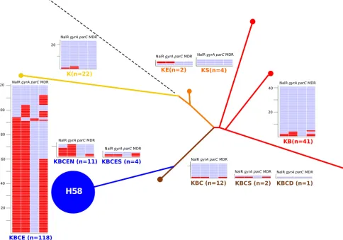
TABLE 4MLPA indel genotyping of 217 AsianS. Typhi isolates
MLPA indel patterna No. ofisolates Country of isolation (no.) No. withdetected by MLPAgyrAmutation
No. with resistance type
Nalidixic acid Multidrug KBCE 118 Vietnam (79), Bangladesh (9), India (18), Pakistan
(5), Laos (6), Nepal (1)
93 99 73
KBCEN 10 India (5), Nepal (3), Pakistan (2) 7 10 2
KBCES 4 Vietnam (4) 2 2 3
KB 41 Vietnam (4), Bangladesh (5), India (3), Pakistan (5), Laos (8), Nepal (8), China (1), Indonesia (7)
3 5 4
KBC 12 Vietnam (1), Bangladesh (1) China (9), Indonesia (1) 0 0 0
KBCS 2 Laos (2) 0 0 0
KBCD 1 Laos (1) 0 0 0
K 22 Bangladesh (1), India (2), Pakistan (6), Nepal (2), China (1), Indonesia (6), Laos (4)
1 2 0
KS 3 China (3) 0 0 0
KE 2 India (1), Bangladesh (1) 0 1 0
No deletions (ancestral) 2 China (2) 0 0 0
Total isolates 217 106 119 82
aFiveS. Paratyphi controls gave an indel pattern of SF and were not included in this table (see Table S1 in the supplemental material).
FIG 4MLPA indel typing of 217 AsianS. Typhi isolates. The phylogenetic tree was determined by detecting SNP variations at 1,485 previously identified loci across the
S. Typhi genomes of 73 isolates using a custom GoldenGate assay inferring the genotype of 217 AsianS. Typhi isolates by MLPA. The color coding and labeling is as shown inFig. 3. The numbers of isolates with a specific indel type are shown, color coded, and associated with a heat map showing the presence (red) or absence (blue) of a NALR phenotype, a mutation in the QRDR of thegyrAgene by MLPA, a mutation in the QRDR of theparCgene by MLPA, and an MDR phenotype.
on May 16, 2020 by guest
http://jcm.asm.org/
[image:7.585.46.530.344.683.2]MLPA detected 106 isolates with an amplicon consistent with a
mutation ingyrAand two isolates with a mutation inparCand a
double mutation ingyrA; all were confirmed by sequencing (Fig.
4,Table 4). Of the 119 isolates with phenotypic NALR, 106 (90%)
tested positive for agyrAmutation by MLPA (Table 4). The MLPA
analysis revealed that the H58 indel patterns KBCE and KBCEN had an increased proportion of NALR isolates compared to other
indel patterns (H58 NALR, non-H58 NALR) (P⬍0.0001, Fisher’s
exact test; seeTable 4). Furthermore, the same H58 indel group
had an increased proportion of MDR strains compared to other
indel patterns (H58 MDR versus non-H58 MDR) (P⬍0.0001,
Fisher’s exact test).
DISCUSSION
Phylogenetically informative subgrouping is vital for understand-ing the local and regional strain circulations of bacterial patho-gens. Diagnostic, research, and reference laboratories use a range of typing methods for recording and reporting bacterial diversity
with a variety of pathogens (22,23). However, these methods
of-ten lack reproducibility, sensitivity, and phylogenetic relevance
(24). The consequence of these limitations is an inability to
per-form robust epidemiological investigations of circulating bacterial pathogens. Genomics offers a solution to these limitations, and DNA sequence-based methodologies can be used to improve
bac-terial subtyping (10,25,26). However, whole-genome sequencing
is currently too specialized and expensive to be employed in areas where typhoid is endemic. We aimed to address this and
devel-oped a simple and reproducibleS. Typhi subtyping method that
can be performed in locations with limited molecular biology equipment. The MLPA method utilizes a limited number of steps and can be conducted with a standardized set of reagents and protocols. The method we developed performs well and can be made commercially available, depending on the demand within the enteric fever research community. It is difficult to perform a direct cost comparison of MLPA and sequencing, as demand im-pacts the cost of the MLPA kit; however, we estimate a cost in the realm of $10 to $20 per isolate for MLPA compared to the current cost of $500 to $1,000 per isolate for next-generation sequencing
(price estimated on 5⫻to 15⫻coverage of a wholeS. Typhi
ge-nome, using a 454 Junior next-generation sequencer with current costs in Vietnam; this could be reduced by using commercial se-quencing facilities).
The MLPA method successfully detected all 11 phylogeneti-cally informative indels, permitting us to infer a genotype for each.
In our test set of 73S. Typhi isolates, MLPA indel patterns
accu-rately predicted SNP-based genotype assignments in 90% of the strains. For the remaining isolates, the MLPA profiles were a com-bination of indels that were similar, but not identical, to those we anticipated from SNP genotypes. Our method also permitted the
incorporation of probes targeting the QRDR regions ofgyrAand
parC. The ability to detect mutations ingyrAandparCis highly
relevant for epidemiological studies ofS. Typhi, as NALRS. Typhi
has been reported in the majority of countries where typhoid is
endemic (15,19,21,27). NALR is a reliable marker of isolates with
a reduced susceptibility to fluoroquinolones, the most common group of antimicrobials used to treat typhoid. When treated with
fluoroquinolones, NALRS. Typhi is associated with an extended
fever clearance time, prolonged antimicrobial therapy, and an
in-creased risk of treatment failure (28). Therefore, knowing the
prevalence ofgyrAmutations in endemic locations is important
for treatment and is of value to epidemiologists, clinicians, and those making guidelines for clinical care.
Among the 106 isolates in which NALR mutations were
de-tected by MLPA, allgyrAandparCmutations were confirmed by
direct PCR amplicon sequencing, indicating 100% specificity for the detection of mutations in these genes using MLPA. Notably,
we detected only two strains containing theparCmutation; these
strains were from India, confirming our previous observation that this mutation is rare and has only been identified in strains
circu-lating in India (15). There were 13 NALR isolates that tested
neg-ative forgyrAandparCmutations using MLPA; direct sequencing
of the amplicons confirmed the MLPA data. We suggest that this 10% discrepancy between MLPA and NALR phenotypic data was caused by either fluctuation in zone size readings during antimi-crobial susceptibility testing by disk diffusion or by novel
muta-tions in the QRDR that were not represented in this assay (15).
The successful use of MLPA to identify point mutations is encour-aging and suggests that this method can be adapted to establish a
simple and robust alternative to SNP typing forS. Typhi and other
monomorphic pathogens, which may be more phylogenetically informative than the use of genomic indels.
The potential limitations of our work include the lack of an incHI1 plasmid probe, a bias toward isolates from Vietnam, and the fact that some indel profiles are shared by several genetically
diverseS. Typhi genotypes, while other indel profiles define much
more homogeneous groups. Hence, the resolution offered by in-del typing is somewhat varied. We suggest that the discrepancies between SNP and MLPA typing are induced by the stability of some indels in different lineages, predicting that some indel loci may not be as phylogenetically informative as others. However, notwithstanding these limitations, our comparison of genome-wide SNP with MLPA typing showed that MLPA was able to
dis-tinguish between five commonS. Typhi isolates and, most
signif-icantly, was able to correctly identify H58 strains from Vietnam and across Asia. Principally, we were able to distinguish H58
strains with and withoutgyrAmutations with 100% accuracy. It
has been shown that H58 strains currently predominate in the majority of endemic locations, and there is an association between
H58 strains and S83FgyrAmutations and an MDR phenotype (3,
13). The MLPA data confirmed that H58S. Typhi is dominant in
Vietnam and is widely dispersed across Asia, and it is associated with both NALR and MDR in these regions. As the use of high-resolution SNP typing and genome sequencing have previously reported similar associations, the ability of MLPA to reproduce this result confirms its reliability and potential utility for epidemi-ological investigations. We advocate a new system for the
subtyp-ing ofS. Typhi and propose that indel detection, either by MLPA
or conventional PCR, be adopted until SNP typing becomes more
routine. We aim to develop a global Web-basedS. Typhi,S.
Para-typhi A, invasive nontyphoidal Salmonella reference network
whereby serotyping is combined with simple genotyping informa-tion, antimicrobial resistance profiling, and the time and date of isolation. The use of a standardized kit-based assay, such as the one presented here, that can be adapted through targeted genome sequencing would allow for routine data sharing and a global re-source for assessing international epidemiology and defining ap-propriate measures for therapy and vaccination.
In conclusion, we have developed a simple, reliable, and epi-demiologically relevant method that can be used in laboratories
with modest molecular biology equipment to subgroupS. Typhi
on May 16, 2020 by guest
http://jcm.asm.org/
and identify common NALR-associated mutations. This MLPA method can distinguish between strains that currently dominate
the global population structure ofS. Typhi, offering a more
sen-sitive and simple alternative to other subgrouping methodologies that are currently being used in low- and middle-income coun-tries.
ACKNOWLEDGMENTS
This work was supported by the Wellcome Trust and fellowship schemes from the NHMRC of Australia (no. 628930, to K.E.H.) and the OAK foundation of the United States. S.B. is supported by a Sir Henry Dale Fellowship jointly funded by the Wellcome Trust and the Royal Society (grant no. 100087/Z/12/Z). The funders had no role in study design, data collection or analysis, decision to publish, or preparation of the manu-script.
REFERENCES
1.Crump JA, Luby SP, Mintz ED.2004. The global burden of typhoid fever. Bull. World Health Organ.82:346 –353.
2.Parry CM, Hien TT, Dougan G, White NJ, Farrar JJ.2002. Typhoid fever. N. Engl. J. Med.347:1770 –1782.
3.Roumagnac P, Weill FX, Dolecek C, Baker S, Brisse S, Chinh NT, Le TAH, Acosta CJ, Farrar J, Dougan G, Achtman M.2006. Evolutionary history ofSalmonellaTyphi. Science314:1301–1304.
4.Kim S, Kim SH, Park JH, Lee KS, Park MS, Lee BK.2009. Clustering analysis ofSalmonella entericaserovar Typhi isolates in Korea by PFGE, ribotying, and phage typing. Foodborne Pathog. Dis.6:733–738. 5.Sen B, Dutta S, Sur D, Manna B, Deb AK, Bhattacharya SK, Niyogi SK.
2007. Phage typing, biotyping & antimicrobial resistance profile of Salmo-nella entericaserotype Typhi from Kolkata. Indian J. Med. Res.125:685– 688.
6.Kidgell C, Reichard U, Wain J, Linz B, Torpdahl M, Dougan G, Achtman M.2002.Salmonellatyphi, the causative agent of typhoid fever, is approximately 50,000 years old. Infect. Genet. Evol.2:39 – 45. 7.Kariuki S, Gilks C, Revathi G, Hart CA. 2000. Genotypic analysis of
multidrug-resistantSalmonella entericaserovar Typhi, Kenya. Emerg. In-fect. Dis.6:649 – 651.
8.Nair S, Schreiber E, Thong KL, Pang T, Altwegg M.2000. Genotypic characterization ofSalmonellaTyphi by amplified fragment length poly-morphism fingerprinting provides increased discrimination as compared to pulsed-field gel electrophoresis and ribotyping. J. Microbiol. Methods
41:35– 43.
9.Thong KL, Cheong YM, Puthucheary S, Koh CL, Pang T.1994. Epide-miologic analysis of sporadicSalmonellaTyphi isolates and those from outbreaks by pulsed-field gel electrophoresis. J. Clin. Microbiol.32:1135– 1141.
10. Baker S, Holt KE, Clements ACA, Karkey A, Arjyal A, Boni MF, Dongol S, Hammond N, Koirala S, Duy PT, Nga TV, Campbell JI, Dolecek C, Basnyat B, Dougan G, Farrar JJ.2011. Combined high-resolution geno-typing and geospatial analysis reveals modes of endemic urban typhoid fever transmission. Open Biol.1:110008. doi:10.1098/rsob.110008. 11. Holt KE, Dolecek C, Chau TT, Duy PT, La TT, Hoang NV, Nga TV,
Campbell JI, Manh BH, Vinh Chau NV, Hien TT, Farrar J, Dougan G, Baker S.2011. Temporal fluctuation of multidrug resistantSalmonella
Typhi haplotypes in the Mekong river delta region of Vietnam. PLoS Negl. Trop. Dis.5:e929. doi:10.1371/journal.pntd.0000929.
12. Holt KE, Parkhill J, Mazzoni CJ, Roumagnac P, Weill FX, Goodhead I, Rance R, Baker S, Maskell DJ, Wain J, Dolecek C, Achtman M, Dougan G. 2008. High-throughput sequencing provides insights into genome variation and evolution inSalmonellaTyphi. Nat. Genet.40:987–993. 13. Holt KE, Phan MD, Baker S, Duy PT, Nga TVT, Nair S, Turner AK,
Walsh C, Fanning S, Farrell-Ward S, Dutta S, Kariuki S, Weill FX, Parkhill J, Dougan G, Wain J.2011. Emergence of a globally dominant IncHI1 plasmid type associated with multiple drug resistant typhoid. PLoS Negl. Trop. Dis.5:e1245. doi:10.1371/journal.pntd.0001245. 14. Schouten JP, McElgunn CJ, Waaijer R, Zwijnenburg D, Diepvens F,
Pals G. 2002. Relative quantification of 40 nucleic acid sequences by
multiplex ligation-dependent probe amplification. Nucleic Acids Res.30:
e57. doi:10.1093/nar/gnf056.
15. Parry CM, Thuy CT, Dongol S, Karkey A, Vinh H, Chinh NT, Duy PT, Thieu Nga TV, Campbell JI, Van Minh Hoang N, Arjyal A, Bhutta ZA, Bhattacharya SK, Agtini MD, Dong B, Canh do G, Naheed A, Wain J, Tinh Hien T, Basnyat B, Ochiai L, Clemens J, Farrar JJ, Dolecek C, Baker S.2010. Suitable disk antimicrobial susceptibility breakpoints de-finingSalmonella entericaserovar Typhi isolates with reduced susceptibil-ity to fluoroquinolones. Antimicrob. Agents Chemother.54:5201–5208. 16. Clinical and Laboratory Standards Institute.2012. Performance
stan-dards for antimicrobial disk susceptibility tests; approved standard— eleventh edition. CLSI document M02-A11. Clinical and Laboratory Stan-dards Institute, Wayne, PA.
17. Phan MD, Kidgell C, Nair S, Holt KE, Turner AK, Hinds J, Butcher P, Cooke FJ, Thomson NR, Titball R, Bhutta ZA, Hasan R, Dougan G, Wain J.2009. Variation inSalmonella entericaserovar Typhi IncHI1 plas-mids during the global spread of resistant typhoid fever. Antimicrob. Agents Chemother.53:716 –727.
18. Holt KE, Baker S, Dongol S, Basnyat B, Adhikari N, Thorson S, Pulickal AS, Song Y, Parkhill J, Farrar JJ, Murdoch DR, Kelly DF, Pollard AJ, Dougan G.2010. High-throughput bacterial SNP typing identifies dis-tinct clusters ofSalmonellaTyphi causing typhoid in Nepalese children. BMC Infect. Dis.10:144. doi:10.1186/1471-2334-10-144.
19. Kariuki S, Revathi G, Kiiru J, Mengo DM, Mwituria J, Muyodi J, Munyalo A, Teo YY, Holt KE, Kingsley RA, Dougan G.2010. Typhoid in Kenya is associated with a dominant multidrug-resistantSalmonella entericaserovar Typhi haplotype that is also widespread in Southeast Asia. J. Clin. Microbiol.48:2171–2176.
20. Stamatakis A.2006. RAxML-VI-HPC: maximum likelihood-based phy-logenetic analyses with thousands of taxa and mixed models. Bioinformat-ics22:2688 –2690.
21. Chau TT, Campbell JI, Galindo CM, Van Minh Hoang N, Diep TS, Nga TT, Van Vinh Chau N, Tuan PQ, Page AL, Ochiai RL, Schultsz C, Wain J, Bhutta ZA, Parry CM, Bhattacharya SK, Dutta S, Agtini M, Dong B, Honghui Y, Anh DD, Canh do G, Naheed A, Albert MJ, Phetsouvanh R, Newton PN, Basnyat B, Arjyal A, La TT, Rang NN, Phuong le T, Van Be Bay P, von Seidlein L, Dougan G, Clemens JD, Vinh H, Hien TT, Chinh NT, Acosta CJ, Farrar J, Dolecek C.2007. Antimicrobial drug resistance ofSalmonella entericaserovar Typhi in Asia and molecular mechanism of reduced susceptibility to the fluoroquinolones. Antimi-crob. Agents Chemother.51:4315– 4323.
22. Boxrud D, Monson T, Stiles T, Besser J.2010. The role, challenges, and support of PulseNet laboratories in detecting foodborne disease out-breaks. Public Health Rep.125(Suppl 2):57– 62.
23. Gerner-Smidt P, Scheutz F.2006. Standardized pulsed-field gel electro-phoresis of Shiga toxin-producingEscherichia coli: the PulseNet Europe Feasibility study. Foodborne Pathog. Dis.3:74 – 80.
24. Baker S, Hanage WP, Holt KE.2010. Navigating the future of bacterial molecular epidemiology. Curr. Opin. Microbiol.13:640 – 645.
25. Croucher NJ, Harris SR, Fraser C, Quail MA, Burton J, van der Linden M, McGee L, von Gottberg A, Song JH, Ko KS, Pichon B, Baker S, Parry CM, Lambertsen LM, Shahinas D, Pillai DR, Mitchell TJ, Dougan G, Tomasz A, Klugman KP, Parkhill J, Hanage WP, Bentley SD.2011. Rapid pneumococcal evolution in response to clinical interventions. Sci-ence331:430 – 434.
26. Harris SR, Feil EJ, Holden MTG, Quail MA, Nickerson EK, Chantratita N, Gardete S, Tavares A, Day N, Lindsay JA, Edgeworth JD, de Len-castre H, Parkhill J, Peacock SJ, Bentley SD.2010. Evolution of MRSA during hospital transmission and intercontinental spread. Science327:
469 – 474.
27. Kumar Y, Sharma A, Mani KR.2009. High level of resistance to nalidixic acid inSalmonella entericaserovar Typhi in central India. J. Infect. Dev. Ctries.3:467– 469.
28. Parry CM, Vinh H, Chinh NT, Wain J, Campbell JI, Hien TT, Farrar JJ, Baker S.2011. The influence of reduced susceptibility to fluoroquinolo-nes inSalmonella entericaserovar Typhi on the clinical response to ofloxa-cin therapy. PLoS Negl. Trop. Dis.5:e1163. doi:10.1371/journal.pntd .0001163.