R E S E A R C H A R T I C L E
Open Access
Comprehensive evaluation of gene
expression signatures in response to
electroacupuncture stimulation at Zusanli
(ST36) acupoint by transcriptomic analysis
Jing-Shan Wu
1, Hsin-Yi Lo
1, Chia-Cheng Li
1, Feng-Yuan Chen
1, Chien-Yun Hsiang
2*and Tin-Yun Ho
1,3*Abstract
Background:Electroacupuncture (EA) has been applied to treat and prevent diseases for years. However, molecular events happened in both the acupunctured site and the internal organs after EA stimulation have not been clarified. Methods:Here we applied transcriptomic analysis to explore the gene expression signatures after EA stimulation. Mice were applied EA stimulation at ST36 for 15 min and nine tissues were collected three hours later for microarray analysis. Results:We found that EA affected the expression of genes not only in the acupunctured site but also in the internal organs. EA commonly affected biological networks involved in cytoskeleton and cell adhesion, and also regulated unique process networks in specific organs, such asγ-aminobutyric acid-ergic neurotransmission in brain and inflammation process in lung. In addition, EA affected the expression of genes related to various diseases, such as neurodegenerative diseases in brain and obstructive pulmonary diseases in lung.
Conclusions:This report applied, for the first time, a global comprehensive genome-wide approach to analyze the gene expression profiling of acupunctured site and internal organs after EA stimulation. The connection between gene expression signatures, biological processes, and diseases might provide a basis for prediction and explanation on the therapeutic potentials of acupuncture in organs.
Keywords:Electroacupuncture, ST36, Zusanli, Microarray
Background
Acupuncture, a traditional therapy in ancient China over thousands of years, has been widely accepted and used in Western society [1, 2]. Acupuncture is believed to balance Yin-Yang, stimulate the circulation of vital energy (qi) and blood, maintain the body health, and prevent the incidence of illness [3]. Electroacupuncture (EA) is a modification of acupuncture that stimulates acupoints with electrical current and displays reproducible in both research and clinical application. Moreover, EA therapy has been used for postoperative analgesia and
anesthesia, for the treatment of diverse disorders of internal organs, and for the release of pain [4, 5].
In traditional Chinese medicine, ST36 (Zusanli) is a commonly acupoint that modulates the biological activities of gastrointestinal system, immune system, cardiovascular system, and muscular system. Transcutaneous EA at ST36 reduces gastric accommodation and improves impaired gastric motility in patients with functional dyspepsia [6]. Transcutaneous neuromodulation at ST36 also improves the frequency of spontaneous defecation and increases the bowel movements in patients with chronic constipation [7]. Chronic EA at ST36 improves baroreflex function and hemodynamic parameters in rats with heart failure [8]. Long-term EA stimulation at ST36 and DU20 (BaiHui) also relieves the increased mean arterial pressure and cardiovascular abnormality in both structure and function in spontaneously hypertensive rats [9]. ST36 displays the * Correspondence:[email protected];[email protected]
2
Department of Microbiology, China Medical University, 91 Hsueh-Shih Road, Taichung 40402, Taiwan
1Graduate Institute of Chinese Medicine, China Medical University, 91 Hsueh-Shih Road, Taichung 40402, Taiwan
Full list of author information is available at the end of the article
anti-nociceptive and anti-hyperalgesic effect. EA at ST36 reduces postoperative analgesic requirements and associ-ated side effects in patients undergoing lower abdominal surgery [10]. Treatment of EA at ST36 and GN34 also ameliorates L5 spinal nerve ligation-induced neuropathic pain in rats [11]. Moreover, EA at ST36 and CV4 (Guanyuan) improves clinical curative effects in patients with sepsis in a prospective randomized controlled trial via the regulation of immune system [12]. EA at ST36 promotes myofiber regeneration and restoration of neuro-muscular junctions in a rabbit gastrocnemius contusion model [13]. Furthermore, EA at ST36 also improves intes-tinal mucosal immune barrier in sepsis by increasing the concentration of secretory IgA, the percentage of CD3+,
γ/δ, and CD4+ T cells, and the ratio of CD4+/CD8+ T cells [14].
ST36 is an acupoint of Foot’s Yang Supreme Stomach Meridian that targets at gastrointestinal tract [15]. We wondered whether EA stimulation at ST36 altered mo-lecular events in other organs. The genome-wide analysis of ST36-stimulated region (skin) and distant visceral organs or tissues, including cerebral medulla, cerebral cortex, hippocampus, lung, spleen, kidney, uterus and thigh muscle, was therefore performed. Mice were stimu-lated by EA at ST36, and gene expression signatures of nine organs or tissues were explored by microarray analysis. The process network and disease connection of gene expression profiles were further analyzed to elucidate the molecular events and effects of organs after ST36 stimulation.
Methods
Animals
Female BALB/c mice (6–8-week-old, 18–22 g) were ob-tained from National Laboratory Animal Center (Taipei, Taiwan) and maintained in an air-controlled pathogen-free animal facility with a 12-h light/dark cycle at 23 ± 2 °C. Food and water were available ad libitum. Mouse experi-ments were conducted under ethics approval from China Medical University Animal Care and Use Committee (Permit No. 101–61-N).
EA stimulation
A total of 10 mice was randomly divided into two groups of 5 mice. For control group, mice were anesthetized with isoflurane without ST36 stimulation. For ST36 group, mice were anesthetized with isoflurane, gently immobi-lized in a plastic restrainer, and applied EA stimulation at ST36 acupoint, which is located at the midpoint of tibialis anterior muscle of hind limbs. Briefly, sterilized acupunc-ture needle (0.24 × 12 mm, 36 gauge, Yu-Kuang Acupunc-ture Co., Taipei, Taiwan) was inserted bilaterally into the acupoint, which was 3–4 mm below the knee midline and laterally 1–2 mm at a depth of 2–3 mm. Electrical stimu-lation pulse with voltage ranging from 3.5 to 5 V, duration
of 0.05 ms, and frequency of 2 Hz was generated from a pulse generator (HANS model, LH202H, Taipei, Taiwan) and applied using two outlets via two needles. The inten-sity of EA stimulation was determined as the minimum voltage causing moderate muscle contraction for 15 min. Three hours after EA stimulation, mice were sacrificed by carbon dioxide inhalation, and organs were removed for RNA extraction.
Total RNA extraction
Total RNAs from acupunctured site, muscle, cerebral cortex, cerebral medulla, hippocampus, lung, spleen, kidney, and uterus were extracted using RNeasy Mini kit (Qiagen, Valencia, CA, USA). The amount and the integrity of total RNA were quantified and evaluated using a spec-trophotometer (Beckman Coulter, Fullerton, CA, USA) and an Agilent 2100 bioanalyzer (Agilent Technologies, Santa Clara, CA, USA), respectively.
Microarray analysis
Microarray analysis was performed as described previously [16, 17]. Briefly, fluorescence-labeled RNA targets were prepared from total RNA using MessageAmp™ aRNA kit (Ambion, Austin, TX, USA) and Cy5 dye (Amersham Pharmacia, Piscataway, NJ, USA). Fluorescent targets were hybridized to the Mouse Whole Genome OneArray (Phalanx Biotech Group, Hsinchu, Taiwan) and scanned by an Axon 4000 scanner (Molecular Devices, Sunnyvale, CA, USA). The Cy5 fluorescent intensity of each spot was analyzed by genepix 4.1 software (Molecular Devices, Sunnyvale, CA, USA). The signal intensity of each spot was normalized by R program in limma package using quantile normalization. Normalized data were analyzed using the “geneSetTest” function implemented in the limma package to detect groups of regulated genes in biological pathways. This function computes a p-value to test the hypothesis that the selected genes tend to be up- or down-regulated. Then, the score of each path-way in EA treatment was defined as score = −log(p) if
Results
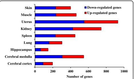
EA affected the expression of genes in distant organs To explore the molecular events happened in local or distant regions after ST36 stimlation, we applied EA stimulation in BALB/c mice for 15 min and collected ST36-stimulated region (skin) and distant visceral organs or tissues, including cerebral medulla, cerebral cortex, hippocampus, lung, spleen, kidney, uterus and thigh muscle, 3 h later for microarray analysis. BALB/c mice were applied for EA stimulation in this study because BALB/c mice are among the most widely used inbred strains for animal experiments. Moreover, BALB/c mice are useful for researches of immunology and neurobiology, the potent biological activities of ST36 acupoint. As ex-pected, EA affected the expression of genes in the skin at ST36 acupoint (Fig. 1). In a total of 29,922 genes, the transcripts of 169 genes and 231 genes were upregulated and downregulated, respectively, by 1.5 fold in EA-treated skin. In addition to skin, EA affected the expression levels of genes in distant organs. EA regulated the expression of 931 genes in uterus, followed by kidney (743 genes), cere-bral medulla (547 genes), muscle (463 genes), spleen (450 genes), lung (303 genes), cerebral cortex (197 genes), and hippocampus (147 genes).
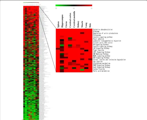
EA affected biological processes in various organs We further analyzed the canonical pathways affected by EA at ST36 acupoint. “geneSetTest” function was per-formed to test a set of signaling and metabolic pathways regulated by EA. Scores of pathways were further visualized by TIGR Multiexperiment Viewer. As shown in Fig. 2, a hierarchical clustering of EA-affected canonical pathways displayed varieties among nine organs or tissues, and the number of signaling and metabolic pathways significantly regulated (score≥ 0.3) by EA in different organs was also
varied. Some pathways, such as oxidative phosphorylation, ribosome, proteasome and serum response factor-mediated pathways, were commonly regulated by EA in organs. However, more pathways were regulated by EA in organs without consistency. About 2/3 pathways in spleen and skin were significantly regulated by EA, while less pathways were regulated by EA in hippocampus.
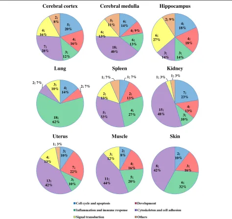
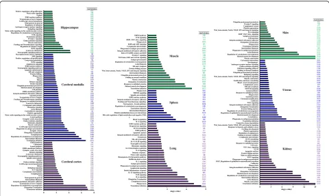
We further analyzed the process network of protein interactions regulated by EA treatment and classified the process networks into five categories, including cell cycle and apoptosis, inflammation and immune response, signaling transduction, cytoskeleton and cell adhesion, and development. EA treatment affected the process networks in these organs in different ratio (Fig. 3). Cyto-skeleton and cell adhesion was the most EA-regulated category in organs, except hippocampus and lung. “ In-flammation and immune response” was the most af-fected category in lung. About 60% of the total number of EA-affected process networks was related to inflam-mation and immune response. Signaling transduction was the most affected category in hippocampus, and approximately 27% of the total number of affected process networks was involved in signaling transduction. In addition, EA treatment affected some unique process networks in organs (Fig. 4). For example, some neuro-physiological processes, such as transmission of nerve impulse and γ-aminobutyric acid-ergic (GABAergic) neurotransmission, were commonly regulated by EA in cerebral cortex, cerebral medulla, and hippocampus, while melatonin signaling, corticoliberin signaling, and long-term potentiation were regulated by EA in cerebral medulla. Moreover, male sex differentiation in kidney, follicle-stimulating hormone-beta signaling pathway in uterus, and blood coagulation in spleens were signifi-cantly affected by EA.
Gene expression connection between EA stimulation and diseases in brain and lung
[image:3.595.58.292.526.664.2]EA stimulation at ST36 regulated the expression of about 300–500 genes in lung and brain. Although the organs with the top two changes in gene expression were uterus and kidney, the ratios of process network categories altered by uterus and kidney were similar to those altered by other organs, except brain and lung. Process network analysis showed that“inflammation and immune response” was the abundant category in lung and neurological processes were unique processes regulated in brain. Therefore, we further analyzed whether genes af-fected by EA were related to those in diseases. As shown in Table 1, EA stimulation commonly regulated the genes in-volved in psychiatry and psychology, mental disorders, mood disorders, and heredodegenerative disorders in brain tissues. EA treatment also regulated the expression of genes related to some unique diseases in brain tissues. For
0 200 400 600 800 1000
Cerebral cortex Cerebral medulla Hippocampus Lung Spleen Kidney Uterus Muscle Skin
Number of genes
Down-regulated genes Up-regulated genes
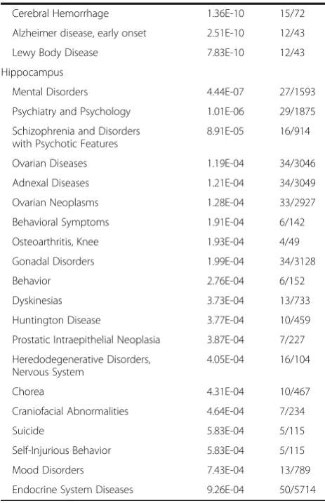
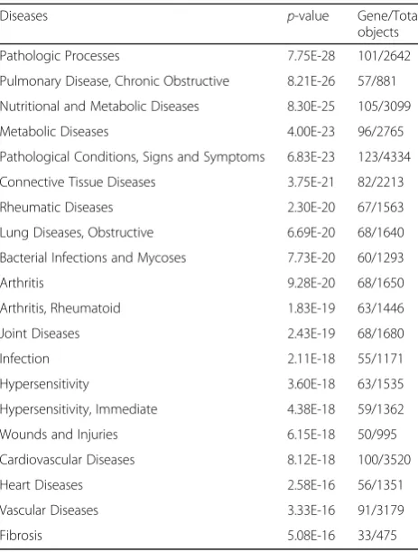
example, genes involved in neurodegenerative diseases, such as Alzheimer’s disease, amyloid neuropathies and parkinsonian disorders, were regulated by EA in cerebral medulla. Genes related to endocrine system diseases, such as ovarian diseases, adnexal diseases, ovarian neoplasms, gonadal disorders, and prostatic intraepithelial neoplasia, were regulated by EA in hippocampus. As shown in Table 2, genes involved in obstructive pulmonary dis-eases, hypersensitivity, such as rheumatic diseases and rheumatoid arthritis, infection, such as bacterial infec-tions and mycoses, and cardiopulmonary diseases, such as cardiovascular diseases, heart diseases and vascular diseases, were affected by EA in lung. These findings suggested that EA stimulation at ST36 acupoint affected the biological process and network in distant organs. Moreover, EA-affected gene expression profiles might be related to diseases states in brain and lung.
Discussion
In this study, we applied transcriptomic analysis to analyze the gene expression signatures in nine organs or tissues responsive to ST36 stimulation. Microarray analysis has been applied to elucidate the effects of various acupoints in specific organs or tissues. For example, acupuncture at GB34 and LR3 acupoints attenuates the decrease of tyro-sine hydroxylase and exhibits the protective effects via affecting the expression of degeneration-related genes in the substantia nigra region in Parkinsonism mouse model [19]. Acupuncture at PC6 acupoint up-regulates the expression of Tph1 gene and down-regulates the ex-pression of Olr883 genes in rat brains, suggesting that the therapeutic effect of acupuncture for ischemic stroke may be closely related to the suppression of post-stroke depression and the regulation of olfactory transduction in middle cerebral artery occlusion rat model [20]. Moreover,
Spleen Hi
pp
oca
mpu
s
Uterus Cer
ebra
l cortex
Cer
ebra
l med
ulla
Kidney Musc
le
Lung Skin
[image:4.595.57.538.88.482.2]EA at PC3 and PC6 acupoints significantly ameliorates the colonic lesions, and affects both the inflammatory pathways in colons and the immunity-associated pathway in spleens in mice with trinitrobenzene sulfuric acid-induced colitis [21]. Gene expression profiles of specific organs or tissues after EA stimulation at ST36 have also been analyzed. For example, gene expression profiles in periaqueductal gray-spinal dorsal horn region of rats after EA stimulation at ST36 and SP6 show that the modula-tion of neural-immune interacmodula-tion in the central nervous system plays an important role during EA analgesia [22]. Gene expression profiling of rat arcuate nucleus region
responsive to EA at ST36 and SP6 shows that the ex-pression levels of genes are effectively regulated by low-frequency EA, compared with high-low-frequency EA. It might explain the mechanisms of therapeutic effects of the low-frequency EA [23]. In addition to brain tissues, EA at ST36 affects the expression of cell adhesion molecules in muscle, which might be related to the glucose-lowering effect of ST36 in rats with type 1 diabetes [24]. Acupuncture at ST36, CV12 (Zhongwan), and BL20 (Pishu) acupoints down-regulates nuclear factor-κB p65, miRNA-155, and miRNA-21 and up-regulates miRNA-146a expression in chronic atrophic gastritis rats, suggesting that these genes
5; 20%
4; 16%
3; 12% 7;
28% 4; 16%
2; 8%
Cerebral cortex
6; 14%
4; 9%
6; 13% 18;
40% 6; 13%
5; 11%
Cerebral medulla
4; 18%
4; 18%
3; 14% 3; 14% 6; 27%
2; 9%
Hippocampus
4; 14%
2; 7%
18; 62% 2; 7%
3; 10%
Lung
1; 7%
2; 13%
4; 27% 5;
33% 2; 13%
1; 7%
Spleen
7; 23%
4; 13% 3; 10% 15;
48% 1; 3% 1; 3%
Kidney
3; 10%
7; 22%
3; 10% 13;
42% 4; 13%
1; 3%
Uterus
2; 8%
4; 16%
5; 20% 11;
44% 3; 12%
Muscle
2; 10%
3; 16%
6; 32% 8;
42%
Skin
[image:5.595.61.536.88.536.2]may play important roles in therapeutic effect of acupunc-ture in treating chronic atrophic gastritis [25]. Moreover, moxibustion at ST36 affects the biological processes in-volved in immunity and metabolism in moxibustioned skin under pathological and physiological conditions, respectively [26]. Since ST36 displays various benefit or therapeutic effects in whole bodies, we performed a global and comprehensive study on the gene expression signatures of nine different organs or tissues after ST36 stimulation. Our data showed that EA at ST36 affected the gene expression of different organs or tissues in various degrees. Moreover, EA at ST36 has a more impact on the regulation of gene expression in uterus and has a lesser impact in hippocampus.
By process network and disease connection analysis, we found that EA at ST36 affected the process networks involved in inflammation and immune responses in lung and affected the expression of genes involved in respiratory diseases, such as obstructive pulmonary diseases and microbial infection. Interestingly, a prospective single-blind randomized placebo-controlled study shows that transcutaneous electrical nerve stimulation at ST36, EX-B-1(Dingchuan), BL13 (Feishu), and BL23 (Shenshu) improves lung function on patients with stable chronic ob-structive pulmonary disease [27]. Another study shows that
EA at ST36 and BL13 improves lung function of rats with chronic obstructive pulmonary disease and displays an anti-inflammatory effect via downregulation of orexin and its receptor [28]. In addition, EA at ST36 displays a potential protective effect on severe thermal injury-induced remote acute lung injury via the limitation of inflammatory re-sponses in rats [29]. Moreover, EA treatment at ST36 and BL13 attenuates lung injury in rats with endotoxic shock-induced acute lung injury through the activation of NF-E2-related factor pathway and the up-regulation of heme oxygenase-1 expression [30]. Acupuncture at ST36 also regulates the disorders of Fas and Bcl-2 mRNA expression, promotes the apoptosis of eosinophils, and consequently inhibits the development of inflammatory reaction of asthma in rats [31].
Acupuncture has shown some benefit effects on Alzheimer’s disease and Parkinson’s disease. Lu et al. [32] showed that acupuncture at ST36 increases blood perfusion and glycol metabolism in certain brain areas in Alzheimer’s disease rat model by Positron Emission Tomography scanning. ST36 stimulation also induces neurogenesis in adult brains via the up-regulations of brain-derived neurotrophic factor, glial cell line-derived neurotrophic factor, basic fibroblast growth factor and neuropeptide Y, and the activation of the function of
0 2 4 6 8
Translation initiation Elongation-Termination Regulation of cytoskeleton rearrangement Transmission of nerve impulse Integrin-mediated cell-matrix adhesion Intermediate filaments Neurogenesis in general G1-S Synaptogenesis Cytoplasmic microtubules IL-6 signaling Hedgehog signaling Mitosis GABAergic neurotransmission Cholecystokinin signaling Meiosis Spindle microtubules Neuropeptide signaling pathways Leucocyte chemotaxis Relaxin signaling Synaptic vesicle exocytosis ESR1-membrane pathway Axonal guidance Chemotaxis CREM pathway Translation initiation Intermediate filaments Regulation of cytoskeleton rearrangement Synaptogenesis Elongation-Termination Synaptic contact Phagosome in antigen presentation GABAergic neurotransmission Muscle contraction Melatonin signaling Cholecystokinin signaling Synaptic vesicle exocytosis Axonal guidance Nitric oxide signaling in the cardiovascular system Platelet aggregation Corticoliberin signaling Actin filaments Histamine signaling Cytoplasmic microtubules Leucocyte chemotaxis Response to unfolded proteins Feeding and Neurohormone signaling Transmission of nerve impulse Amyloid proteins Integrin-mediated cell-matrix adhesion ESR1-membrane pathway Cell junctions Skeletal muscle development Ubiquitin-proteasomal proteolysis Insulin signaling Relaxin signaling Mitosis Meiosis Phagocytosis Protein folding_ ER and cytoplasm Apoptotic mitochondria Neuropeptide signaling pathways ESR1-nuclear pathway Amphoterin signaling WNT signaling Long-term potentiation Positive regulation cell proliferation MIF signaling Macropinocytosis and its regulation Neuropeptide signaling pathways Leucocyte chemotaxis WNT signaling Regulation of telomere length Feeding and Neurohormone signaling Muscle contraction Cholecystokinin signaling Protein C signaling Translation initiation TCR signaling Regulation of cytoskeleton rearrangement Nitric oxide signaling in the cardiovascular system Hedgehog signaling Androgen receptor nuclear signaling Regulation of initiation Neurogenesis in general GABAergic neurotransmission Transmission of nerve impulse ESR1-nuclear pathway S phase Nitric oxide signaling Positive regulation cell proliferation
-log(p-value)
Hippocampus
Cerebral medulla
Cerebral cortex
0 3 6 9
Translation initiation Phagocytosis Complement system Elongation-Termination IL-6 signaling TCR signaling IL-13 signaling pathway Innate inflammatory response Amphoterin signaling Phagosome in antigen presentation Antigen presentation IL-2 signaling Kallikrein-kinin system Hemopoiesis, Erythropoietin pathway Nitric oxide signaling Immune response_ IL-4 signaling G1-S Interleukin regulation Histamine signaling Neutrophil activation IL-12,15,18 signaling NK cell cytotoxicity Chemotaxis Integrin-mediated cell-matrix adhesion Apoptotic nucleus ESR2 pathway ECM remodeling Progesterone signaling ESR1-nuclear pathway IL-6 signaling Blood coagulation a Bile acid regulation of lipid metabolism and negative FXR-…
Platelet-endothelium-leucocyte interactions Intermediate filaments Translation initiation Regulation of cytoskeleton rearrangement Neurogenesis_Axonal guidance Feeding and Neurohormone signaling Integrin-mediated cell-matrix adhesion ESR1-nuclear pathway Spindle microtubules TREM1 signaling Phagocytosis Translation initiation Skeletal muscle development Response to unfolded proteins Elongation-Termination Muscle contraction Folding in normal condition Ubiquitin-proteasomal proteolysis Intermediate filaments Wnt_beta-catenin, Notch, VEGF, IP3 and integrin signaling Protein folding nucleus ER and cytoplasm Actin filaments Regulation of cytoskeleton rearrangement Antigen presentation TGF-beta, GDF and Activin signaling Protein C signaling Role of NADPH oxidase and ROS Integrin-mediated cell-matrix adhesion Phagosome in antigen presentation Cytoplasmic microtubules BMP and GDF signaling Phagocytosis BMP_TGF_beta_signaling TCR signaling CREM pathway -log(p-value) Muscle Spleen Lung
0 3 6 9 12 15
Translation initiation Muscle contraction Actin filaments Intermediate filaments Regulation of cytoskeleton rearrangement Skeletal muscle development Elongation-Termination Cell junctions Integrin-mediated cell-matrix adhesion Phagocytosis Cadherins EMT_Regulation of epithelial-to-mesenchymal transition Phagosome in antigen presentation WNT signaling Male sex differentiation Protein folding nucleus Apoptotic nucleus G2-M TGF_beta_signaling Cell cycle_ Response to unfolded proteins ER and cytoplasm ECM remodeling Cytoplasmic microtubules Folding in normal condition Regulation of angiogenesis Platelet aggregation Cartilage development Response to hypoxia and oxidative stress Wnt_beta-catenin, Notch, VEGF, IP3 and integrin signaling Connective tissue degradation Translation initiation Phagosome in antigen presentation Cytoskeleton_ Skeletal muscle development Regulation of epithelial-to-mesenchymal transition Regulation of cytoskeleton rearrangement Protein folding nucleus Elongation-Termination Integrin-mediated cell-matrix adhesion WNT signaling Cadherins Chromatin modification Amyloid proteins Phagocytosis Response to unfolded proteins Axonal guidance NOTCH signaling ER and cytoplasm Complement system Blood vessel morphogenesis Ossification and bone remodeling Muscle contraction Wnt_beta-catenin, Notch, VEGF, IP3 and integrin signaling Hedgehog signaling Ubiquitin-proteasomal proteolysis Folding in normal condition FSH-beta signaling pathway Cholecystokinin signaling Androgen receptor nuclear signaling Development_ Cell-matrix interactions Muscle contraction Skeletal muscle development Regulation of cytoskeleton rearrangement Actin filaments Phagosome in antigen presentation Intermediate filaments Translation initiation Phagocytosis Elongation-Termination Integrin-mediated cell-matrix adhesion Cell junctions Antigen presentation BMP_TGF_beta_signaling IL-6 signaling Wnt_beta-catenin, Notch, VEGF, IP3 and integrin signaling Platelet aggregation Complement system Protein C signaling Ubiquitin-proteasomal proteolysis
-log(p-value)
Skin
Uterus
Kidney
[image:6.595.57.538.87.374.2]primo vascular system [33]. By database searching and screening for articles on clinical trials, Feng et al. [34] found that ST36 combined with GV20 (Baihui) or GV24 (Shenting) is the most frequent and represent potential combination for vascular dementia treatment. In addition, acupuncture at ST36 improves cognitive deficits and increases pyramidal neuron number of hippocampal CA1 area in vascular dementia rats [35]. Moreover, EA at ST36 alleviates dementia via the modula-tion of interneuron funcmodula-tion and the increases of long-term potentiation of hippocampus in rats [36]. By analyzing the gene expression profiling of cerebral cortex, cerebral medulla, and hippocampus after ST36 stimulation, we found that stimulation at ST36 affected the expression of genes involved in neurodegenerative diseases, such as Alzheimer’s disease and Parkinsonian disorder, and mental disorders, such as dementia. In addition, neuro-physiological processes, such as GABAergic neurotrans-mission and long-term potentiation, were also regulated by ST36 stimulation in brains. The connection between gene expression signatures in brain and neurological
Table 1Top 20 diseases affected by EA stimulation at ST36 in
brain tissues
Diseases p-value Gene/Total
objects
Cerebral cortex
Psychiatry and Psychology 4.95E-16 55/1875
Dementia 6.28E-15 47/1480
Central Nervous System Diseases 7.87E-13 65/2983
Mental Disorders 1.65E-12 45/1593
Brain Diseases 1.79E-12 62/2804
Depressive Disorder 3.92E-12 26/557
Neurodegenerative Diseases 1.14E-11 50/2030
Delirium, Dementia, Amnestic, Cognitive Disorders
1.73E-11 15/164
Chorea 2.58E-11 23/467
Dyskinesias 6.16E-11 28/733
Huntington Disease 1.21E-10 22/459
Diabetes Insipidus, Neurogenic 2.95E-10 5/6
Movement Disorders 4.92E-10 29/859
Nervous System Diseases 7.33E-10 86/5368
Mood Disorders 1.55E-09 27/789
Hyponatremia 2.71E-09 42,498
Intellectual Disability 4.60E-09 22/558
Basal Ganglia Diseases 7.57E-09 26/792
Heredodegenerative Disorders, Nervous System
1.03E-08 30/1045
Behavior and Behavior Mechanisms 1.11E-08 20/484
Cerebral medulla
Mental Disorders 1.15E-32 124/1593
Psychiatry and Psychology 3.08E-32 135/1875
Schizophrenia and Disorders with Psychotic Features
1.99E-29 88/914
Neurodegenerative Diseases 1.50E-19 117/2030
Basal Ganglia Diseases 1.50E-17 64/792
Brain Diseases 3.66E-17 138/2804
Dementia 7.17E-17 91/1480
Movement Disorders 2.13E-16 65/859
Central Nervous System Diseases 1.08E-15 140/2983
Amyloid Neuropathies 3.68E-15 42,655
Tauopathies 1.57E-14 72/1113
Heredodegenerative Disorders, Nervous System
2.17E-13 67/1045
Mood Disorders 1.45E-12 55/789
Neurologic Manifestations 6.85E-12 81/1511
Pathological Conditions, Signs and Symptoms
8.83E-12 169/4334
Alzheimer Disease 1.86E-11 65/110
Parkinsonian Disorders 5.34E-11 35/401
Table 1Top 20 diseases affected by EA stimulation at ST36 in brain tissues(Continued)
Cerebral Hemorrhage 1.36E-10 15/72
Alzheimer disease, early onset 2.51E-10 12/43
Lewy Body Disease 7.83E-10 12/43
Hippocampus
Mental Disorders 4.44E-07 27/1593
Psychiatry and Psychology 1.01E-06 29/1875
Schizophrenia and Disorders with Psychotic Features
8.91E-05 16/914
Ovarian Diseases 1.19E-04 34/3046
Adnexal Diseases 1.21E-04 34/3049
Ovarian Neoplasms 1.28E-04 33/2927
Behavioral Symptoms 1.91E-04 6/142
Osteoarthritis, Knee 1.93E-04 4/49
Gonadal Disorders 1.99E-04 34/3128
Behavior 2.76E-04 6/152
Dyskinesias 3.73E-04 13/733
Huntington Disease 3.77E-04 10/459
Prostatic Intraepithelial Neoplasia 3.87E-04 7/227
Heredodegenerative Disorders, Nervous System
4.05E-04 16/104
Chorea 4.31E-04 10/467
Craniofacial Abnormalities 4.64E-04 7/234
Suicide 5.83E-04 5/115
Self-Injurious Behavior 5.83E-04 5/115
Mood Disorders 7.43E-04 13/789
diseases might provide an explanation on the thera-peutic effects of acupuncture for neurological diseases.
In this study, we found that, in addition to brain and lung, EA stimulation at ST36 affected the expression of genes in the local region, such as acupunctured skin, and in the distant regions, like muscle, uterus, kidney, and spleen. How can the stimulation at body surface affect the gene expression in the internal region far from the acupunctured site? Autonomic nervous system is frequently considered to be a mediator of acupuncture. Vagus nerve is a primary target for exploring the possible effect of acupuncture on internal organs because vagus nerve broadly regulates the functions of internal organs. Acupuncture stimulation raises the vagal tone and conse-quently affects the heart rate and the arterial pressure of cardiovascular system, and the intestinal motility of gastrointestinal system [37]. Acupuncture also exhibits anti-inflammatory effects via vagal modulation of inflamma-tory responses in internal organs. For example, acupuncture at ST36 activates the splenic nerve via vagus nerve activity to induce anti-inflammatory responses in macrophages of spleens in a lipopolysaccharide-induced inflammation rat model [3]. EA also controls systemic inflammation by inducing vagal activation of aromatic L-amino acid decarboxylase, leading to the production of dopamine in the adrenal medulla and the inhibition of cytokine production
[38]. Some neurotransmitters are involved in the transmis-sion of acupuncture stimulation to nerves. Tjen-A-Looi et al. [39] showed that EA at P5 and P6 acupoints restores the blood pressure in phenylbiguanide-induced hypotension and bradycardia cat models through both opioid and GABAer-gic processing mechanisms. They also showed that EA at P5 and P6 modulates the cardiovascular depressor responses during gastric distention in rats via GABAer-gic mechanisms [40]. Our data also showed that gene expression signatures responsive to ST36 stimulation connected to the GABAergic neurotransmission net-work in brain.
Conclusions
In conclusion, we performed a global comprehensive study on the gene expression signatures of nine different organs or tissues after ST36 stimulation. EA at ST36 affected the expression of genes not only in acupunctured site but also in internal organs. Gene expression signatures showed that stimulation at ST36 acupoint commonly affected process networks involved in cytoskeleton and cell adhesion in these organs. However, EA at ST36 also regulated unique process networks in specific organs or tissues. In addition, ST36 stimulation affected the expression of genes related to various diseases. The connection between gene expression signatures and diseases might provide a basis for the pre-diction and the explanation on the therapeutic potentials of acupuncture in various organs.
Abbreviations
EA:Electroacupuncture; GABAergic:γ-aminobutyric acid-ergic
Funding
This work was supported by grants from Ministry of Science and Technology (MOST104–2320-B-039-018-MY3, MOST104–2325-B-039-004, and MOST105– 2320-B-039-017-MY3), China Medical University (CMU104-H-01 and CMU104-H-02), and CMU under the Aim for Top University Plan of the Ministry of Education, Taiwan.
Availability of data and materials
Materials and data in this study are available to other researchers upon request. All microarray data are MIAMI compliant database (Gene Expression Omnibus accession number GSE73939).
Authors’contributions
JSW and HYL carried out animal studies and involved in the interpretation of animal experiment data. CCL and FYC carried out microarray analysis. CYH and TYH involved in conception and design of experiments, obtaining grants and overall coordination of the project, interpretation of data, and preparation of the manuscript. All authors read and approved the final manuscript.
Ethics approval
All procedures on the animal studies were complied with the standards for the care and use of experimental animals. Mouse experiments were conducted with the ethics approval from China Medical University Animal Care and Use Committee (Permit No. 101–61-N).
Consent for publication
Not applicable
Competing interests
[image:8.595.57.289.108.414.2]The authors declare that they have no competing interests.
Table 2Top 20 diseases affected by EA stimulation at ST36 in
lung
Diseases p-value Gene/Total
objects
Pathologic Processes 7.75E-28 101/2642
Pulmonary Disease, Chronic Obstructive 8.21E-26 57/881
Nutritional and Metabolic Diseases 8.30E-25 105/3099
Metabolic Diseases 4.00E-23 96/2765
Pathological Conditions, Signs and Symptoms 6.83E-23 123/4334
Connective Tissue Diseases 3.75E-21 82/2213
Rheumatic Diseases 2.30E-20 67/1563
Lung Diseases, Obstructive 6.69E-20 68/1640
Bacterial Infections and Mycoses 7.73E-20 60/1293
Arthritis 9.28E-20 68/1650
Arthritis, Rheumatoid 1.83E-19 63/1446
Joint Diseases 2.43E-19 68/1680
Infection 2.11E-18 55/1171
Hypersensitivity 3.60E-18 63/1535
Hypersensitivity, Immediate 4.38E-18 59/1362
Wounds and Injuries 6.15E-18 50/995
Cardiovascular Diseases 8.12E-18 100/3520
Heart Diseases 2.58E-16 56/1351
Vascular Diseases 3.33E-16 91/3179
Publisher’s Note
Springer Nature remains neutral with regard to jurisdictional claims in published maps and institutional affiliations.
Author details
1Graduate Institute of Chinese Medicine, China Medical University, 91 Hsueh-Shih Road, Taichung 40402, Taiwan.2Department of Microbiology, China Medical University, 91 Hsueh-Shih Road, Taichung 40402, Taiwan. 3Department of Health and Nutrition Biotechnology, Asia University, Taichung 41354, Taiwan.
Received: 23 March 2017 Accepted: 4 August 2017
References
1. Ernst E. Acupuncture. Lancet Oncol. 2010;11:20.
2. NIH National Center for Complementary and Integrative Health. Website– introduction. https://nccih.nih.gov/about/plans/2011/introduction.htm. Accessed 1 Mar 2017.
3. Lim HD, Kim MH, Lee CY, Namgung U. Anti-inflammatory effects of acupuncture stimulation via the vagus nerve. PLoS One. 2016;11:e0151882. 4. Gao YH, Li CW, Wang JY, Kan Y, Tan LH, Jing XH, Liu JL. Activation of
hippocampal MEK1 contributes to the cumulative antinociceptive effect of electroacupuncture in neuropathic pain rats. BMC Complement Altern Med. 2016;16:517.
5. Cha M, Chae Y, Bai SJ, Lee BH. Spatiotemporal changes of optical signals in the somatosensory cortex of neuropathic rats after electroacupuncture stimulation. BMC Complement Altern Med. 2017;17:33.
6. Xu F, Tan Y, Huang Z, Zhang N, Xu Y, Yin J. Ameliorating effect of transcutaneous electroacupuncture on impaired gastric accommodation in patients with postprandial distress syndrome-predominant functional dyspepsia: a pilot study. Evid Based Complement Alternat Med. 2015;2015:168252.
7. Zhang N, Huang Z, Xu F, Xu Y, Chen J, Yin J, Lin L, Chen JD. Transcutaneous neuromodulation at posterior tibial nerve and ST36 for chronic constipation. Evid Based Complement Alternat Med. 2014;2014:560802.
8. Lima JW, Hentschke VS, Rossato DD, Quagliotto E, Pinheiro L, Almeida E Jr, Dal Lago P, Lukrafka JL. Chronic electroacupuncture of the ST36 point improves baroreflex function and haemodynamic parameters in heart failure rats. Auton Neurosci. 2015;193:31–7.
9. Huo ZJ, Li Q, Tian GH, Zhou CM, Wei XH, Pan CS, Yang L, Bai Y, Zhang YY, He K, Wang CS, Li ZG, Han JY. The ameliorating effects of long-term electroacupuncture on cardiovascular remodeling in spontaneously hypertensive rats. BMC Complement Altern Med. 2014;14:118.
10. Lin JG, Lo MW, Wen YR, Hsieh CL, Tsai SK, Sun WZ. The effect of high and low frequency electroacupuncture in pain after lower abdominal surgery. Pain. 2002;99:509–14.
11. Li C, Ji BU, Kim Y, Lee JE, Kim NK, Kim ST, Koo S. Electroacupuncture enhances the antiallodynic and antihyperalgesic effects of milnacipran in neuropathic rats. Anesth Analg. 2016;122:1654–62.
12. Yang G, Hu RY, Deng AJ, Huang Y, Li J. Effects of electro-acupuncture at Zusanli, Guanyuan for sepsis patients and its mechanism through immune regulation. Chin J Integr Med. 2016;22:219–24.
13. Yu ZG, Wang RG, Xiao C, Zhao JY, Shen Q, Liu SY, Xu QW, Zhang QX, Wang YT. Effects of Zusanli and Ashi acupoint electroacupuncture on repair of skeletal muscle and neuromuscular junction in a rabbit gastrocnemius contusion model. Evid Based Complement Alternat Med. 2016;2016:7074563. 14. Zhu MF, Xing X, Lei S, Wu JN, Wang LC, Huang LQ, Jiang RL.
Electroacupuncture at bilateral Zusanli points (ST36) protects intestinal mucosal immune barrier in sepsis. Evid Based Complement Alternat Med. 2015;2015:639412.
15. Cheng K, Qin Z, Wang J, Zhai L. Lower He-sea sequence and indication specificity analysis regarding Zusanli (ST 36), Shangjuxu (ST 37) and Xiajuxu (ST 39). Zhongguo Zhen Jiu. 2015;35:1167–70.
16. Chou ST, Hsiang CY, Lo HY, Huang HF, Lai MT, Hsieh CL, Chiang SY, Ho TY. Exploration of anti-cancer effects and mechanisms of Zuo-Jin-Wan and its alkaloid components in vitro and in orthotopic HepG2 xenograft immunocompetent mice. BMC Complement Altern Med. 2017;17:121. 17. Chou ST, Lo HY, Li CC, Cheng LC, Chou PC, Lee YC, Ho TY, Hsiang CY.
Exploring the effect and mechanism ofHibiscus sabdariffaon urinary tract infection and experimental renal inflammation. J Ethnopharmacol. 2016;194:617–25.
18. Eisen MB, Spellman PT, Brown PO, Botstein D. Cluster analysis and display of genome-wide expression patterns. Proc Natl Acad Sci U S A. 1998;95:14863–8. 19. Yeo S, An KS, Hong YM, Choi YG, Rosen B, Kim SH, Lim S. Neuroprotective
changes in degeneration-related gene expression in the substantia nigra following acupuncture in an MPTP mouse model of Parkinsonism: Microarray analysis. Genet Mol Biol. 2015;38:115–27.
20. Zhang C, Wen Y, Fan X, Yang S, Tian G, Zhou X, Chen Y, Meng Z. A microarray study of middle cerebral occlusion rat brain with acupuncture intervention. Evid Based Complement Alternat Med. 2015;2015:496932. 21. Ho TY, Lo JY, Chao DC, Li CC, Liu JJ, Lin C, Hsiang CY. Electroacupuncture
improves trinitrobenzene sulfonic acid-induced colitis, evaluated by transcriptomic study. Evid Based Complement Alternat Med. 2014;2014:942196. 22. Wang K, Xiang XH, Qiao N, Qi JY, Lin LB, Zhang R, Shou XJ, Ping XJ, Han JS,
Han JD, Zhao GP, Cui CL. Genomewide analysis of rat periaqueductal gray-dorsal horn reveals time-, region-and frequency-specific mRNA expression changes in response to electroacupuncture stimulation. Sci Rep. 2014;4:6713. 23. Wang K, Zhang R, He F, Lin LB, Xiang XH, Ping XJ, Han JS, Zhao GP, Zhang QH,
Cui CL. Electroacupuncture frequency-related transcriptional response in rat arcuate nucleus revealed region-distinctive changes in response to low- and high-frequency electroacupuncture. J Neurosci Res. 2012;90:1464–73. 24. Tzeng CY, Lee YC, Chung JJ, Tsai JC, Chen YI, Hsu TH, Lin JG, Lee KR, Chang
SL. 15 Hz electroacupuncture at ST36 improves insulin sensitivity and reduces free fatty acid levels in rats with chronic dexamethasone-induced insulin resistance. Acupunct Med. 2016;34:296–301.
25. Zhang J, Huang K, Zhong G, Huang Y, Li S, Qu S, Zhang J. Acupuncture decreases NF-κB p65, miR-155, and miR-21 and increases miR-146a expression in chronic atrophic gastritis rats. Evid Based Complement Alternat Med. 2016;2016:9404629.
26. Yin HY, Tang Y, Lu SF, Luo L, Wang JP, Liu XG, Yu SG. Gene expression profiles at moxibustioned site (ST36): A microarray analysis. Evid Based Complement Alternat Med. 2013;2013:890579.
27. Liu X, Fan T, Lan Y, Dong S, Fu J, Mao B. Effects of transcutaneous electrical acupoint stimulation on patients with stable chronic obstructive pulmonary disease: a prospective, single-blind, randomized, placebo-controlled study. J Altern Complement Med. 2015;21:610–6.
28. Zhang XF, Zhu J, Geng WY, Zhao SJ, Jiang CW, Cai SR, Cheng M, Zhou CY, Liu ZB. Electroacupuncture at Feishu (BL13) and Zusanli (ST36) down-regulates the expression of orexins and their receptors in rats with chronic obstructive pulmonary disease. J Integr Med. 2014;12:417–24.
29. Song XM, Wu XJ, Li JG, Le LL, Liang H, Xu Y, Zhang ZZ, Wang YL. The effect of electroacupuncture at ST36 on severe thermal injury-induced remote acute lung injury in rats. Burns. 2015;41:1449–59.
30. Yu JB, Shi J, Gong LR, Dong SA, Xu Y, Zhang Y, Cao XS, Wu LL. Role of Nrf2/ ARE pathway in protective effect of electroacupuncture against endotoxic shock-induced acute lung injury in rabbits. PLoS One. 2014;9:e104924. 31. Wu ZL, Li CR, Liu ZL, Zhang QR. Effects of acupuncture at“Zusanli”(ST 36)
on eosinophil apoptosis and related gene expression in rats with asthma. Chin Acupunct Moxibustion. 2012;32:721–5.
32. Lu Y, Huang Y, Tang C, Shan B, Cui S, Yang J, Chen J, Lin R, Xiao H, Qu S, Lai X. Brain areas involved in the acupuncture treatment of AD model rats: a PET study. BMC Complement Altern Med. 2014;14:178.
33. Nam MH, Ahn KS, Choi SH. Acupuncture stimulation induces neurogenesis in adult brain. Int Rev Neurobiol. 2013;111:67–90.
34. Feng S, Ren Y, Fan S, Wang M, Sun T, Zeng F, Li P, Liang F. Discovery of acupoints and combinations with potential to treat vascular dementia: a data mining analysis. Evid Based Complementd Alternat Med. 2015;2015:310591.
35. Li F, Yan CQ, Lin LT, Li H, Zeng XH, Liu Y, Du SQ, Zhu W, Liu CZ. Acupuncture attenuates cognitive deficits and increases pyramidal neuron number in hippocampal CA1 area of vascular dementia rats. BMC Complement Altern Med. 2015;15:133.
36. He X, Yan T, Chen R, Ran D. Acute effects of electro-acupuncture (EA) on hippocampal long term potentiation (LTP) of perforant path-dentate gyrus granule cells synapse related to memory. Acupunct Electrother Res. 2012;37:89–101.
37. He W, Wang X, Shi H, Shang H, Li L, Jing X, Zhu B. Auricular acupuncture and vagal regulation. Evid Based Complement Alternat Med. 2012;2012:786839. 38. Torres-Rosas R, Yehia G, Peña G, Mishra P, del Rocio Thompson-Bonilla M,
39. Tjen-A-Looi SC, Li P, Li M, Longhurst JC. Modulation of cardiopulmonary depressor reflex in nucleus ambiguus by electroacupuncture: roles of opioids andγ-aminobutyric acid. Am J Physiol Regul Integr Comp Physiol. 2012;302:833–44.
40. Tjen-A-Looi SC, Guo ZL, Li M, Longhurst JC. Medullary GABAergic mechanisms contribute to electroacupuncture modulation of cardiovascular depressor responses during gastric distention in rats. Am J Physiol Regul Integr Comp Physiol. 2013;304:321–32.
• We accept pre-submission inquiries
• Our selector tool helps you to find the most relevant journal • We provide round the clock customer support
• Convenient online submission • Thorough peer review
• Inclusion in PubMed and all major indexing services • Maximum visibility for your research
Submit your manuscript at www.biomedcentral.com/submit