Development 140, 3311-3322 (2013) doi:10.1242/dev.091082 © 2013. Published by The Company of Biologists Ltd
INTRODUCTION
Studies of stemness status in mouse embryonic stem cells (mESCs) through transcriptomic, proteomic and drug screenings have enabled the identification of many gene families potentially involved in the maintenance of cell pluripotency, among which are the regulators of Ras family (Chen et al., 2006; Ivanova et al., 2006; Wray et al., 2010). Understanding the function of newly discovered genes involved in the control of the pluripotency/differentiation balance in stem cells requires the use of complementary models to address cell biology and developmental questions in fast and efficient ways. In this report, we have characterised the effects of a novel regulated GTPase, muscle ras oncogene homologue (MRAS; also known as muscle and microspikes RAS), in mESCs and in Xenopus laevis. The convenient and efficient manipulation of gene expression in Xenopusembryos, along with studies performed in the mESC model, allow information concerning the basic conserved function of genes during evolution to be examined.
mESCs are maintained in a pluripotent state in the presence of the leukemia inhibitory factor (LIF) cytokine, which induces the JAK1/STAT3/SOCS3 pathway that forms part of the pluripotency circuitry along with Wnt and bone morphogenetic protein 4 (BMP4) secreted factors, and the master genes such as Oct4(Pou5f1),Nanog and Sox2(Boeuf et al., 1997; Duval et al., 2000; Niwa, 2007;
Chambers and Tomlinson, 2009; Trouillas et al., 2009a). Extensive microarray studies, performed in mESCs grown under pluripotent conditions (with LIF) or induced to differentiate (24 hours or 48 hours without LIF) and restimulated for a short period of time by LIF, have led to the identification of three LIF signatures (Pluri, Spe-Lifind andPleio-Lifind) (Mathieu et al., 2012; Trouillas et al., 2009a; Trouillas et al., 2009b). An interesting feature of genes from the Pluricluster is their early downregulation upon LIF withdrawal as compared with the master genes (Schulz et al., 2009; Trouillas et al., 2009b).
Mrasis a member of the R-rassubfamily of the large Rasfamily and encodes a small GTPase (Díez et al., 2011). In mESCs, Mrasis in the Plurigene cluster, with an expression profile similar to that of Esrrband Tcl1(Ivanova et al., 2006; Trouillas et al., 2009b). Mrasexpression is under the control of the KLF5 transcription factor (Parisi and Russo, 2011; Parisi et al., 2010), with elevated transcripts in specific brain areas, heart and the developing bones. The protein is concentrated on plasma membrane-associated structures (Kimmelman et al., 1997; Kimmelman et al., 2002; Matsumoto et al., 1997; Watanabe-Takano et al., 2010). MRAS expression and its GTPase activity are stimulated by nerve growth factor (NGF) in pheochromocytoma PC12 cells, in which it triggers ERK-dependent neuronal differentiation (Kimmelman et al., 2002; Sun et al., 2006). MRAS expression is also stimulated by the BMP2 signalling pathway in myoblast C2C12 cells, in which it induces osteoblast differentiation under the control of the p38 and JNK kinases (Watanabe-Takano et al., 2010). Activated forms of MRAS, such as the MRAS-G22V mutant which is constitutively in the GTP-bound form, mimic inducers (NGF or BMP2) and trigger neuronal or osteoblast differentiation in PC12 or C2C12 models, respectively (Kimmelman et al., 2002; Sun et al., 2006; Watanabe-Takano et al., 2010), induce formation of peripheral microspikes in fibroblasts and participate in the reorganisation of the actin
1University of Bordeaux, CIRID, UMR 5164, F-33000 Bordeaux, France. 2CNRS, CIRID, UMR 5164, F-33000 Bordeaux, France.
*Present address: IRHT, F-68100 Mulhouse, France ‡Present address: INSERM U1029, F-33405 Talence, France §Present address: CTSA, F-92141 Clamart, France
¶Authors for correspondence ([email protected];
Accepted 4 June 2013 SUMMARY
Pluripotent mouse embryonic stem cells (mESCs), maintained in the presence of the leukemia inhibitory factor (LIF) cytokine, provide a powerful model with which to study pluripotency and differentiation programs. Extensive microarray studies on cultured cells have led to the identification of three LIF signatures. Here we focus on muscle ras oncogene homolog (MRAS), which is a small GTPase of the Ras family encoded within the Plurigene cluster. To characterise the effects of Mrason cell pluripotency and differentiation, we used gain- and loss-of-function strategies in mESCs and in the Xenopus laevisembryo, in which Mrasgene structure and protein sequence are conserved. We show that persistent knockdown of Mrasin mESCs reduces expression of specific master genes and that MRAS plays a crucial role in the downregulation of OCT4 and NANOG protein levels upon differentiation. In Xenopus, we demonstrate the potential of Mrasto modulate cell fate at early steps of development and during neurogenesis. Overexpression of Mrasallows gastrula cells to retain responsiveness to fibroblast growth factor (FGF) and activin. Collectively, these results highlight novel conserved and pleiotropic effects of MRAS in stem cells and early steps of development.
KEY WORDS: LIF, Mras, mES cells, Neurogenesis, Pluripotency, Mouse, Xenopus
MRAS GTPase is a novel stemness marker that impacts
mouse embryonic stem cell plasticity and
Xenopus
embryonic
cell fate
Marie-Emmanuelle Mathieu1,2, Corinne Faucheux1,2, Claire Saucourt1,2,*, Fabienne Soulet1,2,‡,
Xavier Gauthereau1,2, Sandrine Fédou1,2, Marina Trouillas1,§, Nadine Thézé1,2, Pierre Thiébaud1,2,¶and
Hélène Bœuf1,2,¶
D
E
V
E
LO
P
M
E
N
cytoskeleton (Matsumoto et al., 1997). In addition, these activated forms are oncogenic in murine mammary epithelial cell lines (Ward et al., 2004). MRAS function is potentially regulated by plexin B1, a dual-function GTPase-activating protein (GAP) for RRAS and MRAS, which remodels axon and dendrite morphology, respectively (Saito et al., 2009).
There is a single Mrasgene from nematodes to mammals and in the ascidian embryo the orthologue has a role in neural differentiation, suggesting an evolutionarily conserved function (Keduka et al., 2009). The Xenopus embryo has contributed substantially to the deciphering of the molecular mechanisms that control embryo patterning, which relies on signalling pathways that are highly conserved between mammals and amphibians (Harland and Grainger, 2011; Heasman, 2006). LIF and its receptors (gp130 and gp190), along with the Plurigenes (such as Esrrb, Sod2, Cd9, Fzd5, Susd2and Smarcd3), are conserved in Xenopus laevis(our unpublished data). In addition, cells from the animal part of the Xenopusblastula embryo (animal cap cells) are similar to mammalian ESCs. They are pluripotent and able to differentiate toward the lineage-specific cell types of the three germ layers (Lan et al., 2009; Okabayashi and Asashima, 2003). Moreover, the molecular mechanisms that govern self-renewal mediated by Oct4are conserved between mammals and Xenopus (Morrison and Brickman, 2006; Abu-Remaileh et al., 2010). This makes Xenopusa suitable model in which the basic conserved functions of these genes can be studied during early embryogenesis and organogenesis, two processes not easily amenable in the mouse embryo. For instance, our recent work using a combined approach of mESCs and Xenopusembryonic cells has demonstrated a conserved function for a histone methyltransferase adaptor protein in smooth muscle cell differentiation (Gan et al., 2011).
In this study, we have carried out a functional analysis of Mrasin mESC and Xenopusmodels, leading to the characterisation of Mras as a novel stemness marker that impacts on pluripotent cells and on neurogenesis.
MATERIALS AND METHODS Ethics statement
This study was carried out in accordance with the European Community Guide for Care and Use of Laboratory Animals and approved by the Comité d’éthique en expérimentation de Bordeaux, No. 33011005-A.
Cell culture
CGR8, E14Tg2aWT and E14Tg2a GeneTrap Mras (feeder-free) ESC lines were grown as described (Trouillas et al., 2009b). The E14Tg2a GeneTrap Mras cell line (E14Dmras) was purchased from the Mutant Mouse Regional Resource Center (MMRRC) under the strain name BayGenomics ESC line CSH312, stock number 014915-UCD. This cell line contains in exon 2 of
Mrasan insertion of the β-geo fusion cDNA in one allele. A half dose of
Mrasexpression was validated with two sets of primers (sequences are listed in supplementary material Table S1). The E14Tg2aWT (E14WT) control cell line was purchased from ATCC.
Heat map analysis
Heat map analysis was performed using the FunGenES website interface (Schulz et al., 2009; Trouillas et al., 2009b).
Cell lysates and western blots
Plated ESCs were rinsed twice at room temperature with PBS and lysed directly in mild RIPA buffer (Trouillas et al., 2009b). Total protein (100 µg) was loaded on 10% SDS-PAGE gels, transferred to nitrocellulose membranes and incubated with OCT4 (Abcam, 18976; 1:500), anti-NANOG (Abcam, 21603; 1:1000) and anti-ERK2 (Santa Cruz Biotech, sc-154; 1:5000) antibodies.
Flow cytometry
Plated cells were washed once at room temperature with PBS, trypsinised and counted. Cells were fixed for 20 minutes in 4% paraformaldehyde, permeabilised with Perm/Wash Buffer (BD Biosciences) for 10 minutes and labelled with 20 µl antibody for 1 hour with the Mouse Pluripotent Stem Cell Transcription Factor Analysis Kit (BD Biosciences). After two washes with the Wash Buffer for 10 minutes, cells were processed on a FACSCanto II flow cytometer (BD Biosciences). The percentage of positive cells and the mean fluorescence intensity (MFI) were obtained with DIVA software (BD Biosciences).
RT-PCR and RT-qPCR
Total RNA from adherent ESCs was prepared with Trizol reagent as described (Trouillas et al., 2009b). The reverse transcription reaction products were used for qPCR with specific primer sets and relative expression was calculated using Hprt(CGR8 cell line) or Erk2(Mapk1) (E14 cell lines) as reference genes. RNA from Xenopusembryo and explants was prepared and analysed according to standard protocols (Sive et al., 2000; Naye et al., 2007). Primers are listed in supplementary material Table S1.
Construction of expression vectors and establishment of stable clones
Expression vector construction
RT-PCR-amplified fragments of Mus musculus Mras (NM_008624) were inserted in fusion with EGFPcDNA in the NheI/XhoI site of the pCXN2 vector (Niwa et al., 1991). A 627 bp Xenopus laevis Mras cDNA was cloned by RT-PCR from stage 20 embryo RNA into the EcoRI/XhoI sites of the pCS2 vector (a gift from R. Rupp) using gene-specific primers: forward, 5⬘-cgcggaattcATGGCAACGAGTGCTGTTCTGAG-3⬘; reverse, 5⬘cgcgct -cgagTCACAATACGACACACTGCAGCCTG-3⬘.
Mouse Mras was cloned from pCAG-EGFP-MRAS into pCS2 by digestion with NheI (end filled)/XhoI and ligation into the EcoRI (end filled)/XhoI sites of pCS2. MRAS G22V and MRAS S27N encoding sequences were cloned from the pEF-BOS-MycMras constructs (Sun et al., 2006) into the BamHI and XbaI sites of pCS2. Plasmid constructs were checked by DNA sequencing.
Derivation of mESC stable clones
To establish ESC clones expressing EGFP or EGFP-MRAS, CGR8 cells maintained in LIF-containing medium were transfected using Lipofectamine (Invitrogen) with 0.5-2 µg pCXN2-derived construct per 6-cm Petri dish. After 2 weeks (for EGFP) and up to 4 weeks (for EGFP-MRAS) of selection in the presence of 300 µg/ml G418, double-positive clones (NeoRand EGFP+) were picked and grown in the presence of G418.
Images were captured on a Zeiss Axiovert 200 microscope with an AxioCam HR camera and AxioVision 3.0 and Axioviewer 3.0 software.
Embryological methods
Xenopus laevisembryos were obtained by standard procedures (Sive et al., 2000) and staged according to Nieuwkoop and Faber (Nieuwkoop and Faber, 1967). Animal cap explants were dissected from stage 8 or stage 11 embryos, treated with FGF2 or activin (R&D Systems) and cultured until the control embryos reached the appropriate stage before RT-PCR analysis. For microinjection experiments, capped mRNAs were synthesised using the mMessenger mMachine SP6 Kit (Ambion). Injections were performed into one blastomere at the 2-cell stage, with 250 pg lacZmRNA as lineage tracer. For knockdown experiments, 20-40 ng morpholinos (MOs) (Gene Tools; supplementary material Table S2) were injected into 2-cell stage embryos. In the animal cap assay, 500 pg MrasmRNA and 50 pg noggin
mRNA were injected into the animal pole of the 2-cell stage embryo in both blastomeres.
Whole-mount in situhybridisation and immunohistochemistry In situhybridisation and immunohistochemistry were performed according to standard protocols (Sive et al., 2000). For serial sections, embryos post-fixed in MEMFA (0.1 M MOPS, 2 mM EGTA, 1 mM MgSO4and 3.7%
formaldehyde) were embedded in Paraplast (Sigma) and cut on a microtome. The 12/101 antibody (Developmental Studies Hybridoma Bank) was used at 1:2 dilution.
D
E
V
E
LO
P
M
E
N
In vitrotranslation
In vitrotranscribed mRNAs (500 ng) were translated in rabbit reticulocyte lysate (Promega) and the reaction products were analysed by 12% SDS-PAGE followed by autoradiography.
Sequence analysis
The Mus musculusMRAS protein sequence (NM_008624) was BLASTed in the SWISS-PROT database and the sequences of various organisms were retrieved and aligned using the MultiAlin tool (Corpet, 1988). Phylogenetic analyses were performed with the following sequences: Xenopus laevis(Xl), NP_001085506; Homo sapiens(Hs), NM_012219; Canis familiaris(Cf), XP_534277; Mus musculus(Mm), NM_008624; Rattus norvegicus(Rn), NM_012981; Gallus gallus(Gg), NM_204489; Danio rerio(Dr), 003201047. Genes structure and intron size comparisons were retrieved from Ensembl.
RESULTS
Mras, a new marker of stemness
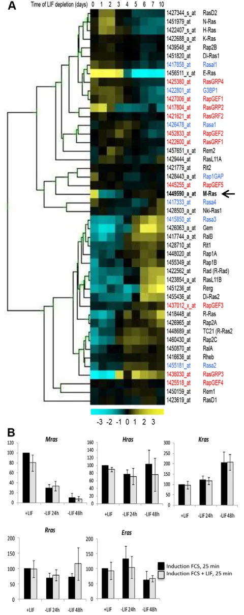
Extensive microarray analyses performed with mESCs grown under different culture conditions (pluripotency or differentiation induction) have enabled the identification of novel markers of pluripotent mESCs and the characterisation of a set of Plurigenes that are highly expressed in undifferentiated mESCs (Schulz et al., 2009; Trouillas et al., 2009a; Trouillas et al., 2009b). Here, we focus on the expression profiles of Ras family members. Probe set numbers of this gene family and related members (including the Ras-Gapand Ras-Gefgenes, 273 probe sets) were retrieved from the Affymetrix 420.2 microarray and their expression profiles analysed in pluripotent cells and after LIF withdrawal from 1 to 10 days. A heat map representation of a selection of genes (46 probe sets) is presented (Fig. 1A).
This analysis showed heterogeneous expression profiles of Ras members, and a particular profile for Mras. Indeed it is the only member that is restricted in its expression to undifferentiated ESCs, with a drastic reduction of expression as early as 1 day after LIF withdrawal. We confirmed, using RT-qPCR analysis, that expression of Mras, but not Hras, Kras,Rrasor Eras, is regulated by LIF (Fig. 1B). We also showed that Mrasexpression is under LIF control only in undifferentiated cells. Indeed, Mrasexpression is not induced by short LIF treatment in committed (24 hours minus LIF) or differentiated (48 hours minus LIF) cells, in contrast to the Lifindgenes, which respond to LIF in ESC-derived committed and differentiated cells and in other mature LIF-responsive cell types (Trouillas et al., 2009a; Trouillas et al., 2009b; Mathieu et al., 2012). These results revealed that Mras constitutes a new marker of stemness.
Persistent downregulation of Mrasleads to a
decrease in the expression of core pluripotency markers
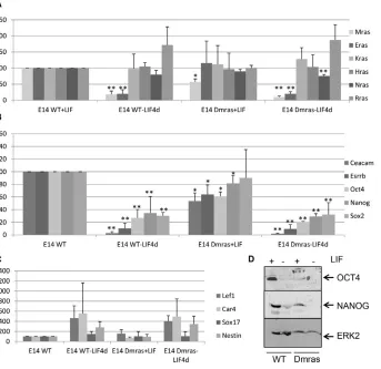
To determine whether Mrascontrols the fate of pluripotent cells, the expression of the core pluripotent genes (Oct4,Sox2and Nanog) and a selection of Plurior differentiation markers was examined in cells in which Mrasexpression was knocked down. We used a clonal heterozygous GeneTrap MrasESC line (E14Dmras), which harbours an insertion of a β-geo cassette in exon 2 (see Materials and methods). Mrasexpression was decreased by 50% in this cell line when compared with the parental E14WT mESC line (Fig. 2A). This mutated cell line, despite being alkaline phosphatase positive in the presence of LIF, displays a reduced self-renewal efficiency (7%, versus 35% for E14WT; supplementary material Fig. S1A).
[image:3.612.54.294.61.664.2]To further characterise the deficit caused by decreased expression of Mras, E14WT and E14Dmras were grown in the presence or absence of LIF for 4 days. Whereas there was no
Fig. 1. Mrashas a unique expression profile among Ras and effector-of-Ras proteins.(A) Heat map analysis of expression profiles of Ras members and regulators (Gefin red and Gapin blue) on a timescale of LIF depletion from days 1 to 10 in mESCs. (B) RT-qPCR analysis of Rasfamily members in cells grown with or without LIF for 24 hours or 48 hours and reinduced for 25 minutes with foetal calf serum (FCS) or FCS + LIF. Mean
of expression with s.d. of triplicates is shown.
D
E
V
E
LO
P
M
E
N
change in the expression of Ras family members (Fig. 2A), significant downregulation in the expression of the Plurigenes Ceacam1and Esrrband the master genes Oct4and to a lesser extent Nanog was observed in the mutated cell line, with a concomitant decrease in OCT4 and to a lesser extent NANOG protein (Fig. 2B,D). Under these conditions, no significant differences in the expression of the differentiation markers tested were detected between WT and Dmras cells (Fig. 2C). However, we have reproducibly observed that the formation of embryoid bodies (EBs) is slower and that they are smaller in Dmras versus WT lines. Furthermore, analysis of differentiation markers at two time points in differentiation (8-day EBs and 13 days after EB plating) shows that there is a decrease in endodermal (Sox17) and mesodermal [brachyury (T)] but not neuronal [Tuj1 (Tubb3) and Map2] expression markers in Dmras versus E14WT only in plated differentiated cells (data not shown).
These results suggest that the decrease in Mrasexpression, which affects the proper balance in the expression of pluripotent genes, leads to an unpoised state of the cells, which have lost, in part, their potential to respond to differentiation cues.
Overexpression of MRAS leads to different cellular phenotypes
MRAS overexpression or its activation was previously shown to induce differentiation in various cell models (Kimmelman et al., 2002; Sun et al., 2006; Watanabe-Takano et al., 2010). We tested the effect of overexpression of MRAS in mESCs grown with or without LIF. MRAS protein expression was detected in 30-50% of the transfected cells (data not shown) the day after transfection, with an average of 10-fold overexpression of the transfected gene in
transient transfection versus endogenous Mras expression (Fig. 3A,B, Fig. 4A).
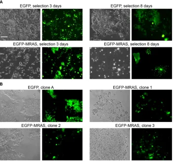
Cells transfected with the EGFP-MRAS fusion protein display specific morphological changes compared with EGFP control cells. Formation of membrane microspikes was observed 3 days after transfection and an increase in the proportion of differentiated cells was observed after 8 days of selection (Fig. 3A). Although differentiated cells could be maintained for more than a month without apparent proliferation, they died after the first passage following trypsin treatment. In addition, after 4 weeks in selective medium with LIF, stable EGFP-MRAS clones, with a slow growth rate compared with the EGFP clones, were recovered and amplified (Fig. 3B, Fig. 4A; supplementary material Fig. S1B,C).
Our data suggest that Mras, like Oct4and Sox2, behaves as a rheostat gene that displays different morphological effects depending upon its level of expression, which is heterogeneous following transfection (Kopp et al., 2008; Niwa et al., 2000).
Stable clones overexpressing MRAS have
sustained expression of OCT4 and NANOG when grown without LIF
[image:4.612.57.400.63.396.2]To determine whether overexpression of MRAS impacts the core pluripotent complex, clones expressing EGFP or EGFP-MRAS were grown with or without LIF for 7 days. Sustained expression of OCT4 was observed in Mras-overexpressing clones grown without LIF (Fig. 4B; supplementary material Fig. S2). In addition, protein expression was analysed by flow cytometry on the EGFP-positive cell fractions. A representative analysis of four clones (EGFP-MRAS-A to -D) displays sustained expression not only of OCT4
Fig. 2. Sustained knockdown of Mras leads to decreased expression of Oct4 and Plurigenes.(A-C) RT-qPCR expression analysis of (A) Mrasand a selection of Ras family members, (B) Pluriand master genes and (C) differentiation markers in E14WT or E14Dmras mESC lines grown with or without LIF for 4 days. The average of three
independent experiments is shown with s.d. *P<0.05, WT versus Dmras in the presence of LIF; **P<0.01, WT or Dmras with LIF versus without LIF. (D) Immunoblot of E14WT and E14Dmras cell lysates cultured with or without LIF for 4 days.
D
E
V
E
LO
P
M
E
N
but also of NANOG upon LIF withdrawal (Fig. 4C). This finding could indicate that cell differentiation has been delayed upon MRAS overexpression. The ERK and PI3K signalling pathways, which are involved in cell differentiation and self-renewal in mESCs, respectively, have been shown to be activated by MRAS in different situations (Kimmelman et al., 2000; Kimmelman et al., 2002). However, we did not observe a constitutive induction of these pathways when Mrasis overexpressed (supplementary material Fig. S3).
Since MRAS-overexpressing clones grew slowly and were difficult to maintain in culture, it was not possible to use them for a more in-depth analysis. Therefore, to explore the functions of Mras in cell fate decision we turned to the Xenopusmodel, which has been fruitful for establishing the functional conservation of pluripotent genes.
Mrasgene structure and protein sequence are
conserved among vertebrates
XenopusMRAS protein shows 84% and 92% identity with the zebrafish and human orthologues, respectively (Fig. 5A). This identity is in agreement with the phylogenetic analysis of the Ras family, which indicates that a single Mrasorthologue is present in the genome from tunicates to mammals (Keduka et al., 2009). Mras gene structure is remarkably conserved among vertebrates and is composed of five exons, interrupted by four introns with identical splice site junctions, having a conserved coding capacity except for the first exon of the zebrafish gene (Fig. 5B). The intronic regions of mammalian Mrasgenes have a very long first intron compared with those of the chicken, Xenopus and zebrafish orthologues (Fig. 5C).
Xenopus Mrasexpression is maternal and zygotic and restricted to the adult brain and ovary
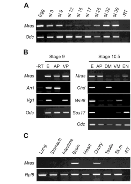
Semi-quantitative RT-PCR analysis indicates that Mras is expressed in unfertilised Xenopuseggs and at all embryonic stages analysed, but that the maternal transcript level decreases after blastula stage (stage 9) and zygotic expression starts at late neurula stage (stage 17) (Fig. 6A). In the blastula embryo, MrasmRNA is found throughout the embryo, with no difference between animal and vegetal poles, whereas in the gastrula embryo (stage 10.5) it is mainly found at the animal pole (Fig. 6B). In adult tissues, Mras is mainly expressed in the brain (as in mammals) and ovary (Fig. 6C).
Rohon-Beard cells and trigeminal neurons are the major sites of expression of Mrasin the Xenopus embryo
We did not detect, through whole-mount in situhybridisation, any localised Mrasexpression in the early Xenopusembryo before stage 24, when a faint labelling was observed in a string of cells along the nerve cord corresponding to Rohon-Beard (RB) cells, which are primary sensory neurons (Roberts, 2000) (Fig. 7A,B). Later in development, labelling also appears in the trigeminal neurons and epiphysis and is clearly detected between somites on each side of the neural tube (Fig. 7C-H).
Mrasregulates neuronal differentiation
[image:5.612.53.405.62.390.2]Mrasoverexpression induces the neuronal differentiation of PC12 mammalian cells (Kimmelman et al., 2002; Sun et al., 2006). Moreover, microarray data analysis indicates that Mrasis re-expressed in ESC-derived neuronal cells (Abranches et al., 2009;
Fig. 3. Overexpression of MRAS leads to morphological changes in cells grown with LIF.(A) mESCs transfected with EGFP or EGFP-MRAS in selective medium with LIF as indicated. (B) Selection of individual clones grown for 4 weeks. Phase contrast (left) and fluorescence (right) images are shown. Scale bar: 100 μm.
D
E
V
E
LO
P
M
E
N
Schulz et al., 2009) (supplementary material Fig. S4). Because Mras is expressed in RB cells of the Xenopus embryo, we investigated the effects of its loss-of-function on neuronal differentiation during development through the use of an antisense morpholino oligonucleotide strategy. A morpholino directed
[image:6.612.54.364.56.354.2]against Mrassequence (MOMras) and a mismatch morpholino (MO mismatch) were tested for their ability to block Xenopus Mrastranslation. Only MOMrasreduced translation of Mras mRNA (supplementary material Fig. S5). Morpholinos were injected into one cell of 2-cell stage embryos and morphant
Fig. 4. Overexpression of MRAS in the absence of LIF leads to sustained expression of OCT4 and NANOG.(A) Relative expression level of Mrasas determined by RT-qPCR. The endogenous level of Mrasin mESCs + LIF or without LIF for 24 hours or transfected with 1 μg pCAG-EGFP (EGFP) (lanes 1-4) is compared with the level of overexpressed Mras after transfection with pCAG-EGFP-MRAS (lanes 5, 6) the day after transfection. The expression of Mrasin stable clones selected for a month in G418 in complete medium with LIF is shown for EGFP clones (lanes 7-9) and EGFP-MRAS clones (lanes 10-12). The mean fluorescence intensity (MFI) of transfected cells is, on average, 15,000 the day after transfection. (B) OCT4 and ERK2 western blot analysis of cell lysates from stable clones expressing EGFP or EGFP-MRAS with and without LIF. (C) FACS analysis of the percentage of cells expressing OCT4, NANOG or SOX2 among WT cells (the mean of three experiments is shown with s.d.) or representative individual clones expressing EGFP or EGFP-MRAS in the presence or absence of LIF for 7 days. The mean MFI is 2894 for clone A, 3000 for clone B, 3000 for clone C and 3822 for clone D, with a background autofluorescence of 100.
Fig. 5. Conservation of vertebrate Mrasat both protein and genomic levels.(A) MRAS protein sequence comparison between dog (Cf, Canis familiaris), human (Hs, Homo sapiens), mouse (Mm, Mus musculus), rat (Rn, Rattus norvegicus), chicken (Gg, Gallus gallus), Xenopus(Xl, Xenopus laevis) and zebrafish (Dr, Danio rerio). Identical amino acids are black, highly conserved in blue and neutral in red. (B) Vertebrate Mrasgene structure with exons represented by black boxes, introns (I-IV) by a thin line and untranslated regions by white boxes. The length of the coding sequence (in nucleotides) is indicated for each exon. The type of splice junction is indicated for each intron. (C) Intron size (in nucleotides) comparison between the different Mras
genes.
D
E
V
E
LO
P
M
E
N
[image:6.612.57.551.452.670.2]embryos were analysed for the expression of the neuronal marker N-tubulin, which marks zones of primary neurogenesis in the embryo. In the stage 15 Xenopusembryo, N-tubulin is expressed in three stripes of cells that correspond to primary motor, inter-and sensory neurons, respectively, the latter of which will become RB sensory neurons (Chitnis et al., 1995). Mrasknockdown by injection of MOMrasresulted mostly in a reduction of the sensory neuron stripe (75%, n=126) and of the trigeminal neurons, with a
milder effect on interneurons and almost no effect on motor neurons (Fig. 8).
The reduction in N-tubulinexpression is specific as neither MO mismatch nor MO standard control (GeneTools) impaired its expression and, moreover, the injection of a MrasmRNA that is insensitive to MO Mrascan rescue N-tubulinexpression (Fig. 8). Additional markers of neuronal differentiation were also investigated: Elrc, Runx1, Pak3 and Islet1 (Perron et al., 1999; Souopgui et al., 2002; Carruthers et al., 2003; Brade et al., 2007). The expression of Elrc(72%, n=77), Pak3(71%, n=42) and Islet1 (81%, n=32) but not of Runx1(3%, n=86) is reduced in MOMras morphants (Fig. 8). Together, these experiments indicate that Mras might function upstream of Elrc, Pak3or Islet1in the regulation of primary neuron formation but downstream or in parallel of Runx1 in the generation of RB sensory neurons.
Mrasacts at a late step of neurogenesis and is required for neuronal differentiation
The early pan-neural marker Sox2is essential for neuroectoderm formation, while Neurogenin is the earliest proneural gene expressed within the neural plate (Kishi et al., 2000; Ma et al., 1996). Mrasdepletion in the Xenopusembryo did not affect the expression of either marker (0%, n=65 for Sox2 and n=54 for Neurogenin), indicating a late role for Mras in neuronal differentiation (Fig. 9).
A constitutively active mutant of Mras(G22V) has been shown to induce neuronal differentiation of PC12 cells, whereas a dominant-negative form of the protein (S27N) impaired neuritogenesis (Sun et al., 2006). We tested whether these two forms of Mrashad an impact on neuronal differentiation. Whereas the constitutively active form of MRAS induced increased expression of N-tubulin(69%, n=86), the dominant-negative form decreased N-tubulinexpression (80%, n=25). Together, these data indicate that Mrasis acting at a late phase of neuronal differentiation and that its activation is a prerequisite to its function during differentiation.
MRAS maintains the competence of gastrula embryo cells for FGF and activin induction and stimulates neural differentiation
[image:7.612.52.261.58.352.2]Embryonic cell patterning of the early Xenopusembryo relies on a limited number of signalling pathways that influence cell fate decisions that can be reproduced in pluripotent animal cap cells of the blastula embryo (Heasman, 2006). We found that FGF2 stimulates Mrasexpression in animal cap cells, whereas activin or BMP treatments have no significant effects (Fig. 10A). Knockdown
Fig. 6. Mrasexpression is biphasic during Xenopus laevis development.RT-PCR analysis of Mrasexpression. (A) Egg and embryonic stages (st). (B) Stage 9 or 10.5 dissected embryos. AP, animal part; VP, ventral part; DM, dorsal mesoderm; VM, ventral mesoderm; EN, endoderm. An1, Vg1, Chordin (Chd), Wnt8and Sox17are used as controls. Total embryo (E) was analysed in parallel. (C) Adult tissues. Sk.m, skeletal muscle. Ornithine decarboxylase(Odc) and Ribosome protein like 8(Rpl8) were used as controls, and a reaction was performed in the absence of reverse transcriptase (–RT).
Fig. 7. Mrasis expressed in Rohon-Beard cells and trigeminal neurons in the Xenopus embryo.(A-H) In situhybridisation with Mrasantisense probe (A,B,E,F) or combined with immunostaining with the muscle-specific antibody 12/101 (C,D,G,H) on stage 24 (A-D) or stage 33 (E-H) embryos. Lateral (A,C,E,G) and dorsal (B,F) views. The plane of the transverse sections shown in D and H are indicated in C and G, respectively. Arrowheads indicate Mrasexpression in Rohon-Beard cells. Ep, epiphysis; No, notochord; Nt, neural tube; RB, Rohon-Beard cells; S, somites; Tn, trigeminal neurons.
D
E
V
E
LO
P
M
E
N
[image:7.612.53.484.570.689.2]with MO Mrasdid not affect mesoderm induction mediated by FGF2 (Fig. 10B) or activin (data not shown), indicating that Mras is not essential for mesoderm induction.
The Xenopusanimal cap assay has been successfully used to demonstrate the involvement of OCT4 and BRG1 in the maintenance of pluripotency (Hansis et al., 2004; Snir et al., 2006). Animal cap cells from the blastula embryo can be induced to form mesoderm when treated with FGF2 or activin, but gastrula stage embryos lose this property (Grimm and Gurdon, 2002; Snape et al., 1987). Animal cap cells from the gastrula stage embryo injected with Mras mRNA still express Xbra in response to FGF2 (Fig. 10C). Both Xenopusand mouse Mrasshowed this property (data not shown). Other marker genes (Cdx4, Pnp and Esr5) previously shown to be FGF targets (Branney et al., 2009) are also re-expressed in late animal cap cells upon Mras expression (Fig. 10C). Similarly, we tested the effect of Mras on the responsiveness of animal caps cells to activin. When MrasmRNA is overexpressed in the embryo, late animal cap cells can still respond to activin treatment and express Xbra, Chordinand Xnr1
(Fig. 10D). Together, these results indicate that Mrascan maintain the mesodermal competence of pluripotent animal cap cells to FGF2 and activin. This property is not limited to mesoderm but can also apply to endoderm, as shown by the expression of the endoderm-specific gene Sox17(Hudson et al., 1997) (Fig. 10D).
When considering the neurectoderm germ layer, the animal cap test cannot be used because both early and late animal cap cells respond to neurogenesis induction by the neural inducer noggin (Lamb et al., 1993). When Mrasis overexpressed in animal caps with noggin, we observed a strong induction of the N-tubulinand Ncam genes, indicating a positive effect of Mras on neuronal differentiation (supplementary material Fig. S6). Together, these results support a conserved role of Mras in neurogenesis and describe a new function of this GTPase at the onset of gastrulation.
DISCUSSION
[image:8.612.52.360.62.324.2]In this study, we have focused on Mras, which encodes a GTPase that is preferentially expressed in undifferentiated mESCs and re-expressed in ESC-derived neuronal cells. The impact of Mraswas
[image:8.612.51.361.565.739.2]Fig. 8. Inhibition of Mrasblocks neuronal differentiation.MO Mras, MO mismatch or MO standard were injected into one blastomere of 2-cell stage Xenopusembryos and morphants were analysed by in situhybridisation for N-tubulin, Elrc, Runx1, Pak3 and Islet1expression. In rescue experiments, 500 pg MrasmRNA was co-injected with MOMras(MOMras+ Mras). The injected side is on the right. Motor (m) inter-(i) and sensory (s) primary neurons and the trigeminal placode (tg) are shown (arrowhead). Arrows indicate change in expression induced by MO Mras. Dorsal views, anterior to bottom.
Fig. 9. Mrasacts at a late step of neurogenesis and is required for neuronal differentiation.MOMras, MO standard, or 500 pg mRNA encoding a constitutively active form (G22V) or a dominant-negative form (S27N) of MRAS was injected into one blastomere of 2-cell stage Xenopusembryos. Embryos were analysed by in situhybridisation for Sox2, Neurogeninor N-tubulinexpression. The injected side is on the right. Arrows indicate change in expression. Dorsal views, anterior to bottom.
D
E
V
E
LO
P
M
E
N
evaluated in two models of pluripotent cells – mESCs and Xenopus blastula embryonic cells (animal cap) – and on the development of the Xenopusembryo.
The involvement of Ras GTPase members in the maintenance of cell pluripotency in mESCs was suggested by the fact that a chemical inhibitor of RasGAP, which maintains Ras members in a GTP-bound active conformation, allows the propagation of mESCs in an undifferentiated pluripotent state in the absence of LIF (Chen et al., 2006). In addition, a recent report has demonstrated the importance of the nucleolar GTP-binding protein nucleostemin (Gnl3) for the self-renewal of mESCs by direct action on the G1 phase of the cell cycle (Qu and Bishop, 2012).
Functional approaches have demonstrated various roles for Ras members in mESCs: ERAS (ES cell-expressed ras) promotes ESC proliferation and the formation of teratomas (Takahashi et al., 2003); the activated KRAS mutant (G12V) alters the expression of pluripotent markers, leading to growth maintenance even with a low dose of LIF (Luo et al., 2007); and activated HRAS mutants induce differentiation toward trophectoderm or extra-embryonic endoderm lineages (Yoshida-Koide et al., 2004; Lu et al., 2008). However, knockout mice failed to reveal any early embryonic function as almost all individual Rasknockouts are viable and
fertile, except for Kras−/−mice, which display lethality at E12 (Ise
et al., 2000; Koera et al., 1997; Nuñez Rodriguez et al., 2006; Umanoff et al., 1995). In this study, we have not observed a compensatory effect in Ras member expression in ESC lines expressing a half dose of Mrasin the presence or absence of LIF. Further studies will be required to determine which genes, if any, might compensate for the absence of individual Rasmembers and to reconcile the lack of an embryonic phenotype in knockout mice with the crucial role played by some Rasmembers in the mESC model.
Our results identify a novel role for MRAS, which was thus far known for its involvement in differentiation processes (Kimmelman et al., 2002; Sun et al., 2006; Watanabe-Takano et al., 2010). We showed that downregulation of Mras expression in mESCs deconstructs the stemness program by decreasing the expression of Oct4and of some Plurigenes, such as Ceacam1and Esrrb, and diminishes the self-renewal capacity of the cells. However, the Dmras cell line and cells in which Mrashas been repressed by shRNA (Parisi et al., 2010) maintain their undifferentiated morphology and are alkaline phosphatase positive, indicating that compensatory effects allow cells to grow and to be amplified in LIF-containing culture medium.
We also showed that overexpression of Mrasleads to changes in cell morphology, with more giant and spiky differentiated cells observed within the first week of cell culture. However, we did not detect any MRAS-dependent induction of the ERK pathway in these early differentiated cells, which did not survive after the first passage in culture (data not shown; supplementary material Fig. S3). Stable clones overexpressing MRAS, which do eventually grow in selective medium, exhibit, in contrast to the wild-type clones, sustained expression of OCT4 and NANOG when cells are deprived of LIF for 7 days, with no effect on SOX2 expression. This indicates that MRAS activates a pathway that alters only the OCT4/NANOG subcomplex and probably not the SOX2/OCT4/ NANOG complex. Oct4is the paradigm of a rheostat effector gene, which modulates the stemness/differentiation balance depending on its level of expression (Niwa et al., 2000). In addition, an OCT4 dose-dependent switch from the Sox2to Sox17promoter triggers mesodermal differentiation (Blin et al., 2010; Stefanovic et al., 2009). Our data suggest that Mrasbehaves as a rheostat gene and that there is a link between MRAS and OCT4 expression. In cells deprived of LIF for a week, Oct4mRNA expression diminishes whether or not Mras is overexpressed (our unpublished data). Therefore, we favour an MRAS-dependent post-translational stabilisation of OCT4 that could lead to sustained expression of the protein in Mras-overexpressing cells grown without LIF. Such post-translational modifications (phosphorylation or ubiquitylation) alter OCT4 stabilisation (Xu et al., 2009; Saxe et al., 2009). Interestingly, Mrasalong with Tcl1, another member of the Pluricluster (Trouillas et al., 2009b), are direct targets of the KLF5 transcription factor, which is involved in the maintenance of cell pluripotency (Parisi et al., 2008; Parisi et al., 2010). MRAS could be a signalling relay of KLF5, acting both on gene transcription and the protein stabilisation machinery.
[image:9.612.53.282.59.337.2]Because stable clones grew slowly, it was not possible to determine whether they remain pluripotent in the absence of LIF using teratoma formation. In addition, we observed a poor capacity of the E14Dmras cell line to form EBs in comparison to WT cells, and found that Brachyury (T) and Sox17expression was decreased in comparison to the WT situation in late differentiated cells, indicating that a proper level of Mrasis required for the cells to maintain their pluripotency and to differentiate efficiently.
Fig. 10. Mrascan maintain the competence of Xenopus gastrula embryo cells for FGF and activin induction but is not required for mesoderm induction. RT-PCR analysis of gene expression in Xenopus animal caps cultured until the control embryo (lane E) reached stage 12.5. (A) Activin, BMP or FGF2 treatment followed by Mrasand Xbraexpression analysis. (B) Animal caps from Mrasmorphants (MO) or control embryos treated with 50 or 100 ng/ml FGF2 (FGF50 or FGF100) and analysed for Xbraexpression. (C,D) Animal caps dissected at stage 8 (Early) or stage 11 (Late) from embryos injected with MrasorBrg1mRNAs or uninjected embryo (Control) were treated with FGF2 (F) or activin (A) or left untreated (–) before expression analysis of Xbra,Cdx4,Pnp,Esr5, Xnr, Chordin (Chd) orSox17 (Sox). Odcwas used as control, and a reaction was performed in the absence of reverse transcriptase (–RT).
D
E
V
E
LO
P
M
E
N
Therefore, we took advantage of the complementary Xenopusmodel to address Mrasfunctions in pluripotency and early development. We used both the in vivoapproach and the model of pluripotent animal cap cells (Sasai et al., 2008). Mras is highly conserved among vertebrates from zebrafish to human at both the gene and protein levels, suggesting a selection pressure and conserved function. As in mESCs, there are clearly two phases of Mras expression in Xenopus, with a maternal expression that decreases between the blastula and early gastrula stages followed by a zygotic expression that increases in the early tadpole, a time when Mras mRNA is found in RB cells and trigeminal neurons. RB neurons are a transient population of primary sensory neurons that is observed in non-mammalian vertebrates, such as zebrafish and Xenopus, and is replaced in the late embryo by dorsal root ganglia (Holland, 2009; Olesnicky et al., 2010; Roberts, 2000). Strikingly, in the mouse embryo, Mrasis also expressed in dorsal root ganglia (Keduka et al., 2009). We conclude that the amphibian and murine Mrasgenes share both strong sequence conservation and expression pattern (Kimmelman et al., 2000).
Loss-of-function of Mras in morphant embryos results in a consistent phenotype that is characterised by the suppression of neuronal differentiation as assessed by N-tubulin,Pak3,Elrc or Islet1 expression. This is supported by experiments in which a dominant-negative form results in fewer neurons. By contrast, a constitutively active form of MRAS stimulates neuronal differentiation, expanding the N-tubulinexpression domain (Fig. 9). Although Mrashas been shown to induce neuronal differentiation, including neurite outgrowth in PC12 cells (Sun et al., 2006), our data constitute the first report of the involvement of Mrasduring vertebrate neurogenesis in vivo. We hypothesise that this property is not restricted to amphibians but is shared by mammalian species because the murine protein is able to rescue morphant embryos as efficiently as the amphibian protein. Mrasacts at a late phase of neurogenesis, downstream of Sox2and Neurogenin. Surprisingly, Runx1 expression is unchanged in Mras morphant embryos. Because Runx1is expressed in RB progenitors and has been shown to be required for their specification independently of Neurogenin (Park et al., 2012), we hypothesise that Mras can act either downstream of Runx1in a common pathway or independently of Runx1in a parallel pathway.
In mESCs, a half dose of Mras leads to downregulation of Ceacam1,Esrrb,Oct4and, to a lesser extent, Nanog, whereas its overexpression in LIF-deprived cells leads to sustained expression of OCT4 and NANOG, suggesting a role in pluripotency maintenance. We have demonstrated that gastrula cells that overexpress Mrascan be induced to form mesoderm when treated with FGF2, suggesting that Mrascan prolong their pluripotency, as shown for Brg1(Smarca4) (Hansis et al., 2004). Mrasis also able to maintain gastrula cell competence to form mesoderm, but also endoderm, under the control of activin. An evaluation of whether OCT4 expression is modulated in Mrasmorphant embryos was not possible owing to the lack of a XenopusOCT4 antibody. However, we have not observed any variation in Oct4 expression at the mRNA level in Mrasmorphants (data not shown).
In conclusion, we have observed in both models two phases of Mras expression that could support two distinct roles. Mras expression in LIF-treated ESCs declines after LIF removal and is re-induced during neural differentiation. In Xenopus, the elevated Mras expression that is observed throughout the embryo until blastula stage could be required for the maintenance of pluripotency, whereas its localised expression in the presumptive neuroectoderm of the gastrula embryo supports its function in neurogenesis. Indeed,
in pluripotent cells neuralised by noggin, Mras expression stimulates neuronal differentiation. Our work, along with the recent report on the function of the GTP-binding protein nucleostemin in the self-renewal of mESCs (Qu and Bishop, 2012), provide new tools and insights for characterising and understanding the links between GTP-binding proteins and stemness and the neurogenic regulators in different models such as mESCs and the Xenopus embryo. In addition, our study reinforces the use of complementary models for a better understanding of gene functions in vertebrates and demonstrates the involvement of Mrasin cell pluripotency and neurogenesis.
Acknowledgements
We thank members of our group for discussions; Sophie Daburon for providing human LIF; Vincent Pitard and Santiago Gonzales for advice on flow cytometry; Charline Maignan for experiments performed with the Dmras cell line; Drs Sachinidis and Pradier for microarray data generated during the FunGenES European program; Drs Kolde and Vilo for the FunGenES website; Austin Smith for the CGR8 cells; Drs Endo, Bellefroid, Grainger, Pandur, Pieler, Perron and Saint Jeannet for plasmids; Dr Goding for reading the manuscript; and Lionel Parra Iglesias for Xenopuscolony care. We acknowledge NIH sponsorship of NHLBI-BayGenomics and the NCRR-MMRRC for providing the ESC gene trap line.
Funding
This work was funded by the European consortium FunGenES [6th Framework, project no. LSHG-CT-2003-503494]; CNRS; University Bordeaux Segalen; Ligue National contre le Cancer; comité de Gironde; Association pour la Recherche sur le Cancer [no. A08/2/1017]; Region Aquitaine; and SFR TransBiomed. M.-E.M., C.S. and M.T. were funded by the Research Ministry, Region Aquitaine and a FunGenES Fellowship, respectively.
Competing interests statement
The authors declare no competing financial interests.
Author contributions
M.-E.M., C.F., C.S., F.S., X.G. and M.T. performed experiments and contributed to data analysis. S.F. performed experiments. N.T. contributed to data analysis and wrote the manuscript. P.T. and H.B. contributed to experimental design, performed experiments, contributed to data analysis and wrote the manuscript. All authors read and approved the final manuscript for publication.
Supplementary material
Supplementary material available online at
http://dev.biologists.org/lookup/suppl/doi:10.1242/dev.091082/-/DC1
References
Abranches, E., Silva, M., Pradier, L., Schulz, H., Hummel, O., Henrique, D. and Bekman, E.(2009). Neural differentiation of embryonic stem cells in vitro: a
road map to neurogenesis in the embryo. PLoS ONE4, e6286.
Abu-Remaileh, M., Gerson, A., Farago, M., Nathan, G., Alkalay, I., Zins Rousso, S., Gur, M., Fainsod, A. and Bergman, Y.(2010). Oct-3/4 regulates stem cell identity and cell fate decisions by modulating Wnt/β-catenin signalling. EMBO J.29, 3236-3248.
Blin, G., Nury, D., Stefanovic, S., Neri, T., Guillevic, O., Brinon, B., Bellamy, V., Rücker-Martin, C., Barbry, P., Bel, A. et al.(2010). A purified population of multipotent cardiovascular progenitors derived from primate pluripotent stem cells engrafts in postmyocardial infarcted nonhuman primates. J. Clin. Invest. 120, 1125-1139.
Boeuf, H., Hauss, C., Graeve, F. D., Baran, N. and Kedinger, C.(1997). Leukemia inhibitory factor-dependent transcriptional activation in embryonic stem cells. J. Cell Biol.138, 1207-1217.
Brade, T., Gessert, S., Kühl, M. and Pandur, P.(2007). The amphibian second heart field: Xenopus islet-1 is required for cardiovascular development. Dev. Biol.311, 297-310.
Branney, P. A., Faas, L., Steane, S. E., Pownall, M. E. and Isaacs, H. V.(2009). Characterisation of the fibroblast growth factor dependent transcriptome in early development. PLoS ONE4, e4951.
Carruthers, S., Mason, J. and Papalopulu, N.(2003). Depletion of the cell-cycle inhibitor p27(Xic1) impairs neuronal differentiation and increases the number of ElrC(+) progenitor cells in Xenopus tropicalis. Mech. Dev.120, 607-616.
Chambers, I. and Tomlinson, S. R.(2009). The transcriptional foundation of
pluripotency. Development136, 2311-2322.
D
E
Chen, S., Do, J. T., Zhang, Q., Yao, S., Yan, F., Peters, E. C., Schöler, H. R., Schultz, P. G. and Ding, S.(2006). Self-renewal of embryonic stem cells by a small molecule. Proc. Natl. Acad. Sci. USA103, 17266-17271.
Chitnis, A., Henrique, D., Lewis, J., Ish-Horowicz, D. and Kintner, C.(1995). Primary neurogenesis in Xenopus embryos regulated by a homologue of the Drosophila neurogenic gene Delta. Nature375, 761-766.
Corpet, F.(1988). Multiple sequence alignment with hierarchical clustering.
Nucleic Acids Res.16, 10881-10890.
Díez, D., Sánchez-Jiménez, F. and Ranea, J. A. G.(2011). Evolutionary expansion of the Ras switch regulatory module in eukaryotes. Nucleic Acids Res. 39, 5526-5537.
Duval, D., Reinhardt, B., Kedinger, C. and Boeuf, H.(2000). Role of suppressors of cytokine signaling (Socs) in leukemia inhibitory factor (LIF) -dependent embryonic stem cell survival. FASEB J.14, 1577-1584.
Gan, Q., Thiébaud, P., Thézé, N., Jin, L., Xu, G., Grant, P. and Owens, G. K.
(2011). WD repeat-containing protein 5, a ubiquitously expressed histone methyltransferase adaptor protein, regulates smooth muscle cell-selective gene activation through interaction with pituitary homeobox 2. J. Biol. Chem.
286, 21853-21864.
Grimm, O. H. and Gurdon, J. B.(2002). Nuclear exclusion of Smad2 is a mechanism leading to loss of competence. Nat. Cell Biol.4, 519-522.
Hansis, C., Barreto, G., Maltry, N. and Niehrs, C.(2004). Nuclear reprogramming of human somatic cells by Xenopus egg extract requires BRG1. Curr. Biol.14, 1475-1480.
Harland, R. M. and Grainger, R. M.(2011). Xenopus research: metamorphosed by genetics and genomics. Trends Genet.27, 507-515.
Heasman, J.(2006). Patterning the early Xenopus embryo. Development133, 1205-1217.
Holland, L. Z.(2009). Chordate roots of the vertebrate nervous system: expanding the molecular toolkit. Nat. Rev. Neurosci.10, 736-746.
Hudson, C., Clements, D., Friday, R. V., Stott, D. and Woodland, H. R.(1997).
Xsox17alpha and -beta mediate endoderm formation in Xenopus. Cell91,
397-405.
Ise, K., Nakamura, K., Nakao, K., Shimizu, S., Harada, H., Ichise, T., Miyoshi, J., Gondo, Y., Ishikawa, T., Aiba, A. et al.(2000). Targeted deletion of the
H-ras gene decreases tumor formation in mouse skin carcinogenesis. Oncogene
19, 2951-2956.
Ivanova, N., Dobrin, R., Lu, R., Kotenko, I., Levorse, J., DeCoste, C., Schafer, X., Lun, Y. and Lemischka, I. R.(2006). Dissecting self-renewal in stem cells with RNA interference. Nature442, 533-538.
Keduka, E., Kaiho, A., Hamada, M., Watanabe-Takano, H., Takano, K., Ogasawara, M., Satou, Y., Satoh, N. and Endo, T.(2009). M-Ras evolved independently of R-Ras and its neural function is conserved between mammalian and ascidian, which lacks classical Ras. Gene429, 49-58.
Kimmelman, A., Tolkacheva, T., Lorenzi, M. V., Osada, M. and Chan, A. M.
(1997). Identification and characterization of R-ras3: a novel member of the RAS gene family with a non-ubiquitous pattern of tissue distribution.
Oncogene15, 2675-2685.
Kimmelman, A. C., Osada, M. and Chan, A. M.(2000). R-Ras3, a brain-specific Ras-related protein, activates Akt and promotes cell survival in PC12 cells.
Oncogene19, 2014-2022.
Kimmelman, A. C., Nuñez Rodriguez, N. and Chan, A. M.(2002). R-Ras3/M-Ras induces neuronal differentiation of PC12 cells through cell-type-specific activation of the mitogen-activated protein kinase cascade. Mol. Cell. Biol.22, 5946-5961.
Kishi, M., Mizuseki, K., Sasai, N., Yamazaki, H., Shiota, K., Nakanishi, S. and Sasai, Y.(2000). Requirement of Sox2-mediated signaling for differentiation of
early Xenopus neuroectoderm. Development127, 791-800.
Koera, K., Nakamura, K., Nakao, K., Miyoshi, J., Toyoshima, K., Hatta, T., Otani, H., Aiba, A. and Katsuki, M.(1997). K-ras is essential for the
development of the mouse embryo. Oncogene15, 1151-1159.
Kopp, J. L., Ormsbee, B. D., Desler, M. and Rizzino, A.(2008). Small increases in the level of Sox2 trigger the differentiation of mouse embryonic stem cells.
Stem Cells26, 903-911.
Lamb, T. M., Knecht, A. K., Smith, W. C., Stachel, S. E., Economides, A. N., Stahl, N., Yancopolous, G. D. and Harland, R. M.(1993). Neural induction by the secreted polypeptide noggin. Science262, 713-718.
Lan, L., Vitobello, A., Bertacchi, M., Cremisi, F., Vignali, R., Andreazzoli, M., Demontis, G. C., Barsacchi, G. and Casarosa, S.(2009). Noggin elicits retinal fate in Xenopus animal cap embryonic stem cells. Stem Cells27, 2146-2152.
Lu, C.-W., Yabuuchi, A., Chen, L., Viswanathan, S., Kim, K. and Daley, G. Q.
(2008). Ras-MAPK signaling promotes trophectoderm formation from
embryonic stem cells and mouse embryos. Nat. Genet.40, 921-926.
Luo, F., Hamoudi, R., Brooks, D. G., Patek, C. E. and Arends, M. J.(2007). Stem cell gene expression changes induced specifically by mutated K-ras. Gene Expr. 14, 101-115.
Ma, Q., Kintner, C. and Anderson, D. J.(1996). Identification of neurogenin, a vertebrate neuronal determination gene. Cell87, 43-52.
Mathieu, M.-E., Saucourt, C., Mournetas, V., Gauthereau, X., Thézé, N., Praloran, V., Thiébaud, P. and Bœuf, H.(2012). LIF-dependent signaling: new pieces in the Lego. Stem Cell Rev.8, 1-15.
Matsumoto, K., Asano, T. and Endo, T.(1997). Novel small GTPase M-Ras participates in reorganization of actin cytoskeleton. Oncogene15, 2409-2417.
Morrison, G. M. and Brickman, J. M.(2006). Conserved roles for Oct4 homologues in maintaining multipotency during early vertebrate
development. Development133, 2011-2022.
Naye, F., Tréguer, K., Soulet, F., Faucheux, C., Fédou, S., Thézé, N. and Thiébaud, P.(2007). Differential expression of two TEF-1 (TEAD) genes during Xenopus laevis development and in response to inducing factors. Int. J. Dev. Biol.51, 745-752.
Nieuwkoop, P. D. and Faber, J.(1967). Normal Table of Xenopus laevis (Daudin). Amsterdam: North-Holland.
Niwa, H.(2007). How is pluripotency determined and maintained? Development 134, 635-646.
Niwa, H., Yamamura, K. and Miyazaki, J.(1991). Efficient selection for high-expression transfectants with a novel eukaryotic vector. Gene108, 193-199.
Niwa, H., Miyazaki, J. and Smith, A. G.(2000). Quantitative expression of Oct-3/4 defines differentiation, dedifferentiation or self-renewal of ES cells. Nat. Genet.24, 372-376.
Nuñez Rodriguez, N., Lee, I. N. L., Banno, A., Qiao, H. F., Qiao, R. F., Yao, Z., Hoang, T., Kimmelman, A. C. and Chan, A. M.-L.(2006). Characterization of R-ras3/m-ras null mice reveals a potential role in trophic factor signaling. Mol. Cell. Biol.26, 7145-7154.
Okabayashi, K. and Asashima, M.(2003). Tissue generation from amphibian animal caps. Curr. Opin. Genet. Dev.13, 502-507.
Olesnicky, E., Hernandez-Lagunas, L. and Artinger, K. B.(2010). prdm1a Regulates sox10 and islet1 in the development of neural crest and Rohon-Beard sensory neurons. Genesis48, 656-666.
Parisi, S. and Russo, T.(2011). Regulatory role of Klf5 in early mouse
development and in embryonic stem cells. Vitam. Horm.87, 381-397.
Parisi, S., Passaro, F., Aloia, L., Manabe, I., Nagai, R., Pastore, L. and Russo, T.
(2008). Klf5 is involved in self-renewal of mouse embryonic stem cells. J. Cell Sci.121, 2629-2634.
Parisi, S., Cozzuto, L., Tarantino, C., Passaro, F., Ciriello, S., Aloia, L., Antonini, D., De Simone, V., Pastore, L. and Russo, T.(2010). Direct targets of Klf5 transcription factor contribute to the maintenance of mouse embryonic stem cell undifferentiated state. BMC Biol.8, 128.
Park, B.-Y., Hong, C.-S., Weaver, J. R., Rosocha, E. M. and Saint-Jeannet, J.-P.
(2012). Xaml1/Runx1 is required for the specification of Rohon-Beard sensory neurons in Xenopus. Dev. Biol.362, 65-75.
Perron, M., Furrer, M. P., Wegnez, M. and Théodore, L.(1999). Xenopus elav-like genes are differentially expressed during neurogenesis. Mech. Dev.84, 139-142.
Qu, J. and Bishop, J. M.(2012). Nucleostemin maintains self-renewal of embryonic stem cells and promotes reprogramming of somatic cells to pluripotency. J. Cell Biol.197, 731-745.
Roberts, A.(2000). Early functional organization of spinal neurons in developing lower vertebrates. Brain Res. Bull.53, 585-593.
Saito, Y., Oinuma, I., Fujimoto, S. and Negishi, M.(2009). Plexin-B1 is a GTPase activating protein for M-Ras, remodelling dendrite morphology. EMBO Rep.10, 614-621.
Sasai, Y., Ogushi, M., Nagase, T. and Ando, S.(2008). Bridging the gap from frog research to human therapy: a tale of neural differentiation in Xenopus animal caps and human pluripotent cells. Dev. Growth Differ.50Suppl. 1, S47-S55.
Saxe, J. P., Tomilin, A., Schöler, H. R., Plath, K. and Huang, J.(2009). Post-translational regulation of Oct4 transcriptional activity. PLoS ONE4, e4467.
Schulz, H., Kolde, R., Adler, P., Aksoy, I., Anastassiadis, K., Bader, M., Billon, N., Boeuf, H., Bourillot, P.-Y., Buchholz, F. et al.; Functional Genomics in Embryonic Stem Cells Consortium(2009). The FunGenES database: a genomics resource for mouse embryonic stem cell differentiation. PLoS ONE4, e6804.
Sive, H. L., Grainger, R. M. and Harland, R. M.(2000). Early Development of Xenopus Laevis. A Laboratory Manual. Cold Spring Harbor, NY: Cold Spring Harbor Laboratory Press.
Snape, A., Wylie, C. C., Smith, J. C. and Heasman, J.(1987). Changes in states of commitment of single animal pole blastomeres of Xenopus laevis. Dev. Biol. 119, 503-510.
Snir, M., Ofir, R., Elias, S. and Frank, D.(2006). Xenopus laevis POU91 protein, an Oct3/4 homologue, regulates competence transitions from mesoderm to neural cell fates. EMBO J.25, 3664-3674.
Souopgui, J., Sölter, M. and Pieler, T.(2002). XPak3 promotes cell cycle
withdrawal during primary neurogenesis in Xenopus laevis. EMBO J.21,
6429-6439.
Stefanovic, S., Abboud, N., Désilets, S., Nury, D., Cowan, C. and Pucéat, M.
(2009). Interplay of Oct4 with Sox2 and Sox17: a molecular switch from stem
cell pluripotency to specifying a cardiac fate. J. Cell Biol.186, 665-673.
D
Sun, P., Watanabe, H., Takano, K., Yokoyama, T., Fujisawa, J. and Endo, T.
(2006). Sustained activation of M-Ras induced by nerve growth factor is essential for neuronal differentiation of PC12 cells. Genes Cells11, 1097-1113.
Takahashi, K., Mitsui, K. and Yamanaka, S.(2003). Role of ERas in promoting tumour-like properties in mouse embryonic stem cells. Nature423, 541-545.
Trouillas, M., Saucourt, C., Guillotin, B., Gauthereau, X., Taupin, J. L., Moreau, J. F. and Boeuf, H.(2009a). The LIF cytokine: towards adulthood. Eur. Cytokine Netw.20, 51-62.
Trouillas, M., Saucourt, C., Guillotin, B., Gauthereau, X., Ding, L., Buchholz, F., Doss, M. X., Sachinidis, A., Hescheler, J., Hummel, O. et al.(2009b). Three LIF-dependent signatures and gene clusters with atypical expression profiles, identified by transcriptome studies in mouse ES cells and early derivatives. BMC Genomics10, 73.
Umanoff, H., Edelmann, W., Pellicer, A. and Kucherlapati, R.(1995). The murine N-ras gene is not essential for growth and development. Proc. Natl. Acad. Sci. USA92, 1709-1713.
Ward, K. R., Zhang, K.-X., Somasiri, A. M., Roskelley, C. D. and Schrader, J. W.
(2004). Expression of activated M-Ras in a murine mammary epithelial cell line
induces epithelial-mesenchymal transition and tumorigenesis. Oncogene23,
1187-1196.
Watanabe-Takano, H., Takano, K., Keduka, E. and Endo, T.(2010). M-Ras is activated by bone morphogenetic protein-2 and participates in osteoblastic determination, differentiation, and transdifferentiation. Exp. Cell Res.316, 477-490.
Wray, J., Kalkan, T. and Smith, A. G.(2010). The ground state of pluripotency.
Biochem. Soc. Trans.38, 1027-1032.
Xu, H., Wang, W., Li, C., Yu, H., Yang, A., Wang, B. and Jin, Y.(2009). WWP2 promotes degradation of transcription factor OCT4 in human embryonic stem cells. Cell Res.19, 561-573.
Yoshida-Koide, U., Matsuda, T., Saikawa, K., Nakanuma, Y., Yokota, T., Asashima, M. and Koide, H.(2004). Involvement of Ras in extraembryonic endoderm differentiation of embryonic stem cells. Biochem. Biophys. Res. Commun.313, 475-481.