Copyright © 2004, American Society for Microbiology. All Rights Reserved.
Multilocus Short Sequence Repeat Sequencing Approach for
Differentiating among
Mycobacterium avium
subsp.
paratuberculosis
Strains
Alongkorn Amonsin,
1Ling Ling Li,
1Qing Zhang,
1John P. Bannantine,
2Alifiya S. Motiwala,
3Srinand Sreevatsan,
3and Vivek Kapur
1*
Department of Microbiology and Biomedical Genomics Center, University of Minnesota, St. Paul,
Minnesota 55108
1; National Animal Disease Center, U.S. Department of Agriculture, Ames, Iowa
50010
2; and Food Animal Health Research Program, Ohio Agricultural Research and
Development Center, and Department of Veterinary Preventive
Medicine, The Ohio State University, Wooster, Ohio 44691
3Received 14 August 2003/Returned for modification 23 October 2003/Accepted 2 January 2004
We describe a multilocus short sequence repeat (MLSSR) sequencing approach for the genotyping of
Mycobacterium avium
subsp.
paratuberculosis
(
M. paratuberculosis
) strains. Preliminary analysis identified 185
mono-, di-, and trinucleotide repeat sequences dispersed throughout the
M. paratuberculosis
genome, of which
78 were perfect repeats. Comparative nucleotide sequencing of the 78 loci of six
M. paratuberculosis
isolates
from different host species and geographic locations identified a subset of 11 polymorphic short sequence
repeats (SSRs), with an average of 3.2 alleles per locus. Comparative sequencing of these 11 loci was used to
genotype a collection of 33
M. paratuberculosis
isolates representing different multiplex PCR for IS
900
loci
(MPIL) or amplified fragment length polymorphism (AFLP) types. The analysis differentiated the 33
M.
paratuberculosis
isolates into 20 distinct MLSSR types, consistent with geographic and epidemiologic correlates
and with an index of discrimination of 0.96. MLSSR analysis was also clearly able to distinguish between sheep
and cattle isolates of
M. paratuberculosis
and easily and reproducibly differentiated strains representing the
predominant MPIL genotype (genotype A18) and AFLP genotypes (genotypes Z1 and Z2) of
M. paratuberculosis
described previously. Taken together, the results of our studies suggest that MLSSR sequencing enables facile
and reproducible high-resolution subtyping of
M. paratuberculosis
isolates for molecular epidemiologic and
population genetic analyses.
Mycobacterium avium
subsp.
paratuberculosis
(
M.
paratuber-culosis
) is a slowly growing, acid-fast, mycobactin J-dependent
bacterium. Infection with this bacterium leads to a chronic
granulomatous enteritis, termed Johne’s disease, in cattle and
other ruminants and occurs worldwide (11). Clinical signs of
the disease include diarrhea, weight loss, fatigue, decreased
milk production, and mortality. Infection with this pathogen
results in considerable economic losses in the dairy production
industry, with estimated annual costs of $40 to $227 per year
per cow, totaling industry-wide annual losses of $1.5 billion
(20, 21). In addition to the serious health and economic
im-pacts of the pathogen to the dairy industry, several reports
suggest a possible link between
M. paratuberculosis
and
Crohn’s disease in humans (5, 7, 10, 30).
Methods for differentiation or subtyping of bacterial strains
provide important information for molecular epidemiologic
analysis and assist in providing an understanding of the
popu-lation genetics of the species. DNA-based molecular subtyping
techniques such as multiplex PCR for IS
900
integration loci
(MPIL) (4, 20), restriction fragment length polymorphism
(RFLP) analysis (6, 8, 23), and amplified fragment length
poly-morphism (AFLP) analysis (20) have been previously applied
to investigate genetic variation in
M. paratuberculosis
.
How-ever, the MPIL, AFLP, and RFLP techniques are generally
unable to resolve
M. paratuberculosis
isolates into meaningful
epidemiologic groups due to the apparently restricted genetic
diversity within the subspecies. Furthermore, the data
gener-ated by these techniques are biallelic and, hence, are able to
provide only limited information regarding the overall genetic
diversity and evolutionary mechanisms within the species.
Short sequence repeats (SSRs) or variable-number tandem
repeats (VNTRs) in bacterial DNA have been used as markers
for the differentiation and subtyping strains of several bacterial
species, including
Mycobacterium tuberculosis
(9, 16),
Yersinia
pestis
(1),
Salmonella enterica
subsp
. enterica
serovar
Typhi-murium (17), and
Bacillus anthracis
(15). SSRs consist of
sim-ple homopolymeric tracts of a single nucleotide
(mononucle-otide repeats) or multimeric tracts (homogeneous or
heterogeneous repeats), such as di- or trinucleotide repeats,
which can be identified as VNTRs in the genome of the
or-ganism (35). The variability of the repeats is believed to be
caused by slipped-strand mispairing (31); the genetic instability
of polynucleotide tracts, especially poly(G-T) (12); and DNA
recombination between homologous repeat sequences (32).
The complete genome sequence of
M. paratuberculosis
strain
K10 (GenBank accession number AE016958) has recently
been characterized in the Department of Microbiology and
Biomedical Genomics Center, University of Minnesota (L. L.
Li et al., unpublished data). Preliminary bioinformatic analyses
led to the identification of numerous SSRs in the
M.
paratu-* Corresponding author. Mailing address: University of Minnesota,
1500 Gortner Ave., St. Paul, MN 55108. Phone: (612) 625-7712. Fax:
(612) 624-6264. E-mail: [email protected].
1694
on May 15, 2020 by guest
http://jcm.asm.org/
berculosis
genome. We evaluated the utility of a multilocus
SSR (MLSSR)-based typing approach for differentiating
among isolates of
M. paratuberculosis.
The results of our
stud-ies suggest that MLSSR is a useful approach for strain
differ-entiation and enables the rapid and facile discrimination of
epidemiologically and geographically distinct strains of
M.
paratuberculosis.
MATERIALS AND METHODS
Bacterial isolates and DNA isolation.A total of 33M. paratuberculosisisolates from different host species and geographic locations were used in this study, as shown in Table 1.M. paratuberculosisisolates were grown on Middlebrook 7H9 broth or 7H11 agar (Difco Laboratories, Detroit, Mich.) with oleic acid-albumin-dextrose-catalase supplement (Becton Dickinson, Sparks, Md.) and mycobactin J (2 mg/100 ml). In some instances the bacterial cultures were incubated at 37°C for 4 to 6 months until colonies were observed. DNA was isolated from the bacterial culture by use of the QIAamp DNA Mini kit (Qiagen Inc., Valencia, Calif.), as described previously (20).
Database search for SSRs and primer design.The whole-genome sequence of
M. paratuberculosisstrain K10 was analyzed for SSRs with Tandem Repeat Finder (version 2.02) software (3). The coordinates of the SSRs were then matched for the regions upstream and downstream to locate the repeats and
open reading frame (ORF) flanking the repeat by use of the DNA sequence viewer and annotation software Artemis (28). Primers specific for sequences flanking these repeat sequences were designed with Primer 3 software (27) to yield an average amplification product of⬃250 bp for each sequence (Table 2).
MLSSR.A total of 78 loci were amplified by PCR with specific primers, and the amplification products were sequenced to identify sequence polymorphisms in each locus among six strains ofM. paratuberculosis(reference strain MAP-K-10 and isolates from cattle [isolates MAP-08 and MAP-09], sheep [isolates MAP-06 and MAP-11], and a human [isolate MAP-14]) (Table 1). These sixM. paratuberculosisisolates were selected because they represent the extent of ge-netic diversity in the species, as previously identified by MPIL and AFLP anal-yses (20).
[image:2.603.47.539.81.428.2]The 25-l PCR amplification reaction mixture for each SSR comprised 1⫻ PCR buffer II (Perkin-Elmer, Applied Biosystems Division, Foster City, Calif.), 2.0 mM MgCl2 (Perkin-Elmer), 200M each deoxynucleoside triphosphate (Roche Diagnostic Co., Indianapolis, Ind.), 0.6mol of each primer (Integrated DNA Technologies, Coralville, Iowa), 0.5 U ofAmpliTaqGold (Perkin-Elmer), 5% dimethyl sulfoxide (Sigma Chemical Co, St. Louis, Mo.), and 1l of DNA. The amplification conditions consisted of an initial denaturation at 94°C for 15 min, followed by 35 cycles of denaturation at 94°C for 45 s, annealing at 60°C for 1 min, and extension at 72°C for 2 min 30 s, with a final extension step at 72°C for 7 min. A 2-l volume of the PCR products was mixed with 2l of loading buffer (0.2% Orange G in 50% glycerol), and the mixture was electrophoresed in 1% agarose with 0.5g of ethidium bromide per ml. The gels were photographed
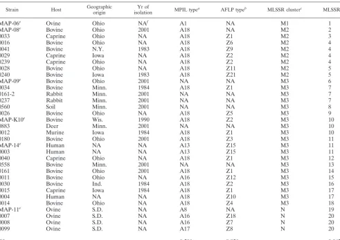
TABLE 1.
M. paratuberculosis
isolates examined in this study
Strain Host Geographicorigin isolationYr of MPIL typea AFLP typeb MLSSR clusterc MLSSR typed
MAP-06
eOvine
Ohio
NA
fA1
NA
M1
1
MAP-08
eBovine
Ohio
2001
A18
NA
M2
2
0033
Caprine
Ohio
NA
A18
Z1
M2
3
0016
Bovine
Ohio
NA
A18
Z6
M2
4
0041
Bovine
N.Y.
1983
A18
Z9
M2
4
0029
Caprine
Iowa
NA
A18
Z2
M2
4
0239
Caprine
Ohio
NA
A18
Z2
M2
4
0028
Bovine
Ohio
NA
A18
Z11
M2
5
0240
Bovine
Iowa
1983
A18
Z21
M2
5
MAP-09
eBovine
Ohio
2001
NA
NA
M3
6
0034
Bovine
Minn.
1984
A18
Z1
M3
7
0161-2
Rabbit
Minn.
2001
NA
NA
M3
7
0237
Rabbit
Minn.
2001
NA
NA
M3
7
0560
Soil
Minn.
2001
NA
NA
M3
8
0026
Bovine
Ohio
NA
A18
Z5
M3
9
MAP-K10
eBovine
Wis.
1990
A18
Z2
M3
10
0883
Deer
Minn.
2001
NA
NA
M3
10
0012
Murine
Iowa
1984
A18
Z1
M3
10
0180
Bovine
Ohio
2001
A18
Z3
M3
11
MAP-14
eHuman
NA
NA
A13
Z15
M3
11
0003
Human
NA
NA
A13
Z15
M3
11
0040
Caprine
Ohio
NA
A18
Z1
M3
12
0558
Bovine
Minn.
2001
NA
NA
M3
13
0161
Bovine
Ohio
2001
A18
Z1
M3
14
0011
Bovine
Ohio
NA
A16
Z12
M3
15
0030
Bovine
Ind.
1984
A18
Z2
M3
16
0015
Caprine
Iowa
1984
A18
Z1
M3
17
0004
Human
NA
NA
A18
Z10
M3
17
0014
Bovine
Ohio
NA
A18
Z4
M3
18
MAP-11
eOvine
S.D.
NA
A8
NA
N
19
0007
Ovine
S.D.
NA
A16
Z18
N
20
0008
Ovine
S.D.
NA
A16
Z7
N
20
0099
Ovine
S.D.
NA
A17
Z8
N
20
D
g0.501
0.920
0.967
aIsolates were previously identified by MPIL analysis (20). bIsolates were previously identified by AFLP analysis (20).
cIsolates were identified by MLSSR analysis based on a genetic distance of 0.43. dIsolates were identified by MLSSR analysis based on 100% similarity. eM. paratuberculosisisolates used to examine polymorphisms of 78 SSR loci. fNA, not available.
gDwas calculated by using the equation described previously (13).
on May 15, 2020 by guest
http://jcm.asm.org/
TABLE 2. Primers for 78 sequence repeat loci used for polymorphism analysis of the region
Repeat and locus Position in genomea SSR Forward primer Forwardprimer
size (bp) Reverse primer
Reverse primer
size (bp)
Mononucleotide
1793091 1793091–1793109 GGGGGGGGGGGG
GGGGGGGG TCAGACTGTGCGGTATGGAA 20 GTGTTCGGCAAAGTCGTTGT 20
2719085 2719085–2719094 GGGGGGGGGG GTGACCAGTGTTTCCGTGTG 20 TGCACTTGCACGACTCTAGG 20
Dinucleotide
3803814 3803814–3803824 GC GC GC GC GC G ACCGAGCCGATAGTCATCAG 20 CAGCAGCAGCGAGTACGA 18
3840791 3840791–3840801 GC GC GC GC GC G CTCGATTCGCGATCAGGT 18 CAACTTGACGCCCTGGTACT 20
607784 607784–607794 CG CG CG CG CG C ATCCAACGCCATGTACTCGT 20 GAGCAGCATCGAGGTGAAAC 20
914968 914968–914978 CG CG CG CG CG C GACTAGGCCCTTGCGGTATC 20 CGCAACGTGCTGTCGTAG 18
3294684 3294684–3294693 GC GC GC GC GC GGCAACGCCTCGTACACC 18 CCTACCCGAGCGGTCACT 18
3363782 3363782–3363791 AC AC AC AC AC AGCGGCTCAATTACACACAA 20 ATCAGGTCCGGAATCACCTT 20
3406364 3406364–3406373 GC GC GC GC GC GTTCTCGATGGACAGCTTGC 20 GGAGGACGAACCACACTCAT 20
3424469 3424469–3424478 CG CG CG CG CG ACCGGTTTCAGACAATGGAG 20 GTACGGCGAGCTACGCTATC 20
3456738 3456738–3456747 GC GC GC GC GC AGCACAAGAAGCACCGGTAT 20 GGATCAACCTCGAGATCCTG 20
3458712 3458712–3458721 CG CG CG CG CG ACCAACTGCAGATCCTCGAT 20 GTAATCCCAGCCGGTTCAT 19
3573587 3573587–3573596 CG CG CG CG CG ATCGCCGACTATCTCAATCC 20 ACCGCCAGATAGTGAATTGC 20
3676817 3676817–3676826 CG CG CG CG CG ACCCGGACACGACGTAGC 18 GTTCGACATCGTGCACCTC 19
3735342 3735342–3735351 GC GC GC GC GC TGTCGAACTTGCTCTTGGTG 20 CGTCCTGCAACATCTTCTCC 20
3754236 3754236–3754245 GC GC GC GC GC ACACGTAGTCGGTGGAGACG 20 GTTGCAATCACACGAACCAC 20
3831350 3831350–3831359 GC GC GC GC GC GCACGCATCTGTTCAACG 18 CACAACAAGATTGGCCTCAG 20
3951841 3951841–3951850 CG CG CG CG CG ACGCCGCACTACGAATATCT 20 ACATCTCGAAGGACGTTTCG 20
4086404 4086404–4086413 GC GC GC GC GC AGTCGCTGGGTTTTCAGGA 19 ATACCGCACGGTGTTCTGAT 20
4159074 4159074–4159083 GC GC GC GC GC TATTCGGGTCCATGCTCAAT 20 ATGTGACGGAGGTCACACTG 20
4176609 4176609–4176618 CG CG CG CG CG TTCATCGACTACCGGCTCTT 20 TGTTGGGGGACCATGTAACT 20
4465488 4465488–4465497 CG CG CG CG CG AAGCTGAACTGGTGGGAGAG 20 GTCGAAAACGCTGTCGTAGG 20
4518335 4518335–4518344 CG CG CG CG CG CTTTGGTTCCCAGCAGCAT 19 GGAGTATTCACCCGACCAAA 20
4717322 4717322–4717331 GC GC GC GC GC TACCAACATCCCGACCTGAC 20 CGTAGGGGATAACCTGCTGA 20
14474 14474–14483 GC GC GC GC GC ATACCTGCGTCCGATACCAG 20 GCTTTCATGATCACCGGTTT 20
70461 70461–70470 CG CG CG CG CG CGATGGCCTACTTCATCGTC 20 CGGCCGAAATAGATGATGTT 20
70930 70930–70939 GC GC GC GC GC CGATCACCCAGCAGTATGTG 20 ATCAGCTCCTTGGTCACCTG 20
78012 78012–78021 GC GC GC GC GC CCATGCTACGAGGTCGAGTT 20 TTGGGGAATAAACGACTTGC 20
191084 191084–191093 GC GC GC GC GC CAGCCGCAACGACTTCTC 18 TTAGGGTTGGCTTCCCATTA 20
219320 219320–210329 GC GC GC GC GC TTGTCCAGCAAGCAGAAGTG 20 ATAGTGCACCCCCAGCAC 18
274406 274406–274415 TG TG TG TG TG AGTGGAGGCTGAGATGTTGG 20 GGTGAACACCTTGCCGTTAT 20
429640 429640–429649 GC GC GC GC GC AAGTACATCCCGCTGCACAC 20 CGATTACCAGGTGCACGAC 19
676774 676774–676783 GC GC GC GC GC GCGAAATAACCGTTCACCTC 20 GACACCACCTGCGAGTAACC 20
686290 686290–686299 CA CA CA CA CA AGATCGCATCAAAGAGCACCT 21 GGTGAGTTGTCCGCATCAG 19
762468 762468–762477 CG CG CG CG CG GTCGACCCGAAGAGTGAGTG 20 GAATTTTTGGGGTCGTGATG 20
899250 899250–899259 CG CG CG CG CG AGAAAATAGCTGCGGTCGAA 20 GGATCGACACCCTGACCTC 19
909353 909353–909362 CG CG CG CG CG CGATCTCATACACCGTCGTC 20 AGGTGAACCGTAAGCGACAC 20
970000 970000–970009 CG CG CG CG CG TCGAGACCTCAAAAGCCTTG 20 GGGGACCTGCTGGTGATAG 19
2892461 2892461–2892470 GT GT GT GT GT GGGTTGGTCTACGTTTGTCG 20 CCACAGCCCCTCGTAGTG 18
2887399 2887399–2887408 CG CG CG CG CG AGCTGTACCGCGACTACCAC 20 AGCAACCGCGAATATGGTC 19
2831533 2831533–2831542 GC GC GC GC GC CTGTTCGCCTACGTGCTGTA 20 GGGGTGGAGACGATGAAATA 20
2808989 2808989–2808998 CG CG CG CG CG GGTGCGCGATAATGAAACTC 20 AAGACCACGCTGGTGAATCT 20
2103815 2103815–2103194 GC GC GC GC GC GACCAGTTCCGGGCTCAC 18 AGGAACTGTTCAACCCGATG 20
2716662 2716662–2716671 CA CA CA CA CA ACCGATCTGGAAGAACATCG 20 GCCCTGGTCATTACACGACT 20
2709505 2709505–2709514 CG CG CG CG CG GAGGTCGTCTTCCGTTACGA 20 CTCGTCGACCAGCAGTATGG 20
2615343 2615343–2615352 GC GC GC GC GC AGGAAGGCGTCGACAAGAT 19 GAGGTGTCGTGCTTGGAGAT 20
2583850 2583850–2583859 CG CG CG CG CG CGGCTTCGTATTGTCGTCTT 20 GGTCATGAGCAGAACCTTCC 20
2582523 2582523–2582532 CA CA CA CA CA CAGATCGCATCAAAGAGCAC 20 GGTGAGTTGTCCGCATCAG 19
2540968 2540968–2540977 GC GC GC GC GC ATTCAACCAACAGCCTCAGC 20 TAGCCGTTACCGGTGTTGAT 20
2519120 2519120–2519129 CG CG CG CG CG CCCACCAGGTCGAAGAAAT 19 CAACTGGAGGACCAGGTGTT 20
2509961 2509961–2509970 GC GC GC GC GC GCATCTCCACCCAGAAATTG 20 GAACACGTCGGTCTTGGTTT 20
2289642 2289642–2289651 GC GC GC GC GC GATCGAAGACCGAAAACGTC 20 AGATCATCGGTGAGCTGGAG 20
2259726 2259726–2259735 GC GC GC GC GC GAAGGGTCGTTCATGTTGCT 20 CAACTTACGGACCTGGGATG 20
2059817 2059817–2059826 GC GC GC GC GC GTCGGGTTCTTCGTCAACAC 20 ATCGGTATCCATCAGGTCCA 20
1960967 1960967–1960976 GC GC GC GC GC GCATTGCGCTACCTGAGTC 19 TCGACGAGAACATCACGAAC 20
1860248 1860248–1860257 CG CG CG CG CG ACCTGCAGACCGACGATTAC 20 ACTTGCTCACCGAGAACAGC 20
1841444 1841444–1841453 CG CG CG CG CG GCAGCTTGTCCAGATCGAA 19 AAAAAGCAGCGACACCAGAC 20
1829129 1829129–1829138 CG CG CG CG CG GAGGACCACGTGAAAATCGT 20 GGATCCTGCACCAGAACCT 19
1787205 1787205–1787214 CG CG CG CG CG TGATCATCATGGAAGCCAAC 20 GCGGGAATGTTGATAAGGAA 20
1758285 1758285–1758294 CG CG CG CG CG GATCATCCAATCGGTGTCCT 20 GCACACTCGTAATCGCTCAA 20
1707736 1707736–1707745 GC GC GC GC GC GACCACCAAAACTGGTTTCC 20 GTCCGGTAGTGGTCGATGTC 20
1684441 1684441–1684450 GC GC GC GC GC CAGGAACTGTGGGATGTCCT 20 CTGTACGGCTATTCGGTGGT 20
1664073 1664073–1664082 CG CG CG CG CG TTCTGGCCGAATTGATCTCT 20 ATCGTTTTTGCCTGAATTGG 20
1526624 1526624–1526633 GC GC GC GC GC GTCACCATCCGGTACATTCC 20 GAGGTGCCCAAGACGTATCT 20
1257495 1257495–1257504 CG CG CG CG CG GGGATCCTGTGGCAGATAAC 20 CAACTGCTGGACACCTGCTA 20
Trinucleotide
4286068 4286068–4286084 GCG GCG GCG GCG
GCG GC GAATCGTCTTGCCTCACTGG 20 TCGAGCAACTGATCTCCACA 20
4310932 4310932–4310948 CCG CCG CCG CCG
CCG CC CGGCAATACCTCGAACAGAT 20 GCTGAAGAGGTCGTGCAGAT 20
Continued on following page
on May 15, 2020 by guest
http://jcm.asm.org/
under UV light with an Eagle Eye II gel documentation system (Stratagene, La Jolla, Calif.). The PCR amplicons were then sequenced with an ABI 3100 automated fluorescent DNA sequencer (Perkin-Elmer) at the University of Min-nesota’s Advanced Genetic Analysis Center (www.agac.umn.edu).
MLSSR data analysis.The sequences of each SSR locus of 33 isolates were aligned, and the numbers of tandem repeats were identified by use of the MegAlign program (DNASTAR Inc., Madison, Wis.). The nucleotide sequences of 11 polymorphic SSR loci were analyzed for each isolate, and allele numbers were assigned to reflect the number of copies or the number of nucleotide substitutions represented in the SSR sequence for each locus. Statistical analysis for genetic diversity and overall relationships among the isolates was performed with the computer programs ETDIV and ETCLUS, which were modified for use with the SSR data (2). MLSSR types were then assigned on the basis of the unique combination of alleles for each locus. Genetic diversity (D) was calculated by using the following equation: 1⫺ ⌺(allele frequency)2(22, 29). The un-weighted pair group method with arithmetic averages-based cluster analysis and bootstrap analysis with 1,000 replications were performed with the program PAUP (version 4.0; Sinauer Associates, Inc. Sunderland, Mass.), and the index of discrimination (D) was determined as described previously (13).
RESULTS
SSRs in
M. paratuberculosis
genome.
Analysis of the
whole-genome sequence of
M. paratuberculosis
strain K10 (4.83 Mbp)
identified 185 SSRs consisting of three or fewer nucleotides
per repeat unit. Of these, 78 mono-, di-, and trinucleotide
repeats with perfect matches between adjacent copies were
identified and were included as candidate polymorphic loci for
further analysis (Table 2). These 78 SSR loci were also selected
for inclusion in our analysis because they were short (1 to 3
bp), as is common in prokaryotes, and each locus had at least
five copies. Dinucleotide repeats were the most frequently
identified SSRs in the
M. paratuberculosis
genome and were
present at 63 distinct loci, with the copy numbers varying
be-tween 5 and 5.5 per repeat. Mono- and trinucleotide repeats
were represented at 2 and 13 loci, respectively.
MLSSR analysis revealed that 11 of the 78 loci were
poly-morphic in the six isolates examined. The ORFs or genes
flanking each locus were also identified (Table 3). For
exam-ple, locus 2 is located within ORF 210_MAP.128, which is
unique to
M. paratuberculosis
. Locus 3 was identified in an
intergenic region between two ORFs: a 5
⬘
ORF encoding
6-aminohexanoate-cyclic dimer and a 3
⬘
ORF encoding alpha/
beta-hydrolase (Table 3). The functional consequences of the
presence of the loci and the influence of the locus copy number
on the expression of the adjacent genes deserve further
inves-tigation.
MLSSR.
The 11 polymorphic SSR loci identified in the
pre-liminary screening were characterized in 27 additional
M.
para-tuberculosis
isolates that were previously characterized by
MPIL and AFLP analyses (20). The analysis identified 20
MLSSR types among the 33
M. paratuberculosis
isolates
recov-ered from different host species and geographic areas (Tables
1 and 4). The
D
value for each SSR locus was calculated on the
basis of the allele frequency and the number of alleles and
revealed an average number of alleles per locus of 3.20, with an
average
D
value of 0.393 and a range of
D
values of 0.100 to
0.700 (21, 28) (Table 3). While the allelic variation observed in
this study focused on the number of copies of the SSRs (Fig.
1A), it is noteworthy that some loci also revealed one or two
base substitutions in some isolates (Fig. 1B). For instance, the
analysis revealed a single polymorphic site each at SSR loci 4
and 10 and four and five polymorphisms at loci 5 and 9,
re-spectively (Fig. 2). It is interesting that the vast majority of the
nucleotide substitutions were found in MAP-06, an isolate
recovered from a sheep.
Genetic relationships among
M. paratuberculosis
isolates
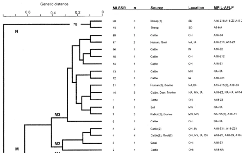
based on MLSSR analysis.
The unweighted pair group method
with arithmetic averages-based cluster analysis of
M.
[image:4.603.44.542.80.286.2]paratu-berculosis
identified 20 distinct MLSSR types among the
iso-lates that were grouped into two major clusters, clusters M and
N (Fig. 3). Cluster M contained 87.88% (29 of 33) of the
isolates in the sample, including isolates recovered from
bo-vine, caprine, murine, deer, rabbit, and human sources. The
isolates in cluster M with the most common MPIL and AFLP
fingerprints, A18 and Z1 and Z2, respectively, were further
TABLE 2—
Continued
Repeat and locus Position in genomea SSR Forward primer Forwardprimer
size (bp) Reverse primer
Reverse primer
size (bp)
440731 440731–440747 TGG TGG TGG TGG
TGG TG CAGCGTGATCTGCGACCT 18 GATCAGCGAACTGCTCACG 19
1028129 1028129–1028145 GGT GGT GGT GGT
GGT GG AGATGTCGACCATCCTGACC 20 AAGTAGGCGTAACCCCGTTC 20
2955362 2955362–2955378 TGC TGC TGC TGC
TGC TG GACAAGTTCGGGTTGACCAC 20 AGTTCCTCGACCCAGTCGT 19
3558075 3558075–3558090 GCC GCC GCC GCC
GCC G CTGGAACGTGTCCGAATTG 19 GTATTCGGTGCGGATCTCCT 20
1653414 1653414–1653429 CGC CGC CGC CGC
CGC C TGAGCAGGAACCAGATCTCC 20 GTGGGGTGGATGAGTACGAC 20
1651920 1651920–1651935 CGC CGC CGC CGC
CGC C AGCATCTTGAGCCCACATCT 20 CCGAAATCAATTCTGGTCGT 20
3182509 3182509–3182523 GCG GCG GCG GCG
GCG CGGTCAGGTCGCAGATTT 18 GGTCAGCGAGAAACCACTTG 20
3562415 3562415–3562429 CTG CTG CTG CTG
CTG CTGGCATTGGGAATGTTCTT 20 CAGCACCATGTAGCCGATCT 20
212174 212174–212188 CGA CGA CGA CGA
CGA CAGAGCGGACTGCATTGAG 19 TCGGTGTTGTCCGGATTC 18
1737056 1737056–1737070 GCG GCG GCG GCG
GCG GCTCGTTGCAGGTCAGGTAG 20 GGCATGATCACCGAAAGC 18
1536798 1536798–1536812 CCG CCG CCG CCG
CCG CTGGAGTGGAAGAGCAGTCC 20 GCTGCGTTACCTCAACACC 19
aThe position in the genome is the coordinate of the SSR locus in theM. paratuberculosisstrain K10 genome (GenBank accession number AE016958).
on May 15, 2020 by guest
http://jcm.asm.org/
divided into three groups, clusters M1, M2, and M3. Cluster
M1 contained one isolate (isolate MAP-06), which was
recov-ered from a sheep and which had the A1 MPIL fingerprint.
Three of the five isolates from caprine sources were assigned to
cluster M2. A total of 13 unique genotypes, including a
major-ity (10 of 15) of the bovine isolates included in this study, were
represented in cluster M3. In addition, the three isolates from
human sources included in the sample used in this study were
also found in cluster M3. Interestingly, two isolates recovered
from humans (isolates MAP-14 and 0003) were clustered into
the same clade as an isolate of bovine origin (isolate 0180).
Isolates that were recovered from a mouse (isolate 0012),
rab-bits (isolates 0237 and 0160 to 0162), a deer (isolate 0883), and
soil (isolate 0560) were also grouped along with the M3
geno-type.
In contrast to cluster M, which consisted of isolates
recov-ered from a variety of animal species, all four isolates that were
included in cluster N were recovered from sheep. Strains of
ovine origin (four of five) also showed a distinct allelic profile
compared with the profiles of strains from cattle, goats, or
humans.
Discriminatory power of subtyping methods.
The
discrimi-natory power (
D
) of MLSSR in comparison with those of other
subtyping methods was determined as described previously
(13). MPIL analysis differentiated the 27
M. paratuberculosis
isolates for which MPIL typing information was available into
6 subtypes with a
D
value of 0.50, indicating only limited
discriminatory power, while MLSSR differentiated 27
M.
para-tuberculosis
into 17 subtypes with a
D
value of 0.96. In contrast,
AFLP analysis differentiated the 24
M. paratuberculosis
isolates
for which AFLP typing information was available into 15
sub-types, with a
D
value of 0.92 (20). In comparison, MLSSR
differentiated the 24
M. paratuberculosis
isolates into 14
sub-types, with higher
D
value of 0.95. Overall, MLSSR
distin-guished 20 subtypes among the 33 isolates in the sample with
a
D
value of 0.96, indicating that it has a relatively high index
of discrimination (Tables 3 and 4).
DISCUSSION
SSRs have been used to type many bacterial pathogens
as-sociated with human and animal infections (32). Within the
genus
Mycobacterium
, VNTR or mycobacterial interspersed
repetitive units have been used for the subtype-specific
[image:5.603.47.541.82.263.2]differ-entiation of several
Mycobacterium
species (19, 26, 35). In the
TABLE 3. SSRs used in MLSSR analysis
Markerlocus Locusname SSR Genome ORFa allelesNo. ofb Dvaluec
1
1793091
GGGGGGGGGGGGGGGGGGGG
GI: 13881617 phosphatidylethanolamine-binding
protein (
Mycobacterium tuberculosis
CDC 1551)
5
0.700
2
2719085
GGGGGGGGGG
No hit
d(210_MAP.128)
3
0.616
3
e607784
CG CG CG CG CG C
IGI: RMMR05031 6-aminohexanoate-cyclic dimer
hydrolase (
Mycobacterium marinum
)
2
0.189
GI: 13883430 hydrolase, alpha/beta-hydrolase fold
family (
M. tuberculosis
CDC1551)
4
3406364
GC GC GC GC GC
GI: 2791627
fixA
(
M. tuberculosis
H37Rv)
2
0.100
5
3735342
GC GC GC GC GC
GI: 13092881 putative
S
-adenosyl-
L-homocysteine
hydrolase (
Mycobacterium leprae
)
4
0.363
6
4286068
GCG GCG GCG GCG GCG GC
IGI: RMMR06009 heat shock protein 70 (
M. marinum
)
2
0.395
7
4310932
CCG CCG CCG CCG CCG CC
GI: 10579910 phytoene dehydrogenase (
Halobacterium
spp.)
2
0.100
8
1028129
GGT GGT GGT GGT GGT GG
GI: 3261715
mfd
(
M. tuberculosis
H37Rv)
4
0.668
9
2955362
TGC TGC TGC TGC TGC TG
GI: 2117199
narG
(
M. tuberculosis
H37Rv)
4
0.553
10
3558075
GCC GCC GCC GCC GCC G
GI: 1781217
nuoG
(
M. tuberculosis
H37Rv)
3
0.279
11
1536798
CCG CCG CCG CCG CCG
GI: 5524340
PstA
(
Mycobacterium avium
)
4
0.363
aGI, NCBI gene identification number; IGI, Integrated Genomics gene identification number. bThe average number of alleles was 3.20.
cDwas calculated by using the equation 1⫺ 兺(allele frequency)2(22). The averageDvalue was 0.393. dNo hit, no nucleotide match with any sequences in the GenBank database.
eLocus 3 is located in an intergenic region of two ORFs (6-aminohexanoate-cyclic dimer and hydrolase [the alpha/beta-hydrolase fold]).
TABLE 4. Profiles of alleles at 11 SSR loci for 20 clones
of
M. paratuberculosis
MLSSR
type Referenceisolate isolatesNo. of
No. of copies of SSRs or no. of nucleotide substitutions at the following locus 1a 2a 3b 4b 5b 6c 7c 8c 9c 10c 11c
1
MAP-06
1
7 11 5 1 1 5 5 3 1 0
0
2
MAP-08
1
7 10 5 5 5 5 4 4 4 5
5
3
0033
1
7 11 5 5 5 4 5 4 4 5
5
4
0016
4
7 11 5 5 5 4 5 4 3 5
5
5
0028
2
7 10 5 5 5 4 5 4 3 5
5
6
MAP-09
1
11 11 5 5 5 5 5 5 5 5
3
7
0237
3
⬎
14 9 5 5 5 5 5 5 5 5
5
8
0560
1
⬎
14 11 5 5 5 5 5 5 5 5
5
9
0026
1
8 10 5 5 5 5 5 5 5 5
5
10
MAP-K10
3
⬎
14 10 5 5 5 5 5 5 5 5
5
11
MAP-14
3
7 10 5 5 5 5 5 5 5 5
5
12
0040
1
7 9 5 5 3 5 5 5 5 5
5
13
0558
1
7 9 5 5 5 5 5 5 5 5
5
14
0161
1
8 11 5 5 5 5 5 4 5 5
5
15
0011
1
9 11 5 5 5 5 5 4 5 5
5
16
0030
1
9 11 5 5 5 5 5 5 5 5
5
17
0004
2
7 11 5 5 5 5 5 5 5 5
5
18
0014
1
7 11 5 5 5 5 5 6 5 5
5
19
MAP-11
1
⬎
14 11 4 5 4 4 5 3 4 6
4
20
0007
3
⬎
14 10 4 5 4 4 5 3 4 6
4
aThe numbers of G mononucleotides present (⬎14, 14 or more). bThe numbers of copies of the dinucleotide repeat.
cThe numbers of copies of the trinucleotide repeat.
on May 15, 2020 by guest
http://jcm.asm.org/
[image:5.603.44.283.476.699.2]present study we have identified polymorphic SSRs by genomic
analysis of
M. paratuberculosis
and used this information to
develop a highly discriminatory method for the typing of
M.
paratuberculosis
isolates.
The SSRs discovered during our preliminary screening of
the
M. paratuberculosis
genome were similar to the repeats that
have previously been described in other bacteria, including
Haemophilus
,
Mycoplasma
, and
Mycobacterium
spp. (24, 32,
33). It has previously been recognized that regions of mono-,
, and trinucleotide tandem repeats are often the most
di-verse in a bacterial genome, while complex longer repeats
generally have lower levels of diversity (14). This is thought to
result from slipped-strand mispairing (or replication slippage
events) of the DNA polymerase that occurs with greater
fre-quency on the SSRs, a hypothesis that remains to be tested for
the SSRs that we have identified in
M. paratuberculosis
(32).
Several important attributes of a strain differentiation assay
determine its utility in a clinical or epidemiologic setting.
Es-pecially for organisms such as
M. paratuberculosis
that have
restricted levels of genetic diversity, the discriminatory power
of an assay is a particularly important attribute. Assays such as
MPIL and RFLP analysis have been shown to have only
mod-erate abilities to differentiate among epidemiologically distinct
isolates of
M. paratuberculosis
and therefore have limited
[image:6.603.131.455.72.497.2]ap-plicabilities in molecular epidemiologic studies (4, 5, 33). The
recently described AFLP technique has been shown to have a
greater resolving power than the other two approaches but
suffers from the limitation that allelic variation is indexed at
FIG. 1. Sequence analysis of two representative SSR loci. (A) Locus 8 with (GGT)
5repeats. Strain MAP-K10 contains five copies of GGT,
while MAP-08 and MAP-11 contain four and three copies of GGT, respectively. (B) Locus 10 with (GCC)
5repeats. Strain MAP-K10 contains five
copies of GCC, while MAP-11 and MAP-07 have a A-to-C substitution in a single copy of GCC.
on May 15, 2020 by guest
http://jcm.asm.org/
anonymous biallelic markers (20). In contrast, the MLSSR
assay described herein is far more discriminatory, being able to
differentiate 33
M. paratuberculosis
from distinct geographic
localities and host species into 20 subtypes on the basis of
allelic variation at the 11 SSR loci examined, with a notably
high
D
value of 0.96. Consistent with its high discriminatory
power, MLSSR enabled the differentiation of seemingly
mono-morphic
M. paratuberculosis
strains that were indistinguishable
by MPIL and AFLP analyses (20). An important advantage of
the MLSSR approach is that it also indexes variations at known
genetic loci and has the ability to identify multiple alleles per
locus. Together, these attributes not only allow an increase in
the strain-resolving power of the assay but also enable an
understanding of the genetic mechanisms driving strain
diver-sification and evolution within the species.
Another key attribute of a strain differentiation assay is its
ability to identify epidemiologically and genetically related
strains of a bacterial species. In this context, MLSSR analysis
clearly showed that some isolates that are of sheep origin
(cluster N) are genetically distinct from those of bovine,
ca-prine, and human origin (cluster M), a finding consistent with
those of previous studies (4, 6, 20). It is noteworthy, however,
that the five isolates of sheep origin examined during this study
were represented by three distinct MLSSR types (MLSSR
types 1, 19 and 20), and four isolates clustered together in
cluster N. Interestingly, all four of these phylogenetically
linked
M. paratuberculosis
isolates were recovered from sheep
in South Dakota, suggesting that they are both genetically and
epidemiologically related and well distinguishable from the
other isolates in the collection. The same isolates were also
grouped into four distinct MPIL genotypes (A1, A8, A16, and
A17) and three AFLP genotypes (Z7, Z8, and Z18), suggesting
that they are indeed genetically distinct from the other isolates
in the collection. However, by the MPIL and the AFLP
ap-proaches, these isolates do not cluster together as closely as
they do by MLSSR analysis (20). Hence, these results suggest
that MLSSR analysis may enable molecular epidemiologic
in-vestigations that will lead to a better understanding of strain
transmission and the spread of
M. paratuberculosis
in natural
populations and across host species.
In contrast to the relatively close clustering of the sheep
M.
paratuberculosis
strains in the samples examined, far greater
diversity was observed in isolates of bovine origin. The analysis
[image:7.603.106.482.78.427.2]showed that while a majority of the
M. paratuberculosis
isolates
FIG. 2. Allelic variation at 11 SSR loci among 33
M. paratuberculosis
isolates. The aligned nucleotide sequences of each of the alleles at the
11 SSR loci discovered and characterized during this investigation, along with adjacent conserved sequences, are shown. The SSRs and
polymorphic sites are boxed. The locations of single-nucleotide polymorphisms are indicated with asterisks below the aligned sequences.
on May 15, 2020 by guest
http://jcm.asm.org/
of bovine origin clustered together in the M3 subgroup, 60%
(three of five) of the caprine isolates were represented by the
closely related cluster M2, suggesting that caprine isolates bear
greater genetic resemblance to cattle strains than to isolates of
ovine origin, a finding that is consistent with the findings of
previous studies (34). Similarly, deer and cattle strains also
appeared to be more closely related to each other by MLSSR
analysis, suggesting a sharing of strains of
M. paratuberculosis
in wildlife species that graze or that may otherwise come into
close contact with cattle, as hypothesized previously (25).
Our studies demonstrate that MLSSR analysis offers several
advantages over other methods for differentiating among
M.
paratuberculosis
isolates. First, as described above, the
tech-nique has a high discriminatory power for known multiallelic
genetic loci, an essential attribute for the effective
differentia-tion of genetically distinct isolates. Second, MLSSR results are
based upon DNA sequencing and, hence, are unambiguous
and reproducible and can likely be obtained for most loci of all
M. paratuberculosis
isolates, even those recovered from sheep
or wildlife species, as demonstrated by our studies described
herein. However, we note the formal possibility that mutations
or deletions at the primer sites may render some strains
un-typeable at some loci, such as loci 10 and 11 in MAP-06. Third,
MLSSR analysis is based on the amplification of SSR loci by
PCR and thus not only is rapid but also may be performed
directly with bacterial colonies without DNA extraction.
Fourth, due to the considerable advances in automated DNA
sequencing technologies and the falling prices of DNA
se-quencing, the MLSSR method is amenable to adaptation for
high-throughput analysis and can be performed relatively
in-expensively as well. Finally, a key advantage of the approach is
that the data are sequence based and, hence, enable accurate
interlaboratory comparisons to be made and the information
used in the development of SSR databases for further
molec-ular epidemiologic studies, which are greatly required in this
field (18). While it must be recognized that sequence errors
due to strand slippage during either PCR or sequencing
reac-tions may result in an erroneous assignment of genotype, the
occurrence of such slippage errors is minimized by increasing
the amount of sequence coverage at the locus (by confirming
both the forward and the reverse sequences or testing
dupli-cate samples), as is routinely practiced in our laboratory.
In conclusion, we have described here the development of
MLSSR-based typing for the subtype-specific differentiation of
M. paratuberculosis
isolates. Our preliminary analyses suggest
that this approach will be of considerable utility in enabling
detailed molecular epidemiologic and population genetic
anal-yses of this important animal pathogen.
ACKNOWLEDGMENTS
Research in the laboratory of V. Kapur is funded by grants from the
U.S. Department of Agriculture, the National Institutes of Health, and
the Minnesota Agricultural Experimental Station. Research in the
laboratory of S. Sreevatsan is funded through a seed grant from the
Ohio Agricultural Research and Development Center’s research
en-hancement competitive grants program.
REFERENCES
[image:8.603.99.489.72.319.2]1. Adair, D. M., P. L. Worsham, K. K. Hill, A. M. Klevytska, P. J. Jackson, A. M. Friedlander, and P. Keim.2000. Diversity in a variable-number tan-dem repeat fromYersinia pestis. J. Clin. Microbiol.38:1516–1519. 2. Amonsin, A., J. F. Wellehan, L. L. Li, P. Vandamme, C. Lindeman, M.
FIG. 3. Dendrogram depicting genetic relationships among 33
M. paratuberculosis
isolates on the basis of the 11 SSR loci determined by
MLSSR analysis. The dendrogram was generated by the unweighted pair-group method with arithmetic averages with the PAUP program. The
results of the bootstrap analysis are represented as percentages and are indicated adjacent to the major nodes when the branch order was supported
by
⬎
50% of the 1,000 replicate trees. Genetic distance is indicated at the top of the dendrogram. Isolate identifications, sources, geographic
locations, and MILP and AFLP types are shown to the right of the dendrogram.
on May 15, 2020 by guest
http://jcm.asm.org/
Edman, R. A. Robinson, and V. Kapur.1997. Molecular epidemiology of
Ornithobacterium rhinotracheale. J. Clin. Microbiol.35:2894–2898. 3. Benson, G.1999. Tandem repeats finder: a program to analyze DNA
se-quences. Nucleic Acids Res.27:573–580.
4. Bull, T. J., J. Hermon-Taylor, I. Pavlik, F. El-Zaatari, and M. Tizard.2000. Characterization of IS900 loci inMycobacterium aviumsubsp. paratubercu-losisand development of multiplex PCR typing. Microbiology146(Pt 9):
2185–2197.
5. Bull, T. J., E. J. McMinn, K. Sidi-Boumedine, A. Skull, D. Durkin, P. Neild, G. Rhodes, R. Pickup, and J. Hermon-Taylor.2003. Detection and verifica-tion ofMycobacterium avium subsp.paratuberculosisin fresh ileocolonic mucosal biopsy specimens from individuals with and without Crohn’s dis-ease. J. Clin. Microbiol.41:2915–2923.
6. Cousins, D. V., S. N. Williams, A. Hope, and G. J. Eamens.2000. DNA fingerprinting of Australian isolates ofMycobacterium aviumsubsp. paratu-berculosisusing IS900 RFLP. Aust. Vet. J.78:184–190.
7. El-Zaatari, F. A., M. S. Osato, and D. Y. Graham.2001. Etiology of Crohn’s disease: the role ofMycobacterium avium paratuberculosis. Trends Mol. Med.
7:247–252.
8. Francois, B., R. Krishnamoorthy, and J. Elion.1997. Comparative study of
Mycobacterium paratuberculosisstrains isolated from Crohn’s disease and Johne’s disease using restriction fragment length polymorphism and arbi-trarily primed polymerase chain reaction. Epidemiol. Infect.118:227–233. 9. Gascoyne-Binzi, D. M., R. E. Barlow, R. Frothingham, G. Robinson, T. A.
Collyns, R. Gelletlie, and P. M. Hawkey.2001. Rapid identification of lab-oratory contamination withMycobacterium tuberculosisusing variable num-ber tandem repeat analysis. J. Clin. Microbiol.39:69–74.
10. Grimes, D. S.2003.Mycobacterium aviumsubspeciesparatuberculosisas a cause of Crohn’s disease. Gut52:155.
11. Harris, N. B., and R. G. Barletta.2001.Mycobacterium aviumsubsp. para-tuberculosisin veterinary medicine. Clin. Microbiol. Rev.14:489–512. 12. Henderson, S. T., and T. D. Petes.1992. Instability of simple sequence DNA
inSaccharomyces cerevisiae. Mol. Cell. Biol.12:2749–2757.
13. Hunter, P. R., and M. A. Gaston.1988. Numerical index of the discrimina-tory ability of typing systems: an application of Simpson’s index of diversity. J. Clin. Microbiol.26:2465–2466.
14. Keim, P., L. B. Price, A. M. Klevytska, K. L. Smith, J. M. Schupp, R. Okinaka, P. J. Jackson, and M. E. Hugh-Jones.2000. Multiple-locus vari-able-number tandem repeat analysis reveals genetic relationships within
Bacillus anthracis. J. Bacteriol.182:2928–2936.
15. Kim, W., Y. P. Hong, J. H. Yoo, W. B. Lee, C. S. Choi, and S. I. Chung.2002. Genetic relationships ofBacillus anthracisand closely related species based on variable-number tandem repeat analysis and BOX-PCR genomic finger-printing. FEMS Microbiol. Lett.207:21–27.
16. Kremer, K., D. van Soolingen, R. Frothingham, W. H. Haas, P. W. Hermans, C. Martin, P. Palittapongarnpim, B. B. Plikaytis, L. W. Riley, M. A. Yakrus, J. M. Musser, and J. D. van Embden.1999. Comparison of methods based on different molecular epidemiological markers for typing ofMycobacterium tuberculosiscomplex strains: interlaboratory study of discriminatory power and reproducibility. J. Clin. Microbiol.37:2607–2618.
17. Lindstedt, B. A., E. Heir, E. Gjernes, and G. Kapperud.2003. DNA finger-printing ofSalmonella entericasubsp.entericaserovar Typhimurium with emphasis on phage type DT104 based on variable number of tandem repeat loci. J. Clin. Microbiol.41:1469–1479.
18. Maiden, M. C., J. A. Bygraves, E. Feil, G. Morelli, J. E. Russell, R. Urwin, Q. Zhang, J. Zhou, K. Zurth, D. A. Caugant, I. M. Feavers, M. Achtman, and B. G. Spratt.1998. Multilocus sequence typing: a portable approach to the identification of clones within populations of pathogenic microorganisms. Proc. Natl. Acad. Sci. USA95:3140–3145.
19. Mazars, E., S. Lesjean, A. L. Banuls, M. Gilbert, V. Vincent, B. Gicquel, M. Tibayrenc, C. Locht, and P. Supply.2001. High-resolution minisatellite-based typing as a portable approach to global analysis ofMycobacterium tuberculosismolecular epidemiology. Proc. Natl. Acad. Sci. USA98:1901– 1906.
20. Motiwala, A. S., M. Strother, A. Amonsin, B. Byrum, S. A. Naser, J. R. Stabel, W. P. Shulaw, J. P. Bannantine, V. Kapur, and S. Sreevatsan.2003. Molecular epidemiology ofMycobacterium aviumsubsp.paratuberculosis: evidence for limited strain diversity, strain sharing, and identification of unique targets for diagnosis. J. Clin. Microbiol.41:2015–2026.
21. National Animal Health Monitoring System.1997. Johne’s disease on U. S. dairy operations. Report N245.1087. USDA, APHIS, VS, CEAH, National Animal Health Monitoring System, Fort Collins, Colo.
22. Nei, M.1973. Analysis of gene diversity in subdivided populations. Proc. Natl. Acad. Sci. USA70:3321–3323.
23. Pavlik, I., A. Horvathova, L. Dvorska, J. Bartl, P. Svastova, R. du Maine, and I. Rychlik.1999. Standardisation of restriction fragment length polymor-phism analysis forMycobacteriumavium subspeciesparatuberculosis. J. Mi-crobiol. Methods38:155–167.
24. Peterson, S. N., C. C. Bailey, J. S. Jensen, M. B. Borre, E. S. King, K. F. Bott, and C. A. Hutchison III.1995. Characterization of repetitive DNA in the
Mycoplasma genitaliumgenome: possible role in the generation of antigenic variation. Proc. Natl. Acad. Sci. USA92:11829–11833.
25. Riemann, H., M. R. Zaman, R. Ruppanner, O. Aalund, J. B. Jorgensen, H. Worsaae, and D. Behymer.1979. Paratuberculosis in cattle and free-living exotic deer. J. Am. Vet. Med. Assoc.174:841–843.
26. Roring, S., A. Scott, D. Brittain, I. Walker, G. Hewinson, S. Neill, and R. Skuce.2002. Development of variable-number tandem repeat typing of My-cobacterium bovis: comparison of results with those obtained by using exist-ing exact tandem repeats and spoligotypexist-ing. J. Clin. Microbiol.40:2126– 2133.
27. Rozen, S., and H. Skaletsky.2000. Primer 3 on the WWW for general users and for biologist programmers. Methods Mol. Biol.132:365–386. 28. Rutherford, K., J. Parkhill, J. Crook, T. Horsnell, P. Rice, M. A.
Rajan-dream, and B. Barrell.2000. Artemis: sequence visualization and annota-tion. Bioinformatics16:944–945.
29. Selander, R. K., D. A. Caugant, H. Ochman, J. M. Musser, M. N. Gilmour, and T. S. Whittam.1986. Methods of multilocus enzyme electrophoresis for bacterial population genetics and systematics. Appl. Environ. Microbiol.
51:873–884.
30. Stabel, J. R.1998. Johne’s disease: a hidden threat. J. Dairy Sci.81:283–288. 31. Strand, M., T. A. Prolla, R. M. Liskay, and T. D. Petes.1993. Destabilization of tracts of simple repetitive DNA in yeast by mutations affecting DNA mismatch repair. Nature365:274–276.
32. van Belkum, A., S. Scherer, L. van Alphen, and H. Verbrugh.1998. Short-sequence DNA repeats in prokaryotic genomes. Microbiol. Mol. Biol. Rev.
62:275–293.
33. van Belkum, A., S. Scherer, W. van Leeuwen, D. Willemse, L. van Alphen, and H. Verbrugh.1997. Variable number of tandem repeats in clinical strains ofHaemophilus influenzae. Infect. Immun.65:5017–5027.
34. Whittington, R. J., A. F. Hope, D. J. Marshall, C. A. Taragel, and I. Marsh.
2000. Molecular epidemiology ofMycobacterium aviumsubsp. paratubercu-losis: IS900restriction fragment length polymorphism and IS1311 polymor-phism analyses of isolates from animals and a human in Australia. J. Clin. Microbiol.38:3240–3248.
35. Wiid, I. J., C. Werely, N. Beyers, P. Donald, and P. D. van Helden.1994. Oligonucleotide (GTG)5as a marker forMycobacterium tuberculosisstrain identification. J. Clin. Microbiol.32:1318–1321.