Copyright © 2000, American Society for Microbiology. All Rights Reserved.
A Multiplex Approach to Molecular Detection of
Brucella abortus
and/or
Mycobacterium bovis
Infection in Cattle
SRINAND SREEVATSAN,1JACK B. BOOKOUT,1* FIDEL RINGPIS,1VEERA S. PERUMAALLA,2 THOMAS A. FICHT,2L. GARRY ADAMS,2SUE D. HAGIUS,3PHILIP H. ELZER,3BETSY J. BRICKER,4
GIRISH K. KUMAR,5M. RAJASEKHAR,6SRIKRISHNA ISLOOR,6ANDRAJ R. BARATHUR1 ClinCyte, LLC, San Diego, California 921211; Department of Veterinary Pathobiology, College of Veterinary Medicine,
Texas A&M University, College Station, Texas 77843-44672; Department of Veterinary Science, School of Veterinary Medicine, Louisiana State University, Baton Rouge, Louisiana 708033; Division of Zoonotic Disease Research,
National Animal Disease Center, U.S. Department of Agriculture, Ames, Iowa 500104; Department of Genetics and Plant Breeding, University of Agricultural Sciences, Bangalore, India 5600655; and
Institute of Animal Health and Veterinary Biologicals, Hebbal, Bangalore, India 5600246
Received 19 October 1999/Returned for modification 29 December 1999/Accepted 13 April 2000
A multiplex amplification and detection platform for the diagnosis of Mycobacterium bovis and Brucella
abortusinfection simultaneously in bovine milk and nasal secretions was developed. This system (designated the bovine pathogen detection assay [BPDA]-PCR) consists of duplex amplification of species-specific targets
(a region of the BCSP31K gene ofB. abortus and a repeat-sequence region in the hsp65 gene of M. bovis,
respectively). This is followed by a solid-phase probe capture hybridization of amplicons for detection. On the basis of spiking experiments with normal milk, the analytical sensitivity of the assay was 800 CFU
equiva-lents/ml of milk forB. abortusand as low as 4 CFU equivalents per ml of milk forM. bovis. BPDA-PCR was
validated with 45 liver samples from lemmings experimentally infected withB. abortus. The assay sensitivity,
based on culture status as a “gold standard,” was 93.9%. In this experiment, BPDA-PCR also identified five culture-negative liver samples as positive (41.7%). Field studies for the evaluation of BPDA-PCR were per-formed with samples from dairy animals from geographically distinct regions (India, Mexico, and Argentina).
A high prevalence of shedding of B. abortus(samples from India) andM. bovis(samples from Mexico) was
identified by BPDA-PCR. In samples from India, B. abortus shedding was identified in 86% of milk ring
test-positive animals (n ⴝ 15) and 80% of milk ring test-negative cows (n ⴝ 5). In samples from Mexico,
M. boviswas identified by PCR in 32.6% of pools (nⴝ46) of milk that each contained milk from 10 animals
and in 56.2% of nasal swabs (n ⴝ 121) from cattle from tuberculin test-positive herds. In contrast, the
Argentine cattle (nⴝ70) had a modest prevalence ofM. bovisshedding in nasal swabs (2.9%) and milk (1.4%)
and ofB. abortusin milk (11.4%). On the basis of these analyses, we identify BPDA-PCR as an optimal tool for
both screening of herds and testing of individual animals in a disease eradication program. A combination of the duplex assay, screening of milk samples in pools, and the proposed algorithm provides a highly sensitive, cost-effective, and economically viable alternative to serological testing.
Animal health and human health are inextricably linked. For millennia, humans have depended on animals for nutrition, socioeconomic development, and companionship (1). Yet an-imals can transmit many different but potentially devastating diseases to humans.
Brucellosis is a zoonosis of both public health and economic importance in many developing countries (5, 24). Six species of Brucellaare presently known:B. abortus,B. suis,B. melitensis, B. ovis,B. neotomae, andB. canis(1, 24). Brucellosis has been effectively controlled in many developed or industrialized na-tions, thereby reducing the number of cases of brucellosis among humans (5). Despite the preventive and control mea-sures that exist in developed nations, there is still a high po-tential for transmission and spread of brucellae via animal products imported from developing nations (1). Thus, animal brucellosis poses a barrier for trade of animals and animal products and could seriously impair socioeconomic progress in the developing world (19, 20, 37).
Official estimates put losses due to brucellosis in Latin
America equivalent to about US$600 million annually, which explains the priority given by animal health services to reduc-ing the incidence of the disease (5). The World Health Orga-nization reports an annual incidence of brucellosis in people of less than 1 to 78 cases per 100,000 population in the Middle East, with six countries reporting an annual total incidence of over 90,000 cases. Among the Latin American countries, Ar-gentina reports the largest incidence, followed by Mexico and Peru (5, 37). Although brucellosis is a notifiable disease in many countries, official figures do not fully reflect the number of cases reported annually, and the true incidence has been estimated to be between 10 and 25 times higher than what the reported figures indicate (5).
Currently, identification of brucella-infected animals is based on either the milk ring test, enzyme-linked immunosor-bent assay (ELISA), or some PCR-based tests (on an experi-mental basis only) (2, 32, 33, 36). In addition, reports of bru-cellosis in free-ranging bison in Yellowstone National Forest and the need to diagnose such infections rapidly and unam-biguously (to prevent potential spillover of infection to domes-tic animal populations) reemphasize the necessity for the de-velopment of a molecular biology-based assay.
One-third of the world’s human population is infected with Mycobacterium tuberculosis, and 3 million human deaths annu-* Corresponding author. Mailing address: ClinCyte, LLC, 11055
Flintkote Ave., STE H, San Diego, CA 92121. Phone: (858) 457-9669. Fax: (858) 457-1827. E-mail: [email protected].
2602
on May 15, 2020 by guest
http://jcm.asm.org/
ally are attributable to the organism (1, 6). The pulmonary form of zoonotic tuberculosis (TB) caused byMycobacterium bovisin humans is indistinguishable from the TB caused byM. tuberculosis(strict sense) (7, 8, 31). Both species belong to the M. tuberculosiscomplex group, which includesMycobacterium microti and Mycobacterium africanum (31). The four species are closely related at the genetic level (with 95% DNA-DNA homology) and are considered host specialists (31) on the basis of their ability to cause disease in limited host populations. However, M. bovis (considered the ancestral variant of the other three species) has the widest host range including ani-mals and humans (1, 31). Hence, animal TB poses a potential source of TB in humans. In countries where bovine TB is uncontrolled, most human cases occur in young persons and result from the drinking or handling of contaminated milk or milk products (1, 7, 8). Little is known about the frequency of involvement ofM. bovisin nonpulmonary TB in the developing nations because of limited laboratory facilities for the culture and identification (or typing) of tubercle bacilli (7; F. Miolan-Suazo, M. Salman, J. Payeur, C. Ramirez, and J. Rhyan, Proc. U.S. Anim. Health Assoc. Meeting, Tuberculosis session, p. 51–61, 1997).
Information on the incidence of human disease due toM. bovisin developed and developing countries is scarce. A review of zoonotic TB from 1954 to 1970 estimated that the propor-tion of cases due toM. bovisaccounted for 3.1% of all forms of TB: 2.1% of pulmonary forms and 9.4% of extrapulmonary forms (7). The use of direct smear microscopy as the only method for diagnosis of suspected TB, a requirement of any national TB program, may partly explain the relatively low rate of notification of disease due toM. bovisin developing coun-tries (7). Direct smear microscopy (in addition to its low sen-sitivity) does not permit differentiation between species of the M. tuberculosis complex; in addition, culture and species as-signment, even when carried out, are time-consuming and not confirmatory for the accurate identification ofM. bovis(7, 16). Currently, animal TB is diagnosed by a skin test that includes a caudal fold, comparative cervical, single cervical, or double-strength cervical test (1, 14; Miolan-Suazo et al., Proc. USAHA Meeting). The skin test has a low sensitivity (65.6 to 70%) (25; Miolan-Suazo, Proc. USAHA Meeting). Although some re-ports suggest that skin tests have a high specificity (98.8%) (25), false-positive results due to exposure to atypical myco-bacteria, corynemyco-bacteria,Fasciola hepatica(liver fluke), and/or nocardia species are problematic in some countries. Alternate tests including ELISA, lymphocyte stimulation tests, gamma interferon assay (35), and blood tuberculosis battery tests are useful but are complementary to skin tests rather than real alternatives (25; Miolan-Suazo et al., Proc. USAHA Meeting). In the absence of a highly sensitive and specific diagnostic assay, prevention and control of these zoonoses is dependent on the control and eradication of infection in animals, because the transmission of these diseases is primarily from animals to humans and is seldom between humans. This in turn is depen-dent upon a rapid, yet sensitive and specific means of detection of brucellae orM. bovisorganisms in potentially infective ma-terials such as milk or nasal swabs. In this study we describe a duplex amplification assay that can identify one or both organ-isms in a single reaction. The assay described herein is de-signed for milk samples and nasal swabs but can easily be expanded to test other samples such as lymph node aspirates, aborted fetal tissues, placental fluids or placenta from aborted cows, lungs or lung lesions collected at necropsy, and nasal aspirates.
MATERIALS AND METHODS
Bacterial strains. (i)B. abortus.Purified genomic DNAs from two bovine isolates (isolates NADC 8-1070 and NADC 8-0840), two vaccine strains (strains RB51 and S19), and one type strain (strain 544) ofB. abortus(3) were obtained from the collection of the National Animal Disease Center (NADC), Ames, Iowa, for development of the assay.
An aliquot of milk spiked with 109CFU of strain 2308 (killed by pasteuriza-tion) per ml was obtained from the brucellosis laboratory of the Department of Veterinary Science, Louisiana State University, Baton Rouge. Liver samples from 46 experimentally infected lemmings were also provided for the evaluation of the assay. The lemmings had been infected intraperitoneally with 103CFU of either strain 2308 or strain RB51 (10). Clearance of infection was monitored by culturing the lemming liver samples collected aseptically after euthanasia.
(ii)M. tuberculosiscomplex.Whole-cell lysates enriched with genomic DNA from 15 M. bovisstrains and prepared with a mini-bead beater (15) were a generous gift from the Tuberculosis Laboratory of the Centers for Disease Control and Prevention, Atlanta, Ga. This set included 11 field isolates ofM. bovis, characterized on the basis ofoxyR285polymorphism (30), and four strains of M. bovis BCG from the Trudeau Medical Collection (strains TMC1010, TMC1002, TMC1029, and TMC1012). SixM. tuberculosis(strict sense) isolates that had been characterized on the basis of IS6110fingerprints and for mutations in certain drug resistance-associated genes (rpoB,katG, andpncA) were obtained from the Public Health Research Institute, New York, N.Y., and were analyzed in this study.
A freeze-dried vaccine strain of BCG was used in spiking and serial dilution experiments to define assay parameters.
Negative milk samples.A total of 10 raw milk samples were obtained from a local herd in Colorado with no history of brucellosis or tuberculosis. The 10 samples were PCR tested for true-negative status. These samples were used in all spiking experiments and in the entire study as negative controls for PCR with milk.
The optical densities (ODs) of 63 negative samples (including negative milk samples from a local disease-free herd [n⫽10], samples from a disease-free herd from Argentina [n⫽16], PCR-negative controls [n⫽27], and amplicons gen-erated fromEhrlichia equi[n⫽2], and Babesia microti[n⫽2], and drug resistance-associated gene-sequence regions fromM. tuberculosis[two amplicons each;katG,rpoB, andpncA]) were used to define a negative cutoff value (neg-ative mean⫾3 standard deviations).
DNA extraction (milk).In this study, DNA was extracted by using a 1-ml aliquot of milk with a Boom extraction modification of a QIAamp Blood and Tissue DNA Extraction protocol (34). Briefly, 1 ml of milk was centrifuged at 6,000⫻gfor 10 min. The clear whey portion was suctioned out with a transfer pipette and discarded. The remaining milk solids and butterfat were used for further processing and DNA extraction. Preheated sterile, double-distilled, deionized water was used to bring the volume of the samples to 200l, and the mixture was vigorously vortexed to release the pellet from the bottom of the tube. A total of 25l of proteinase K (a 20-mg/ml stock) was added, and the mixture was vortexed to mix. Subsequently, 200l of preheated lysis buffer was added to each tube, and the contents were vortexed again until the mixture was homoge-neous. The mixture was then incubated at 70°C for 30 min. A second aliquot of proteinase K was added, and the mixture was incubated at 70°C for an additional 30 min. After incubation, 210l of ethanol was added, the mixture was vortexed, and the samples were processed to DNA through QIAamp columns (catalog no. 29106; Qiagen, Valencia, Calif.) as described in the product insert (QIAamp Blood and Body Fluids protocol; Qiagen). DNA was eluted in 200l of sterile, double-distilled, deionized water in all cases except where specified.
B. abortus(31-kDa protein or BCSP31K gene region).The gene that encodes the 31-kDa protein is well conserved within the genusBrucella, with stretches of nucleotide sequences that are species specific. A 311-bp region of the BCSP31K gene (GenBank accession no. M20404) with fourB. abortus-specific regions was identified. The primers located at nucleotides 844 to 864 (5⬘-ACGCAGTCAG ACGTTGCCTAT-3⬘) and nucleotides 1154 to 1131 (5⬘-TCCAGCGCACCATC TTTCAGCCTC 3⬘) amplify aB. abortus-specific region (311 bp). AB. abortus -specific probe was designed by using this amplicon sequence and a series of GenBank database searches. The design involved combination of two regions within the amplicon so as to be able to capture both sense and antisense strands of the amplicon. The probe used was 5⬘-CGACGATGGTGCCAGAAGATTT GCGCCTTCTG-3⬘(nucleotides 1035 to 1048 and 910 to 927). Since database searches indicated that both the amplification primers and the probe were unique toB. abortus, this region was used for assay development.
M. tuberculosiscomplex (putative host cell receptor binding protein [a seg-ment ofhsp65gene with a region of direct-repeat sequences]).Thehsp65gene is well conserved within theM. tuberculosiscomplex group and has stretches of specific nucleotide sequences that can be used as probes. A direct-repeat seg-ment inhsp65gene (GenBank accession no. S46909) that contains four regions specific for theM. tuberculosiscomplex was identified. The primers for one such region located at nucleotides 272 to 293 (5⬘-GGCTGGTTACACCTTCGATG CG-3⬘) and nucleotides 618 to 601 (5⬘-AGCCGCCGAAACCCATCT-3⬘) am-plify anM. tuberculosiscomplex-specific region (347 bp). From this amplicon sequence twoM. tuberculosiscomplex-specific regions were identified by data-base searches. These regions could serve as a probe for the duplex assay. AnM.
on May 15, 2020 by guest
http://jcm.asm.org/
tuberculosiscomplex-specific probe was designed to capture both sense and antisense strands of the amplicon. The probe sequence identified was 5⬘-ACAT CGGCAACAACAACATCGGC-TGCCGGTGTTGCCGCT-3⬘ (nucleotides 406 to 426 and 506 to 488). The probes have more than one homologous site within the 347-bp amplicon and may enhance the probability of successful hybridization even under very high stringency conditions. Since BLAST database searches indicated that both the amplification primers and probes were unique toM. tu-berculosiscomplex, this region was used for the assay development.
Internal standard (bovine2-microglobulin).An internal amplification
con-trol was designed and used with all samples to assess the integrity of DNA (during transport at room temperature) and the absence (or presence) of PCR inhibitors in the sample. A 226-bp stretch of the bovine2-microglobulin se-quence (GenBank accession no. X69804) was used as an internal standard. Specific forward and reverse primer sequences at nucleotides 135 to 155 (5⬘ -ACCTGAACTGCTATGTGTATG-3⬘) and nucleotides 360 to 340 (5⬘-TCT CGATCCCACTTAACCTATC-3⬘), respectively, were identified by database searches, as described above for the other two targets. A2-microglobulin probe (nucleotides 207 to 240; 5⬘-TTAAATCGGAGCAGTCAGACCTGTCTTTCAG CAA-3⬘) was also designed for the solid-phase detection assay.
Duplex amplification.Experiments for amplification of eitherM. bovisalone, B. abortus alone, or the two organisms in combination were performed with genomic DNA purified from the respective bacterial cultures. All four primers used in the amplification reactions were biotinylated. The amplification condi-tions and master-mixture components (MgCl2concentration and primer concen-trations) were optimized to amplify all DNAs as singlets or in 15 different combinations ofB. abortusandM. bovisas duplexes. The master-mixture com-position used for a 50-l reaction mixture was 10⫻PCR buffer (with 15 mM MgCl2; Perkin-Elmer, Branchburg, N.J.), deoxynucleoside triphosphates (final concentration, 200M; Pharmacia),M. tuberculosiscomplex-specific sense and antisense primers (final concentration, 1M), B. abortus-specific primers (final concentration, 0.25 M), 2.5l of genomic DNA from pure bacterial cultures (10l of DNA was used for extracts from all primary clinical samples including nasal swabs, lymph nodes, vaginal swabs, and milk), andTaq polymer-ase (2.5 U per reaction mixture). The thermal cycler parameters used for am-plification were initial denaturation at 95°C for 2 min, followed by 35 cycles of denaturation at 94°C for 15 s, annealing at 62°C for 20 s, and extension at 72°C for 20 s. At the end of 35 cycles, a final extension step at 72°C for 7 min was performed.
Solid-phase hybridization and detection.The species-specific probes were designed to target a well-conserved region within the amplicon of interest. The specificities of the probes were identified by a series of GenBank database searches. Each probe was resuspended in freshly prepared 1 M ammonium acetate solution (pH 7.5).M. tuberculosiscomplex-specific probe was used at a concentration of 50 ng/100l, while theB. abortus-specific probe was used at 75 ng/100l. For screening for both pathogens in one well, both probes were added in one solution at the concentrations indicated above. For the detection of individual pathogens, only one specified probe was used per coating solution. Each well of the microtiter plates was coated with 100l of the solution that contained the probes, and the plates were incubated at 37°C overnight. After overnight incubation, the plates were washed twice with phosphate-buffered saline (PBS)–Tween 20 and were then blocked with PBS–1% bovine serum albumin for 2 h at 37°C. The plates were washed twice with PBS–Tween 20, air dried, and stored in airtight sachets with desiccant at 4°C until they were used. On the basis of a series of optimization experiments with different salts (gua-nidium thiocyanate or sodium thiocyanate [NaSCN]) and concentrations (1 and 2.5 M) for hybridization, 1 M NaSCN (pH 5.0⫾0.2) was identified as the ideal neutralization-hybridization buffer for the duplex detection protocol. Briefly, 25-l aliquots of the denatured amplicons were loaded into precoated microtiter plate wells that contained 100l of hybridization buffer (1 M NaSCN). The plates were incubated at 37°C for 1 h. The plates were then washed five times with PBS–Tween 20. A 100-l aliquot of a 1:2,500 dilution of neutravidin-peroxidase (Pierce) was added to each well, and the plate was incubated for 15 min at 37°C. The plates were washed seven times with PBS–Tween 20, and 100 l of tetramethylbenzidine (Moss Inc., Pasadena, Md.) was added to each well.
The plates were incubated in the dark for 10 min at room temperature. The reaction was stopped with 5% sulfuric acid (100l/well), and the plates were read at 450 nm with an automated ELISA Reader (Bartels Prima System; Baxter Scientific).
The background and nonspecific reactivities were tested by using negative controls that consisted of the amplification reaction mixture, amplicons that targeted other drug resistance-associated regions inM. tuberculosis(katG,pncA, rpoB), and 16S rRNA amplicons from other organisms such asB. microtiand E. equi. These served as within-species and other extraneous negative controls. Field studies.The sources of the field samples for these studies are described in Table 1. Animals from brucella buffered antigen plate agglutination test- or caudal fold test-positive and -negative herds from a variety of geographic local-ities were sampled (Table 1) and were analyzed by the bovine pathogen detection assay (BPDA)-PCR. The samples included milk (n⫽136), nasal swabs (n⫽ 149), lymph nodes aspirates (n⫽7), and other specimens (n⫽9).
The samples originated from Mexico, Argentina, and India. All samples were processed to DNA at local laboratories with appropriate biosafety facilities. The DNA was analyzed at the ClinCyte laboratory in San Diego, Calif.
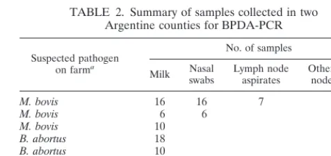
(i) Herd sampling in Argentina.A total of 128 bovine samples were obtained from farms that were classified as either “brucellosis positive” (defined by reac-tivity in a buffered plate antigen assay), “TB positive” (defined by delayed hy-persensitivity to an intradermal injection of purified protein derivative), or dis-ease-free (defined by a negative skin test and a negative buffered plate antigen assay result). The samples included milk (n⫽70), nasal swabs (n⫽28), retro-pharyngeal lymph node aspirates (m⫽7), and historical samples from a slaugh-terhouse (vaginal and lymph node swabs) (n⫽9). Fourteen pools of five indi-vidual milk samples each were also analyzed. Table 2 gives the stratified sampling scheme used for this study.
(ii) Samples from TB-positive herds in Mexico.Two bovine TB-suspect herds (considered heavily infected, as indicated by caudal fold test results) were sam-pled. Nasal swabs (n⫽121) and milk samples (n⫽460) were collected. The milk samples were pooled into batches of 10 milk samples each, and the DNA was analyzed at the ClinCyte laboratory.
(iii) Bovine brucellosis-positive milk samples from India.Milk was sampled from 20 dairy cows either positive (n⫽15) or negative (n⫽5) for brucellosis as determined by the milk ring test. DNA was extracted in a local laboratory and was analyzed by BPDA-PCR at the ClinCyte laboratory.
[image:3.612.53.552.85.147.2]Milk collection technique.All milk samples were obtained from animals dur-ing their routine milkdur-ing time. This approach prevented disruption of farmers’ day-to-day on-farm operations and simplified sample collection. The samples from each animal were obtained from all four quarters of the mammary gland.
TABLE 1. Source of field samples used to validate BPDA
Geographic locality Animal Specimen type No. of samples Comment
United States Laboratory animalsa Liver 45 Experimentally infected (B. abortus)
Mexico Bovine Milk 46b Bovine TB suspectc
Mexico Bovine Nasal swabs 121 Bovine TB suspectc
India Bovine Milk 20 B. abortussuspectd
Argentina Bovine Milk, nasal swabs, lymph node aspirates 128 Infected and disease-free herdse aOperators were blinded as to the infection status of the lemming liver samples for this part of the study.
bPools of 10 samples each.
cSuspicion of bovine TB was based on caudal fold tuberculin test or comparative cervical test positivity. dA subset of Indian samples (n⫽15) was positive by the milk ring test. The other five samples were negative.
eArgentine samples came from TB-positive herds (three herds) or herds with brucellosis (two herds). One disease-free herd was sampled as a control.
TABLE 2. Summary of samples collected in two Argentine counties for BPDA-PCR
Suspected pathogen on farma
No. of samples
Milk Nasalswabs Lymph nodeaspirates Other (lymphnode swabs)
M. bovis 16 16 7
M. bovis 6 6
M. bovis 10
B. abortus 18
B. abortus 10
“Negative” farm 10 6
Total samples collected 70 28 7 9
aSuspicion ofM. bovisinfection was based on purified protein
derivative-positive status, and that forB. abortusinfection was based on brucella BAPA test-positive status.
on May 15, 2020 by guest
http://jcm.asm.org/
[image:3.612.311.549.590.702.2]Collections were done after thorough disinfection of the teat area with povidone iodine. The milk sampler’s (veterinarian’s) gloves were also disinfected and wiped dry between samplings. Two to three strippings of milk from each teat were drawn into a sterile 50-ml tube, and the tubes were capped and stored on ice until further use. The entire collection process involved only two technicians (one who collected the milk and one who assisted) to rapidly obtain samples from the 16 to 24 animals in the entire milking shed at a time.
Nasal swab collections.Animals were individually restrained in a trevis or chute with a nose ring, which kept their necks extended. Both nasal passages of each animal were swabbed. Sterile applicators were applied deep into the nos-trils, the nostril was vigorously scrubbed two to three times, and the applicator was placed in a sterile tube. The swabs were rehydrated with 500l of sterile water and were vortexed before an aliquot was taken for DNA extraction.
Lymph node aspirates.Retropharyngeal lymph node aspirates were collected while the animals were restrained in a chute. An 18-gauge (1.5-in) needle at-tached to a 10-ml syringe was inserted into the lymph node, and an aspirate was obtained. Aspirates were transferred to a sterile 15-ml tube and transported on ice. The tubes were vortexed, and a 200-l aliquot was taken for DNA extraction.
RESULTS
Duplex amplification (initial setup and optimization).
Ex-periments for amplification of eitherM. bovisalone,B. abortus alone, or the two organisms in combination were performed with the following genomic DNAs purified from the respective bacterial cultures: DNAs from 10M. bovisstrains (including 2 TMC strains, 1 BCG vaccine strain, and 7 field isolates), DNAs from 2 M. tuberculosis strains (patient isolates), and DNAs
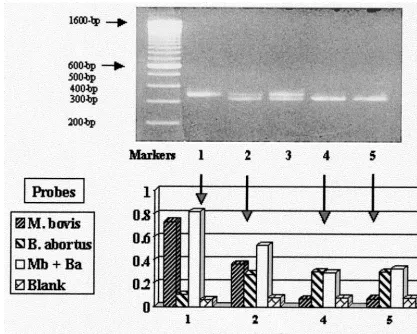
from 5 B. abortus strains (including 2 bovine isolates, type strain 544, and 2 vaccine strains). The amplification conditions and master mixture components were optimized to amplify all DNAs as singlets or in different combinations ofB. abortusand M. bovisas duplexes (Fig. 1).
The negative cutoff value of 0.200 for the assay was based on the average ODs for all negative samples⫹3 standard devia-tions.
Analytical sensitivity estimations. (i) Serially dilutedM.
bo-visandB. abortusamplicons.M. bovisandB. abortusamplicons were generated and column purified (Hi-Pure; Boehringer Mannheim) to remove excess primers. The copy number in each amplicon was determined by using the estimated DNA concentration (calculations were based on the absorbance at an OD of 260 nm and amplicon size). Equal numbers of copies of each amplicon were mixed and serially diluted (2⫻105, 2⫻ 104, 2⫻103, 2⫻102, and 2⫻101copies per reaction mixture), and the amplicons were then reamplified. The reamplified amplicons were analyzed by agarose gel electrophoresis and solid-phase probe-capture hybridization and detection as described above. The analytical sensitivity in this amplicon dilution experiment was 20 copies (on the basis of the lowest copy number used in the analysis) for amplicons of both pathogens.
FIG. 1. (Top panel) Agarose gel electrophoretic pattern of amplicons generated from genomic DNA ofM. bovisalone (lane 1),M. bovisandB. abortus(clinical strain 8-1070) mixed (lane 2),M. bovisandB. abortus(vaccine strain RB51) mixed (lane 3), orB. abortusalone (strain 8-1070 in lane 4 and RB51 in lane 5). (Bottom panel) Related ODs at 450 nm recorded for each amplicon when probed with either theM. bovis(Mb) probe alone (
,
), theB. abortus(Ba) probe alone ( ), or a combination of both probes (䊐). Also shown ( ) are the ODs at 450 nm of blank wells (negative controls). Clear increases in ODs are noted for each individual amplicon with species-specific probes with no evidence of cross-reactivity (lanes 1, 4, and 5), while reactivity with both probes was seen when both amplicons were present (lane 2). Blank wells were negative throughout.
on May 15, 2020 by guest
http://jcm.asm.org/
[image:4.612.94.512.73.407.2](ii) Serial dilution ofB. abortusgenomic DNA in milk.One milliliter of normal raw cow milk was spiked with from 200 ng to 0.002 pg ofB. abortusDNA (i.e., at equivalents of 2⫻108 to 2 genomes). The copy number (or genome equivalent) es-timates were calculated on the basis of the fact that 100 fg of B. abortusDNA is equivalent to 20 organisms (P. H. Elzer, personal communication, 1999). One aliquot of milk was not spiked and was used as a negative control. The DNA was reextracted from the milk by the protocol described above. Forty microliters of the reextracted DNA was amplified as described above and was analyzed on a microtiter plate. By this analysis the analytical sensitivity of the assay was 40 genome equivalents per reaction mixture by microtiter plate detection.
(iii) Serial dilution ofB. abortusorganisms in milk.Strain
2308 of B. abortus was spiked into a sample of milk at 109 CFU/ml, and the mixture was pasteurized. An aliquot of this was serially diluted (108 to 0 CFU/ml) in raw milk from a healthy cow, and DNA was extracted in 200 l of distilled deionized water. Ten microliters of this DNA was PCR ampli-fied and was detected by microtiter plate analysis. The results of this analysis indicated that the assay could detect down to 1⫻102CFU equivalents ofB. abortusper reaction mixture (or 2⫻103CFU equivalents/ml of milk).
Since the results obtained with whole-cell dilutions and genomic DNA dilutions are more accurate than those obtained with amplicons, the analytical sensitivity of the assay was esti-mated to be 40 to 100 CFU/reaction mixture (or 800 to 2,000 CFU/ml of milk).
(iv) Dilution ofM. bovisBCG in milk.An aliquot ofM. bovis
BCG was diluted in PBS–Tween 20 (0.05% Tween 20), and the mixture was resuspended to match a McFarland 0.1 standard (bacterial count equivalent to 4⫻107CFU/ml). A total of 100
l of this suspension was placed in 900l of milk (correspond-ing to 4⫻107CFU/ml of milk), and the mixture was serially diluted to 10⫺3CFU/ml. The DNA was extracted from these dilutions as described above. Subsequently, amplification and detection were performed as described above.
Microtiter plate detection identified less than 1 CFU ofM. bovis/ml of milk by our assay format. This indicates a high analytical sensitivity for the detection ofM. bovis, possibly due to the presence of four to eight copies of the direct repeat of the amplification target per organism.
Assay performance with liver samples from lemmings
ex-perimentally infected withB. abortus2308 or RB51.The assay
performance was tested with a set of 45 liver samples from infected lemmings. In total, 33 positive and 12 culture-negative liver samples were tested. Nine of 12 culture-negative samples were from RB51 (an avirulent vaccine strain)-infected lem-mings. These samples were interpreted as being from animals that had “cleared” the infection. Of the 33 culture-positive liver samples, 31 were positive by PCR. Therefore, by using culture as a “gold standard,” the sensitivity of BPDA-PCR was 93.9% (31 of 33 samples). The two PCR-negative, culture-positive samples were from lemmings infected with the aviru-lent RB51 strain. Among the 12 culture-negative liver samples, 5 (3 from the strain 2308-inoculated group and 2 from the strain RB51-inoculated group) were identified as positive by PCR, while the other 7 (all RB51 group) culture-negative samples were classified as negative by PCR. These experiments show that the performance of BPDA-PCR may be comparable to that of culture in that BPDA-PCR identified five culture-negative samples from experimentally lemmings as positive. These animals may have incompletely cleared the infection and may thus be carrying damaged organisms that could not be revived by the conventional culturing method used.
Addition-ally, identification of infected animals by culture is often diffi-cult because brucellae are fastidious and slow growers.
Duplex identification ofB. abortusand/orM. bovisin milk
samples from brucellosis-positive animals sampled in India.
DNA was extracted from the milk of 20 cows from a herd in Bangalore, India, known to be infected withB. abortusand was analyzed by BPDA-PCR. Of these 20 milk samples 15 were milk ring test positive (i.e., positive forB. abortusantibodies). The PCR analysis identified 13 of the 15 milk ring test-positive animals and 4 of the 5 milk ring test-negative animals asB. abortusshedders. The ODs at 450 nm ranged from 0.287 to 0.838 and 0.121 to 0.209 for positive and negative samples, respectively. The other negative milk ring test-samples and two milk ring test-positive samples were negative by PCR (Table 1). In addition, upon analysis of these samples by individual probe (M. tuberculosiscomplex- or B. abortus-specific) hy-bridization assays, two samples were found to be positive for bothB. abortusandM. bovis. All other samples were positive only forB. abortusby BPDA-PCR. These studies further val-idate the high degree of sensitivity of BPDA-PCR in identify-ing infected, sheddidentify-ing animals that may be missed by the milk ring test. BPDA-PCR analysis was also able to identify two animals concurrently infected withM. bovisandB. abortus.
Duplex PCR identification ofB. abortusand/orM. bovisin
milk samples from brucellosis-suspect animals sampled in
Argentina. (i) BPDA-PCR performance with suspected M.
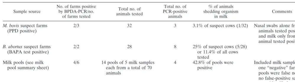
bovis-positive samples.Thirty-two animals from three puri-fied protein derivative-positive farms were tested. The types of samples tested were milk (n⫽32), nasal swabs (n⫽22), and retropharyngeal lymph node biopsy specimen (n ⫽ 7). Only 1 of 32 milk samples and 2 nasal swabs (from two differ-ent farms) were positive forM. bovis(positive OD range, 0.241 to 2.842; negative OD range, 0.067 to 0.192). All lymph node biopsy specimens were negative by tests with both multiple and individual probes. All samples from one farm suspected of being positive were negative.
(ii) BPDA-PCR performance with suspectedB. abortus
-pos-itive samples.Twenty-eight milk samples from
brucellosis-pos-itive animals (according to the results of the buffered antigen plate agglutination [BAPA] test) in two herds were tested. Seven BAPA test-positive animals tested positive (positive OD range, 0.219 to 0.518; negative OD range, 0.078 to 0.189) by BPDA-PCR and were defined asB. abortusshedders by anal-ysis with individual probes (Tables 2 and 3).
(iii) BPDA-PCR performance with milk sample pools.
Four-teen milk sample pools (with each pool made up of five sam-ples) were analyzed by BPDA-PCR. From retrospective anal-ysis of individual milk samples, it was expected that 6 of the 14 milk sample pools should be positive. BPDA-PCR analysis correctly identified four of six milk sample pools as positive. The two pools in which organisms were not detected were low-positive pools, in that each of these had a sample that was borderline positive (ODs of 0.219 and 0.295, respectively, when individual samples were tested withB. abortus-specific probes). The results indicate that pooling of samples from five animals for BPDA-PCR may miss some level shedders or low-level-positive animals. Additional studies with pools of samples from three animals or other combinations will be required to define low-level shedders adequately.
(iv) Internal standard analysis.A total of 125 samples from
Argentina were tested for amplification of a 226-bp region of bovine2-microglobulin to determine if any samples contained PCR inhibitors or if the DNA extraction was functionally in-tact. All samples analyzed by agarose gel electrophoresis of the amplicon products were shown to have single, nondiffuse bands with high concentrations of amplicons for the2
on May 15, 2020 by guest
http://jcm.asm.org/
globulin region of interest. The amplification of the 226-bp region from all samples indicates no PCR inhibition or DNA degradation; therefore, any BPDA-PCR-negative sample was considered to be from a nonshedder or to contain organisms at a level below the detection limits of the assay.
Identification of M. bovis in milk samples or nasal swabs
from skin test-positive herds sampled in Mexico.Two sets of
samples from Mexico were analyzed. One set consisted of milk samples from animals from caudal fold tuberculin test- and comparative cervical test-positive farms. Forty-six milk sample pools were tested. Each pool was a mixture of milk from 10 individual animals. Of the 46 milk sample pools, 15 were pos-itive (pospos-itive OD range, 0.241 to 1.124) with the BPDA-PCR multiplex probe- andM. tuberculosis complex-specific probe-coated microtiter plates. Retrospective analysis of all 10 indi-vidual milk samples from one positive pool identified two pos-itive animals. The second set of samples analyzed included nasal swabs (n⫽121) obtained from a second geographically distinct area in Mexico. Of the 121 samples, 68 (56.2%) were BPDA-PCR-positive forM. bovis(OD range, 0.406 to 2.882). Animals from these farms were also bovine TB suspect on the basis of intradermal test results.
DISCUSSION
Economics of disease eradication and the need for an
accu-rate tool.The presence of infection caused by the two zoonotic
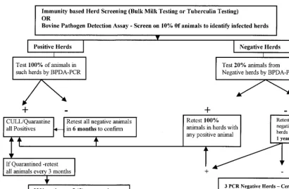
pathogens, B. abortus and M. bovis, in animal populations poses an economic threat to countries around the world (1, 5, 6, 7, 24). This disease has contributed to nontariff trade barri-ers by impeding the safe, free trade of cattle and cattle prod-ucts implemented by international and other national trade agreements (6). Losses due to reduced sales and lowered pro-duction and the trade barrier that these diseases have created reemphasize the need for a rapid, sensitive, specific, and effi-cient molecular biology-based diagnostic tool. This would eventually lead to reduced rates of M. bovis and B. abortus infection and therefore an increase in the trade, movement, and marketability of cattle in South, Central, and North Amer-ica. Countries in these regions look to the United States for technologies and protocols that they can use to aid them with their screening for such zoonoses and to initiate profitable livestock trading across international borders. On the basis of the prevalence data from the three countries surveyed (5, 6, 36; Miolan-Suazo et al., Proc. USAHA Meeting), an algorithm for an eradication program is proposed (Fig. 2). The algorithm
includes the need for a stepwise, multistage, periodic testing process for a successful eradication program. The algorithm allows the testing of milk samples in pools made up of milk from 5 to 10 animals in each pool in high-incidence areas, thus reducing the cost of screening each animal. As demonstrated in the samples from Mexico, analysis of pools made up of 5 to 10 milk samples is feasible and will help identify positive sam-ples as a part of the proposed eradication algorithm.
Current diagnostic practices and their pitfalls.Diagnosis of
brucellosis primarily uses the milk ring test or the BAPA test. The milk ring test or the BAPA test has been widely applied for identification of infected herds in the United States and other countries, followed by ELISA, the complement fixation test, or the standard tube agglutination test to confirm infec-tion in individual animals (2, 5, 33). The milk ring test is an agglutination test with fair specificity (79.4 to 86.7%) and sen-sitivity (85.4 to 99.3%) (32). False-positive results by the milk ring test occur immediately after parturition and during mas-titis. Additionally, batch-to-batch variations in milk ring test antigen levels lead to variability associated with the lower sen-sitivity of the assay (P. H. Elzer, personal communication). The results indicate that the milk ring test may be less sensitive than PCR. On the other hand, while ELISAs have proved to have higher sensitivities and specificities, like the milk ring test they measure antibodies that reflect exposure of the animal to bru-cellae and do not necessarily indicate present infection or the ability of the animal to spread the disease (5). Despite some of these drawbacks, the two serological tests have been used in successful eradication schemes. Additionally, these tests can be used as an initial screen to identify exposed or infected herds in an eradication program.
[image:6.612.51.550.84.222.2]PCR with random or selected primers has shown promising results, but large-scale field validations and evaluations are lacking, especially for detection of chronic infections. For PCR, various targets (species-specific, biotype-specific, and genus-specific targets) have been investigated, including the genes for Omp2 (12), the 31-kDa (BCSP31K) antigen (22), the 16S rRNA, the 16S-23S spacer region, and an insertional ele-ment, namely, IS711 (3, 4). Among these, the gene that en-codes a 31-kDa protein has proved to be a promising target because of the presence of species-specific signature regions in the gene (22). In the past there has been considerable interest in the use of rapid molecular biology-based assays to identify and determine the species of brucellae (3, 4, 5). By using this strategy with species-specific signature regions on Omp2 gene TABLE 3. Summary of shedding status withinM. bovis- orB. abortus-infected herds in two argentine countiesa
Sample source No. of farms positiveby BPDA-PCR/no. of farms tested
Total no. of animals tested
Total no. of PCR-positive
animals
% of animals shedding organism
in milk Comments
M. bovissuspect farms
(PPD positive) 2/3 32 3 3.1% of suspect cows (1/32) Nasal swabs alone from twoanimals tested positive, and milk only from one animal tested positive
B. abortussuspect farms
(BAPA test positive) 2/2 28 8 25% of suspect cows (5/28)or 11.4% of all cows tested
Milk pools (see milk
pool summary sheet) 4/6 14 pools of 5 milk sampleseach from a total of 70 animals
4 42.8% of pools were
positive Included milk samples fromone “negative” farm; two pools were false negative; no false-positive samples were identified
aAll animals tested from specific-pathogen-suspect farms were positive by conventional testing. The sampling did not include any healthy animals within infected
herds and therefore may not reflect the true shedding status (sampling bias). However, these data can be used as an estimate to project the sampling load (testing and retesting) and total assay load.
on May 15, 2020 by guest
http://jcm.asm.org/
(12), a PCR assay has been developed and patented (T. A. Ficht, B. A. Sowa, and L. G. Adams, May 1994, U.S. patent 5310649). The antigen detection methods are potentially use-ful but have not been validated. Combinations of these with PCR, such as immuno-PCR, have considerable potential but require evaluation (5).
Recent developments in the molecular genetics of mycobac-teria have identified species-specific markers in MPT40 (27), oxyR(30), andpncA(9) that enable specific identification ofM. bovis. Among these, only theoxyRpolymorphism at nucleotide 285 has proved to be an accurate and consistent M. bovis -specific marker (9). Other targets that have been investigated include the 16S rRNA gene region with a TB complex-specific signature sequence, targets identified by randomly amplified polymorphic DNA analysis (18, 26, 28, 29), and mycolic acid profiles identified by the high-performance liquid chromatog-raphy (13). The high-performance liquid chromatogchromatog-raphy method requires a considerable bacterial inoculum, thereby requiring mycobacterial culture, which takes 3 to 6 weeks, before application.
It may be argued that IS6110has more copies and would form a good target for the identification of M. tuberculosis complex organisms. However, numerous ancestral isolates from the M. tuberculosiscomplex group (especially M. bovis from cattle, goats, or other animal sources and someM. tuber-culosisisolates from certain geographic localities that have now spread globally) with no copies of IS6110exist (31). While the use of oxyR for the specific identification ofM. bovisis very attractive,oxyRis a pseudogene in theM. tuberculosiscomplex; therefore, it is expected under the classical evolutionary theory that this pseudogene may mutate spontaneously (and at a higher rate than other structural genes) over time and may not be detected in some isolates by the use of specific probes. Thus,
thehsp65region with a direct-repeat sequence, which is pres-ent in multiple copies, was used in the prespres-ent study.
Performance of BPDA-PCR: experimental and field studies.
[image:7.612.94.505.69.337.2]The experiments performed in the present study with purified genomic DNA optimized the assay forB. abortusidentification at⬍2,000 CFU/ml and forM. bovisidentification at⬍10 CFU/ ml. Although the detection level for M. bovis is as low as 4 CFU/ml, we believe that this could be an overestimate due to the indirect nature of CFU equivalent estimation by the use of McFarland standards. Overall, experimental studies with ei-ther organism proved that the assay was very sensitive and specific on the basis of the data obtained for normal and spiked milk samples and liver samples from experimentally infected lemmings. The performance of BPDD-PCR in field studies with infected herds from countries that report the occurrence of the two zoonotic diseases further validates its use as an optimal tool for surveillance for bovine brucellosis and TB. One of the concerns with PCR is the occurrence of false-negative results due to the presence of substances in the sam-ple that interfere with amplification. The extraction process used in the study efficiently eliminates PCR inhibitors. To test this, all Argentine specimens and a subset of samples from Mexico were tested for the presence of a 2-microglobulin segment that is highly conserved and that is expected to be present in host cellular debris of any secretion or excretion. All samples tested positive for2-microglobulin, thus increasing the degree of confidence in the technical interpretation of a negative test result as “negative.” On the other hand, false-positive results may occur as a result of the cross-reactivity of PCR primers and probes or because of contamination during any stage of collection, processing, and detection. Therefore, a series of database searches was performed to identify specific regions for primer and probe design. Additionally, the study FIG. 2. Proposed algorithm for a stepwise repeat sampling and testing protocol for a disease eradication scheme. The proportion of animals to be tested will vary with the prevalence of disease in a given geographic locality (higher numbers in lower-prevalence areas; however, low-prevalence areas will not require many iterations of the same tests).
on May 15, 2020 by guest
http://jcm.asm.org/
used a hygienic milk collection protocol and laboratory proce-dures to minimize cross contamination of samples. This is reflected by the fact that all negative samples (from disease-free herds) and controls yielded ODs below the cutoff value.
The surveillance and monitoring algorithm proposed in this study emphasizes a multistage identification system because PCR identifies animals actively shedding the organism and contaminating the environment. Thus, a negative PCR result should be interpreted with caution, especially with a chronic granulomatous disease such as bovine TB.
However, PCR results are similar to culture results. A pos-itive PCR test indicates the presence of the pathogen, and this is often correlated with infectivity. In the context of infections with M. bovis, a positive PCR result represents a status in which the animal has been infected, is shedding mycobacteria or brucellae, and may infect other animals, but a negative result does not rule out infection. A similar argument can be made about culture, since not all attempts to culture an organ-ism are successful, and not all samples provide sufficient ma-terial to demonstrate positivity. However, BPDA-PCR is ex-tremely sensitive, and this sensitivity lessens the probability of false-negative results. Sample volumes have been optimized in this study to reduce sampling error, and our data show that
⬍10 organisms (BCG serial dilution studies) may be detectable in 1 ml of a milk sample pool. Others have also shown that PCR detects as few as one organism (5 fg of DNA or less) in a specimen aliquot (13, 34).
Miller et al. (24) demonstrated that PCR is effective in diagnosing TB in cattle with various tissues collected at nec-ropsy. They found a 93% success rate compared with the re-sults of necropsy examination for the detection of ruminant TB with paraffin-embedded tissues (23). A recent study by Vitale et al. (34) addressed the performance of PCR versus those of necropsy examination for infection and conventional test re-sult. Using PCR analysis of tissues from slaughtered animals as a gold standard in an evaluation of 100 cattle, they showed 100% sensitivity and specificity for milk samples and lymph node biopsy specimens (collected at necropsy). The lack of PCR-positive lymph node biopsy specimens by BPDA-PCR in our study may be associated with the fact that the representa-tive nodes were not sampled. In most instances appropriate lymph nodes are not superficially accessible for optimal diag-nosis of infection by PCR or culture. Additionally, it is sug-gested that multiple nodes must be sampled forM. tuberculosis complex-specific PCR to improve the diagnostic value (6, 25). Technical difficulties and the stress imposed on the animals, which may reduce milk production, make it somewhat imprac-tical for the use of lymph node biopsy specimens for antemor-tem diagnostic assays.
Although historical data for the detection ofB. abortus bac-teremia for as long as 251 days in nonvaccinated animals and 60 days in vaccinated animals exists (21), most animals in advanced carrier states also shed organisms in their milk. Blood sampling is useful for early detection of the disease process, while BPDA-PCR analysis of milk will efficiently de-tect most (⬎90%) chronically infected animals in the herd. Thus, the optimal sampling strategy may be to use milk alone for the diagnosis of brucellosis and nasal swabs in addition to milk for the diagnosis of bovine TB. Studies by Vitale et al. (34) and Cornejo et al. (6) for bovine TB and those by Leal-Klevezas et al. (17) and Fekete et al. (11) for bovine brucellosis support the idea that the use of milk and/or nasal swab spec-imens may be optimal for the efficient diagnosis of the diseases caused by these two zoonotic pathogens. BPDA-PCR does identify but cannot differentiate vaccine strains ofB. abortus (S19 and RB51) when they are present. Given that only
calf-hood vaccination with strains S19 and RB51 is practiced and that strain RB51 is not excreted or cleared from the body in milk, the possibility of false-positive results by BPDA-PCR due to vaccination is significantly reduced.
The use of PCR for the diagnosis of bovine infections has not been evaluated in large-scale field or herd studies so that guidelines for its use in an eradication program can be recom-mended. Although there are limited data from studies with animal isolates (6, 34), to our knowledge, this is the first study to test the ability of a multiplex molecular biology-based assay to detect any animal disease in the field. Such an assay, when available for large-scale disease screening and surveillance, is very appealing because of its ability to identify the presence of multiple pathogens in herds as well as in individual animals. This multiplex format further translates into a lower cost per pathogen tested per herd. In conclusion, the ability of BPDA-PCR to identify either pathogen in field samples from herds from a variety of geographic regions known to be infected or exposed to the pathogens demonstrates that the test is robust and accurate. Use of a combination of the duplex PCR assay, screening of milk samples in pools, and the proposed algorithm provides a highly sensitive and economically viable alternative to immunodiagnostic methods.
ACKNOWLEDGMENTS
We thank Robert Cooksey (Tuberculosis Laboratory research, Na-tional Center for Infectious Diseases, Centers for Disease Control and Prevention) for providing DNA lysates from well-characterized M. bovisandM. bovisBCG strains for this study and for thorough critique of the manuscript. We also thank Pablo Fiero and Oscar Sbodio (SANCOR, Sunchales, Argentina) for help in collecting samples from farms with either brucellosis or bovine TB.
REFERENCES
1.Acha, P. N., and B. Szyfres.1987. Zoonoses and communicable diseases common to man and animals, 2nd ed., p. 24–45. Scientific publication series no. 503. Pan American Health Organization, Washington, D.C.
2.Breitmeyer, R. E., D. W. Hird, and T. E. Carpenter.1992. Serologic and bacteriologic test results after adult vaccination with strain 19 in three dairy herds infected with brucellosis. J. Am. Vet. Med. Assoc.200:806–811. 3.Bricker, B. J., and S. M. Halling.1994. Differentiation ofBrucella abortusbv.
1, 2, and 4,Brucella melitensis,Brucella ovis, andBrucella suisbv. 1 by PCR. J. Clin. Microbiol.32:2660–2666.
4.Bricker, B. J., and S. M. Halling.1995. Enhancement of brucella AMOS PCR assay for differentiation ofBrucella abortusvaccine strains S19 and RB51. J. Clin. Microbiol.33:1640–1642.
5.Corbel, M. J.1997. Brucellosis: an overview. Emerg. Infect. Dis.3:213–221. 6.Cornejo, B. J., A. Sahagun-Ruiz, F. Suarez-Guemes, C. G. Thornton, T. A. Ficht, and L. G. Adams.1998. Comparison of 18-carboxypropylbetaine and glass bead DNA extraction methods for detection ofMycobacterium bovisin bovine milk samples and analysis of samples by PCR. Appl. Environ. Mi-crobiol.64:3099–3101.
7.Cosivi, O., J. M. Grange, C. J. Daborn, M. C. Raviglione, T. Fujikura, D. Cousins, R. A. Robinson, H. F. A. K. Huchzermeyer, I. de Kantor, and F.-X. Meslin.1998. Zoonotic tuberculosis due toMycobacterium bovisin develop-ing countries. Emerg. Infect. Dis.4:59–70.
8.Dankner, W. M., N. J. Waecker, M. A. Essey, K. Moser, M. Thompson, and C. E. Davis.1993.Mycobacterium bovisinfections in San Diego: a clinico-epidemiologic study of 73 patients and a historical review of a forgotten pathogen. Medicine72:11–37.
9.Del los Monteros, L. E. E., J. C. Gala´n, M. Gutie´rrez, S. Samper, J. F. G. Marı´n, C. Martı´n, L. Domı´nguez, L. de Rafael, F. Baquero, E. Go´mez-Mampaso, and J. Bla´zquez.1998. Allele-specific PCR method based on pncAandoxyRsequences for distinguishingMycobacterium bovisfrom My-cobacterium tuberculosis: intraspecific M. bovis pncA sequence polymor-phism. J. Clin. Microbiol.36:239–242.
10. Elzer, P. H., R. W. Phillips, M. E. Kovach, K. M. Peterson, and R. M. Roop II.1994. Characterization and genetic complementation of aBrucella abortus high-temperature-requirement A (htrA) deletion mutant. Infect. Immun.10: 4135–4139.
11. Fekete, A., J. A. Bantle, S. Halling, and M. R. Sanborn.1990. Preliminary development of a diagnostic test forBrucellausing polymerase chain reac-tion. J. Vet. Diagn. Investig.69:216–227.
12. Ficht, T. A., H. S. Husseinen, J. Derr, and S. W. Bearden.1996.
on May 15, 2020 by guest
http://jcm.asm.org/
specific sequences of theomp2locus ofBrucellatype strains. Int. J. Syst. Bacteriol.46:329–331.
13.Floyd, M. M., V. A. Silcox, W. D. Jones, Jr., W. R. Butler, and J. O. Kilburn. 1992. Separation ofMycobacterium bovisBCG fromMycobacterium tubercu-losisandMycobacterium bovisby using high-performance liquid chromatog-raphy of mycolic acids. J. Clin. Microbiol.30:1327–1330.
14.Francis, J., R. J. Seiler, I. W. Wilkie, D. O’Boyle, M. J. Lumsden, and A. J. Frost.1978. The sensitivity and specificity of various tuberculin tests using bovine PPD and other tuberculins. Vet. Rec.103:420–425.
15. Frothingham, R.1995. Differentiation of strains inMycobacterium tubercu-losiscomplex by DNA sequence polymorphisms, including rapid identifica-tion ofM. bovisBCG. J. Clin. Microbiol.33:840–844.
16. Grange, J. M., M. D. Yates, and I. N. de Kantor.1996. Guidelines for speciation within theMycobacterium tuberculosiscomplex. WHO publication WHO/MC/ZOO/96.4. World Health Organization, Geneva, Switzerland. 17. Leal-Klevezas, D. S., I. O. Martinez-Vasquez, A. Lopez-Merino, and J. P.
Martinez-Soriano.1995. Single-step PCR for the detection ofBrucellaspp. from blood and milk of infected animals. J. Clin. Microbiol.33:3087–3090. 18. Linton, C. J., H. Jalal, J. P. Leeming, and M. R. Millar. 1994. Rapid discrimination ofMycobacterium tuberculosisstrains by random amplified polymorphic DNA analysis. J. Clin. Microbiology32:2169–2174.
19. Luzzi, G. A., R. Brindle, P. N. Socket, J. Solera, P. Klenerman, and D. A. Warrel.1993. Brucellosis: imported and laboratory-acquired cases, and an overview of treatment trials. Trans. R. Soc. Trop. Med. Hyg.87:138–141. 20. Madkour, M. M.1989. Brucellosis. Butterworths, London, United Kingdom. 21. Manthei, C. A., and B. I. L. Deyoe.1981. Brucellosis, p. 104–121.InH. E. Amstutz (ed.), Bovine medicine and surgery. Veterinary Publications Inc., Santa Barbara, Calif.
22. Matar, G. M., I. A. Khneisser, and A. M. Abdelnoor.1996. Rapid laboratory confirmation of human brucellosis by PCR analysis of a target sequence on 31-kDaBrucellaantigen DNA. J. Clin. Microbiol.34:477–478.
23. Miller, J., A. Jenny, J. Rhyan, D. Saari, and D. Suarez.1997. Detection of Mycobacterium bovisin formalin-fixed, paraffin-embedded tissues of cattle and elk by PCR amplification of an IS6110sequence specific for Mycobac-terium tuberculosiscomplex organisms. J. Vet. Diagn. Investig.9:244–249. 24. Nicoletti, P.1980. The epidemiology of brucellosis. Adv. Vet. Sci. Comp.
Med.24:69–98.
25. O’Reilly, L. M., and C. J. Daborn.1995. The epidemiology ofMycobacterium bovisinfections in animals and man: a review. Tubercle Lung Dis.76(Suppl. 1):1–46.
26. Parra, C. A., L. P. Londono, P. D. Portillo, and M. E. Patarroyo.1991.
Isolation, characterization, and molecular cloning of a specific Mycobacte-rium tuberculosisantigen gene: identification of a species-specific sequence. Infect. Immun.59:3411–3417.
27. Portillo, P. D., L. A. Murillo, and M. E. Patarroyo.1991. Amplification of species-specific DNA fragment ofMycobacterium tuberculosisand its possi-ble use in diagnosis. J. Clin. Microbiol.29:2163–2168.
28. Rodriguez, J. G., J. C. Fissanoti, P. D. Portillo, M. E. Patarroyo, M. I. Romano, and A. Cataldi.1999. Amplification of a 500-base-pair fragment from cultured isolates ofMycobacterium bovis. J. Clin. Microbiol.37:2330– 2332.
29. Rodriguez, J. G., G. A. Meija, P. D. Portillo, and M. E. Patarroyo.1995. Species-specific identification ofMycobacterium bovisby PCR. Microbiology 141:2131–2138.
30. Sreevatsan, S., P. Escalante, X. Pan, D. A. Gillies III, S. Siddiqui, C. N. Khalaf, B. N. Kreiswirth, P. Bifani, L. G. Adams, T. Ficht, V. S. Perumaalla, M. D. Cave, J. D. A. Van Embden, and J. M. Musser.1996. Identification of a polymorphic nucleotide inoxyRspecific forMycobacterium bovis. J. Clin. Microbiol.34:2007–2010.
31. Sreevatsan, S., X. Pan, K. E. Stockbauer, N. D. Connel, B. N. Krieswirth, T. S. Whittam, and J. M. Musser.1997. Restricted structural gene polymor-phism in theMycobacterium tuberculosiscomplex indicates evolutionarily recent global dissemination. Proc. Natl. Acad. Sci. USA94:9869–9874. 32. U.S. Animal Health Association, Committee on Brucellosis.1997. Report.
U.S. Animal Health Association (www.usaha.org/speeches/caring97.html). 33. Vanzini, V. R., N. Aguirre, C. I. Lugaresi, S. T. de Echaide, V. G. de
Canavesio, A. A. Guglielmone, M. D. Marchesino, and K. Neilsen.1998. Evaluation of an indirect ELISA for the diagnosis of bovine brucellosis in milk and serum samples in dairy cattle in Argentina. Prev. Vet. Med.36: 211–217.
34. Vitale, F., G. Capra, L. Maxia, S. Reale, G. Vesco, and S. Caracappa.1998. Detection ofMycobacterium tuberculosiscomplex in cattle by PCR using milk, lymph node aspirates, and nasal swabs. J. Clin. Microbiol.36:1050– 1055.
35. Weyants, V., J. Godfroid, B. Limbourg, C. Saegerman, and J.-J. Letesson. 1995. Specific bovine brucellosis diagnosis based on in vitro antigen-specific gamma interferon production. J. Clin. Microbiol.33:706–712.
36. World Health Organization. 1997. Brucellosis. Fact sheet N173. World Health Organization, Geneva, Switzerland.
37. World Health Organization.1986. Sixth report of the Joint FAO/WHO Expert Committee on Brucellosis. WHO Technical report series Rep. 740. World Health Organization, Geneva, Switzerland.