JOURNAL OFCLINICALMICROBIOLOGY, June 2003, p. 2654–2658 Vol. 41, No. 6 0095-1137/03/$08.00⫹0 DOI: 10.1128/JCM.41.6.2654–2658.2003
Copyright © 2003, American Society for Microbiology. All Rights Reserved.
Ischemic Stroke and Splenic Rupture in a Case of
Streptococcus bovis
Endocarditis
Claudia Sto¨llberger,
1* Josef Finsterer,
2Angelika Pratter,
1Wolfgang Kopsa,
3Julius Preiser,
4and Andreas Valentin
1Medizinische Abteilung,1Department of Neurology,2Department of Radiology,3and Department of Pathology,4Krankenanstalt Rudolfstiftung, 1030 Vienna, Austria
Received 23 September 2002/Returned for modification 23 October 2002/Accepted 23 November 2002
A 58-year-old man with an acute stroke suffered from splenic rupture.Streptococcus boviswas found in blood cultures, and gram-negative cocci were found in the infarcted spleen. Hemorrhagic transformation of the stroke occurred. Echocardiography showed aortic endocarditis. Cardiac surgery was not performed because of concern about cerebral bleeding. The patient died due to cerebral rehemorrhage after 3 weeks.
Splenic rupture in cases of ischemic stroke may be due to trauma, hematological disorder, malignancy, vasculitis, or sys-temic infection. Splenic rupture may also be caused by splenic infarction due to embolism. Cases of splenic rupture and stroke that are both due to embolism from infective endocar-ditis, as in the following report, have not been described pre-viously.
Case report.A 58-year-old man was hospitalized, because of an acute stroke in the supply area of the right middle cerebral artery with left-sided hemiparesis, 20 h after the onset of symp-toms. For the previous 10 months, the patient had suffered from recurrent pharyngitis and tympanitic effusions. Eight weeks before admission, sinusitis ethmoidalis with fever had occurred and was treated with oral amoxicillin at 1,000 mg/day for 1 week. Despite antibiotic therapy, subfebrile tempera-tures, malaise, and night sweats persisted. At that time, labo-ratory tests revealed a blood sedimentation rate of 80 mm/h, a leukocyte count of 8.9/nl, an erythrocyte count of 3.67/pl, a hemoglobin level of 121 g/liter, a hematocrit of 0.34, and mi-crohematuria. Five weeks before admission, the patient com-plained of sudden-onset dyspnea and fatigue. A chest X ray and computed tomography of the lung, performed 3 days be-fore admission, showed infiltrates in both lungs, which were interpreted as pneumonia. The patient’s history revealed that he had suffered from pleuritis and pulmonary embolism after cholecystectomy 22 years before and from arterial hyperten-sion for the past 3 years. He smoked 20 cigarettes/day. He was on regular medication with terazosin, fosinopril, and hydro-chlorothiazide.
At admission, clinical neurologic examination showed left-sided central facial palsy, weakness of the left upper limb (Medical Research Council grade 1) and left lower limb (Med-ical Research Council grade 0), and left-sided hemihypesthe-sia. Clinical cardiological examination revealed pulmonary rales, a systolic murmur along the left sternal border extending to the carotid arteries, and pretibial edema. Blood pressure was 130/60 mm Hg, body temperature was 38°C, and body
weight was 90 kg. The electrocardiogram was normal except for sinus tachycardia of 118/min. The leukocyte count was 8.7/nl (normal counts, 6.0 to 9.0/nl), the erythrocyte count was 3.15/pl (normal counts, 4.2 to 5.5/pl), the hemoglobin level was 93 g/liter (normal levels, 136 to 172 g/liter), the hematocrit was 0.27 (normal, 0.4 to 0.5), the C-reactive protein level was 115 mg/liter (normal,ⱕ6 mg/liter), the sodium level was 128 mmol/ liter (normal, 135 to 150 mmol/liter), the serum iron concen-tration was 2.15mol/liter (normal, 11 to 29mol/liter), trans-ferrin saturation was 0.04 (normal, 0.16 to 0.46), the␥-glutamyl transpeptidase level was 155 U/liter (normal, 6 to 28 U/liter), the alkaline phosphatase level was 384 U/liter (normal, 60 to 170 U/liter), the cholinesterase level was 2,765 U/liter (normal, 3,500 to 8,500 U/liter), the albumin level was 0.55 (normal, 0.58 to 0.70), the gamma globulin level was 0.23 (normal, 0.10 to 0.19), and the blood sedimentation rate was 68 mm/h (normal,
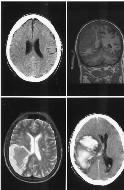
⬍20 mm/h). Cerebral computed tomography showed a diffuse hypodense lesion in the posterior supply area of the right middle cerebral artery (Fig. 1).
The patient received pentoxifylline (800 mg/day), acetylsal-icylic acid (100 mg/day), and low-molecular-weight heparin (10,000 IU/day). After two blood cultures had been taken, levofloxacin (500 mg/day), amoxicillin (6 g/day), and clavulanic acid (600 mg/day) were started. The patient received two packs of red blood cells. Because the patient became confused and hypotensive and developed bloody diarrhea during the follow-ing 2 days, he was transferred to the intensive care unit 60 h after admission. Emergency transthoracic echocardiography showed small cardiac cavities, suggesting hypovolemia. The cardiac valves were not adequately visualized in the emergency situation. Because of a simultaneous fall of the hematocrit to 0.16, acute hemorrhage was suspected. Abdominal ultrasound and computed tomography showed blood within the peritoneal cavity. The patient underwent emergency laparatomy 3 days after admission, at which time a splenic rupture was found. Splenectomy was performed. Postoperatively, the heparin dose was reduced to 5,000 IU/day, and metronidazole (1,500 mg/ day) was added to the antibiotic therapy regimen. The patient was extubated on the 1st postoperative day. The postoperative course was complicated by recurrent pulmonary edema, inter-preted as due to hypertension. The two blood cultures were
* Corresponding author. Mailing address: Steingasse 31/18, 1030 Vienna, Austria. Phone and fax: 43 1 713-98-70. E-mail: claudia [email protected].
2654
on May 15, 2020 by guest
http://jcm.asm.org/
FIG. 1. (Upper left) Cerebral computed tomography 21 h after onset of symptoms shows a hypodense area with loss of differentiation between gray and white matter in the posterior supply area of the right middle cerebral artery. (Upper right and lower left) Seven days later, a coronal T1-weighted (upper right) and an axial T2-weighted (lower left) (where T stands for relaxation time) cerebral magnetic resonance image show
hemorrhagic transformation of the ischemic area. (Lower right) Thirteen days later, cerebral computed tomography shows a space-occupying hemorrhage with blood in the ventricles.
2655
on May 15, 2020 by guest
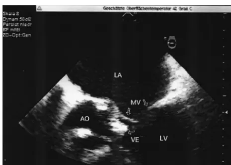
[image:2.603.84.501.45.682.2]positive for Streptococcus bovis. No other organism was iso-lated from the blood cultures. The strain was sensitive to pen-icillin, aminopenpen-icillin, amoxpen-icillin, cefazolin, erythromycin, clindamycin, and vancomycin and was resistant to tobramycin, tetracycline, and levofloxacin. On the 5th postoperative day, a single generalized tonic-clonic seizure occurred, followed by respiratory insufficiency. The patient had to be reintubated and mechanically ventilated. Secondary hemorrhage in the area of the recent ischemic stroke was found upon cerebral magnetic resonance imaging (Fig. 1). On the 6th postoperative day, a high blood pressure amplitude of 180/40 mm Hg led to the suspicion of aortic insufficiency. Transthoracic and transesoph-ageal echocardiography showed mobile vegetations on the aortic cusps and severe aortic insufficiency (Fig. 2). Aortic endocarditis was diagnosed on the basis of clinical, echocar-diographic, blood chemistry, and bacteriological findings. His-tological examination of the resected spleen revealed a splenic infarct with a destroyed arterial wall and intravascular fibrin thrombi, containing gram-positive cocci consistent withS. bovis
and surrounding inflammatory cellular infiltrates with neutro-philic granulocytes (Fig. 3). Acute cardiac surgery was consid-ered but was refused at the time because of concern about further cerebral bleeding and was planned for in 5 weeks. The further course was complicated by pneumonia. Repeated blood and sputum cultures did not show growth of any bacte-ria. Colonoscopy, performed to look for an entry portal ofS. bovis, revealed an ulcus of the rectal mucosa, sigmoid divertic-ula, and a colonic polyp at 25 cm. Twenty-one days after the operation, the pupils widened acutely and became areactive
bilaterally. A computed tomography scan of the brain showed a new massive hemorrhage (Fig. 1). The patient died on the next day. The autopsy confirmed the diagnosis of aortic valve endocarditis.
The stroke and splenic infarction were most probably due to embolization of infectious material from the aortic cusps dur-ing the period of untreated infection. BecauseS. bovis bacte-remia is often associated with bowel pathology, the most prob-able means of infection was migration ofS. bovis, a member of the human gut flora in 10 to 16% of healthy people, through an intestinal lesion into the bloodstream (10). The colonic polyp, the ulcus in the rectal mucosa, or the diverticula may have served as an entry portal.S. boviscan persist for years in the human body, despite antibiotic therapy (10). This organism, which is known to occur more often in patients without pre-existing cardiac pathologies than in those with pathologies, finally affected the aortic valve, destroyed the cusps, and led to aortic insufficiency (1, 5, 12). Generally, high rates of valve destruction, embolic episodes, and neurological complications are reported for patients with S. bovisendocarditis (1, 5, 6). The hemorrhagic transformation in both organs was most probably due to induction of pyogenic arterial wall necrosis and mycotic aneurysms by S. bovis (3, 9). Possibly the anti-thrombotic therapy with acetylsalicylic acid and heparin en-hanced the propensity to bleeding (4). Ischemic and hemor-rhagic strokes occur in 25 to 35% of patients with endocarditis and are clustered within the period of untreated infection (2, 7, 8). Secondary intracerebral hemorrhage has been identified as a predictor of mortality in patients with endocarditis and neu-rological deficits (2). Accordingly, repeated bleeding into the ischemic stroke area with consequent irreversible brain dam-age was considered responsible for the fatal outcome for our patient.
S. bovisendocarditis is a severe illness because of the fre-quent involvement of multiple valves and the frefre-quent occur-rence of hemodynamically relevant valvular insufficiency, ne-cessitating cardiac surgery for 70 to 73% of patients (1, 5). Whether surgery would have changed the fatal course in our patient remains speculative. Patients with recent strokes un-dergoing cardiac surgery pose a difficult management problem. There is always the risk that cardiopulmonary bypass and he-parinization may cause the neurological condition to deterio-rate (11). For our patient, the situation was even more difficult because of hemorrhagic transformation of the ischemic in-farcts (11). In the case presented, however, embolic complica-tions might have been prevented, and a more favorable out-come might have been achieved, if endocarditis had been diagnosed earlier and if infection had been treated appropri-ately. Typically, only embolic events lead to hospitalization and diagnosis (7, 8).
[image:3.603.46.279.67.233.2]We conclude that splenic rupture in a stroke patient should always lead to a search for infective endocarditis as the com-mon pathomechanism. Endocarditis should be considered as
FIG. 2. Transesophageal echocardiogram 6 days after splenectomy shows thickened aortic cusps (AO) with a mobile vegetation (VE) within the left ventricular outflow tract. LA, left atrium; LV, left ventricle; MV, mitral valve.
FIG. 3. (Top) Pathological specimen of the resected spleen shows a large infarct and multiple hemorrhagic areas. (Bottom) Histological examination shows a splenic infarct with a destroyed arterial wall and intravascular fibrin thrombi, containing bacteria and surrounding inflam-matory cellular infiltrates with neutrophilic granulocytes.
2656 NOTES J. CLIN. MICROBIOL.
on May 15, 2020 by guest
http://jcm.asm.org/
on May 15, 2020 by guest
http://jcm.asm.org/
an alternative diagnosis if treatment of subfebrile chronic in-fections does not lead to recovery. Even previous pulmonary disease and pathological findings upon chest X rays should not exclude endocarditis.
REFERENCES
1. Ballet, M., G. Gevigney, J. P. Gare´, F. Delahaye, J. Etienne, and J. P. Delahaye.1995. Infective endocarditis due toStreptococcus bovis. A report of
53 cases. Eur. Heart J.16:1975–1980.
2. Bitsch, A., R. Nau, R. A. Hilgers, R. Verheggen, G. Werner, and H. W. Prange.1996. Focal neurologic deficits in infective endocarditis and other
septic diseases. Acta Neurol. Scand.94:279–286.
3. Corbi, P., H. Manic, E. Donal, F. Roblot, J. P. Richer, D. Coisne, and P. Menu.1999. Mycotic aneurysm of the splenic artery. A rare complication of surgically treated infectious endocarditis at the same time as the causal
cardiac lesion. Arch. Mal. Coeur Vaiss.92:1221–1224.
4. Delahaye, J. P., P. Poncet, V. Malquarti, J. Beaune, J. P. Gare, and J. M. Mann.1990. Cerebrovascular accidents in infective endocarditis: role of
anticoagulation. Eur. Heart J.11:1074–1078.
5. Duval, X., V. Papastamopoulos, P. Longuet, C. Benoit, C. Perronne, C. Leport, and J. L. Vilde.2001. DefiniteStreptococcus bovisendocarditis:
characteristics in 20 patients. Clin. Microbiol. Infect.7:3–10.
6. Gergaud, J. M., J. P. Breux, P. Roblot, R. Gil, and B. Becq-Giraudon.1995. Complications neurologiques de l’endocardite infectieuse. Ann. Med.
In-terne (Paris)146:413–418.
7. Hart, R. G., J. W. Foster, M. F. Luther, and M. C. Kanter.1990. Stroke in
infective endocarditis. Stroke21:695–700.
8. Heiro, M., J. Nikoskelainen, E. Engblom., E. Kotilainen, R. Marttila, and P. Kotilainen.2000. Neurologic manifestations of infective endocarditis. A 17-year experience in a teaching hospital in Finland. Arch. Intern. Med.
160:2781–2787.
9. Krapf, H., M. Skalej, and K. Voigt.1999. Subarachnoid hemorrhage due to
septic embolic infarction in infective endocarditis. Cerebrovasc. Dis.9:182–
184.
10. Mu¨hlemann, K., S. Graf, and M. G. Ta¨uber.1999.Streptococcus bovisclone
causing two episodes of endocarditis 8 years apart. J. Clin. Microbiol.37:
862–863.
11. Parrino, P. E., I. L. Kron, S. D. Ross, K. S. Shockey, A. M. Kron, M. A. Towler, and C. G. Tribble.1999. Does a focal neurologic deficit
contraindi-cate operation in a patient with endocarditis? Ann. Thorac. Surg.67:59–64.
12. Selton-Suty, C., B. Hoen, F. Delahaye, F. Lacassin, V. Goulet, J. Etienne, S. Brianc¸on, and C. Leport.1996. Comparison of infective endocarditis in patients with and without previously recognized heart disease. Am. J.
Car-diol.77:1134–1137.
2658 NOTES J. CLIN. MICROBIOL.