(http://iopscience.iop.org/1468-6996/13/4/043002)
Download details:
IP Address: 202.184.111.75
The article was downloaded on 01/11/2012 at 05:17
Please note that terms and conditions apply.
Sci. Technol. Adv. Mater.13(2012) 043002 (13pp) doi:10.1088/1468-6996/13/4/043002
TOPICAL REVIEW
Progress of key strategies in development
of electrospun scaffolds: bone tissue
Sumit Pramanik, Belinda Pingguan-Murphy
and Noor Azuan Abu Osman
Department of Biomedical Engineering, Faculty of Engineering, Centre for Applied Biomechanics, University of Malaya, Kuala Lumpur 50603, Malaysia
E-mail:[email protected]
Received 22 April 2012
Accepted for publication 26 June 2012 Published 8 August 2012
Online atstacks.iop.org/STAM/13/043002 Abstract
There has been unprecedented development in tissue engineering (TE) over the last few years owing to its potential applications, particularly in bone reconstruction or regeneration. In this article, we illustrate several advantages and disadvantages of different approaches to the design of electrospun TE scaffolds. We also review the major benefits of electrospun fibers for three-dimensional scaffolds in hard connective TE applications and identify the key strategies that can improve the mechanical properties of scaffolds for bone TE applications. A few interesting results of recent investigations have been explained for future trends in TE scaffold research.
Keywords:tissue engineering, hard tissue, electrospinning, nanofiber, polymer, scaffolding
1. Introduction
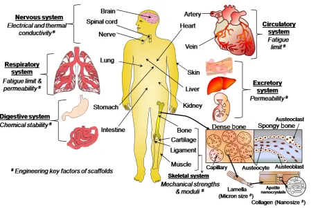
There has been enormous progress in tissue engineering (TE) in recent years. TE scaffolding materials have been extensively explored for the regeneration of various parts of the human body, including skin [1], muscle [2], teeth [3], bone [4,5], cartilage [4], ligament [6], spinal cord [7], nerve [8], genome [9] and artificial organs, particularly cardiac tissues or heart [10], lung [11], kidney [12], liver [13], urinary bladder [14], blood vessels [15], stomach [16], intestine [17], breast [18], ear [19], eye [20] and nose [21]. TE includes the combined growth of living cells, tissues or organs, as well as methods and materials that can restrain the shape of a particular tissue for better functions. When a group of cells (e.g. osteoblasts, osteocytes, osteoclasts and osteoprogenitor cells) perform the same function together, they are called tissue (e.g. bone tissue), which can relate to different organs. The essential human organs, including bone, with different engineering key factors of scaffolds for corresponding functional systems are illustrated in figure 1. In the skeletal system, the extracellular matrix becomes calcified in bone tissue unlike other connective tissues. The hard tissue or bone provides an internal support to the
body via attached tendons and muscles and has several metabolic functions, especially in calcium homeostasis. The remodeling and reorganization of bone tissue have various causes, including mechanical stimuli, metabolic causes (i.e. lack of dietary calcium, illness and aging), endocrine changes and effects of drugs. Over the past two decades, many techniques have been developed to design suitable scaffolds for repairing and regenerating various tissues. However, no suitable scaffold has been designed or developed yet for the repair or regeneration of bone tissue because of its complex composition, peculiar structure and extraordinary mechanical and biological properties. Therefore, in this review, we aim to outline key strategies that can improve the mechanical properties of scaffolds for bone TE applications. We also focus on electrospun scaffolds made of nanofibers to emphasize their benefits for bone tissue repair or regeneration.
1.1. Key engineering properties of primary tissues
Figure 1. Schematic of essential human organs listing functionalities required from implanted scaffolds.
tissues and the effect of electrospun materials on the TE scaffolds are illustrated in table1.
Depending on the applications and functions, an electrospun TE scaffold can be temporary or permanent. Usually, a temporary scaffold is highly porous and fully biodegradable, without side effects of by-products, whereas a permanent scaffold is highly biocompatible, mechanically strong, nondegradable, and remains inside the body for a long time.
1.2. Different tissue engineering approaches
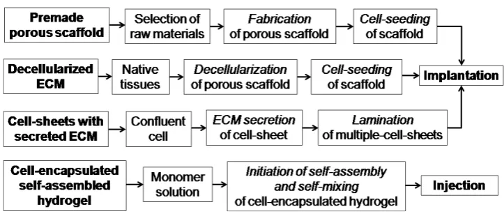
Depending on the source of scaffolds, raw materials and applications, TE can be approached by using four main types of material: premade porous scaffold [33], decellularized extracellular matrix (ECM) [34], cell-sheets with secreted ECM [35] and cell-encapsulated self-assembled hydrogels [36] (see figure2).
1.2.1. Premade porous scaffolding. In this approach, a porous scaffold is produced from different synthetic raw materials using various scaffold fabrication techniques. Then, living cells are seeded on the porous scaffold, and the resulting scaffold is implanted into the body for growing host tissues. The main advantages of this approach are low cost, availability of diverse raw materials, easy handling and simple technique. However, this strategy results in undesired responses from host tissues owing to the lack of biocompatibility, tissue adherence, surface chemistry, mechanochemical stability, and mismatch of degradation rate kinetics with the formation of new tissue.
1.2.2. Decellularized ECM scaffolding. The decellularized ECM technique is similar to the premade porous scaffolding; only the source of the scaffold is different. Here, native tissues are collected from decellularized ECM of similar tissue from another part of the body. The ECM includes the structural components of the niche such as soluble cue, matrix cue, stem cells, basement membrane and niche cells. It provides a physical platform for cell attachment, migration and division. The ECM acts as a reservoir of growth factors and potentiates their actions. It also sends biochemical signals to the cells that are modulated via molecular interactions with ECM biomolecules, such as heparin sulfate proteoglycans (HSPGs), or through adjacent cells. Here, the decellularized ECM exhibits several attractive characteristics as a TE scaffold that favors the ECM tissues for long-term in vivo
applications. Some of the main advantages of this technique over premade scaffolding are lower toxicity, cacogenicity and bioincompatibility. However, this approach has failed in many cases owing to the immunogenicity of the used biomaterials and the cell necrosis at the bulk scaffold related to oxygen deficiency and diffusion of nutrients. Another demerit of this approach is that the donor tissue is likely to elicit immunogenic responses and contain large variation over different batches [37].
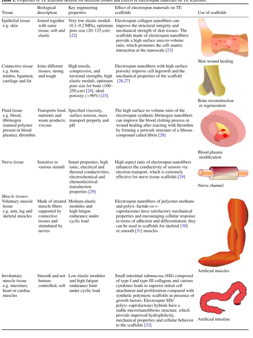
Table 1.Properties of TE scaffolds desired for different tissues and effects of electrospun materials on TE scaffolds.
Biological Key engineering Effect of electrospun materials on TE
Tissue description properties scaffolds Use of scaffolds
Epithelial tissue Joined together Very low elastic moduli Electrospun collagen nanofibers can e.g. skin with same (0.1–0.2 MPa), optimum improve the structural integrity and
tissue; soft and pore size (20–125µm) mechanical strength of skin tissues. The elastic [22] scaffolds made of electrospun nanofibers
provide a high surface area-to-volume ratio, which promotes the cell–matrix interaction at the nanoscale [23]
Skin wound healing Connective tissue Joins different High tensile, Electrospun nanofibers with high surface
e.g. bone, tissues; strong compressive, and porosity improve cell ingrowth and the tendon, ligament, and tough torsional strengths, high mechanical properties of the scaffold cartilage and fat elastic moduli, optimum [26,27]
pore size for bone (100– 250µm) [24], ideal porosity (>90%) [25]
Bone reconstruction or regeneration Fluid tissue Transports food, Specified viscosity, The high surface-to-volume ratio of the
e.g. blood, nutrients and surface tension, mass electrospun synthetic fibrinogen nanofibers fibrinogen waste products; transport property and can improve the blood clotting process in (natural polymer viscous pH wound healing after reacting with thrombin
present in blood by forming a network structure of a fibrous
plasma), thrombin compound called fibrin [28]
Blood plasma modification Nerve tissue Sensitive to Smart properties; high High aspect ratio of electrospun nanofibers
various stimuli ionic, electrical and enhances the conductivity of sensors via thermal conductivities, electron transport, which is extremely electrochemical and effective for nerve tissue scaffolds [29] chemoelectrical
transduction properties [29]
Nerve channel
Muscle tissues:
Voluntary muscle Made of striated Medium elastic Electrospun nanofibers of polyester urethane tissue muscle fibers modulus and and poly(l-lactide-co-ε
-e.g. arm, leg and supported by high fatigue caprolactone) have satisfactory mechanical skeletal muscles connective endurance under properties and encouraging cellular response
tissues and cyclic load in terms of adhesion and differentiation; they stimulated by can be used in scaffolds for skeletal [30]
nerves or smooth [31] muscles
Artificial muscles Involuntary Smooth and not Low elastic modulus Small intestinal submucosa (SIS) composed
muscle tissue human- and high fatigue of type-I and type-III collagens and various e.g. intestines, controlled; soft endurance limit cytokines leads to superior initial cell heart or cardiac under cyclic load attachment and proliferation compared with
muscles synthetic polymeric scaffolds in presence of
growth factors. Electrospun SIS/ poly(ε-caprolactone) hybrids have a stable micro/nanofibrous structure, which provide improved hydrophilicity,
mechanical properties and cellular behavior to the scaffolds [32]
Figure 2. Different TE regeneration techniques.
(e.g. poly(N-isopropylacrylamide)), until confluence. The confluent cell-sheet is then isolated using thermally regulated hydrophobic polymer coatings. Such approach can be repeated to obtain a thicker matrix of multiple-cell-sheet in a thermoresponsive culture dish by laminating a few single-cell sheets. After that, the cell-sheets are recovered from the dish using a low-temperature treatment. Finally, the multiple-cell-sheets are transferred and implanted into the body to observe the ingrowth properties of the host tissues. In some cases, this method is more advantageous than the decellularized ECM scaffolding due to the lower immunogenicity of the biomaterials used to form new tissues.
1.2.4. Cell-encapsulated self-assembled hydrogel.
Cell-encapsulation is a method of entrapping living cells within a homogenous solid mass or a semipermeable membrane. The biomaterials used for encapsulation are usually natural and synthetic hydrogels, which are made by covalent or ionic crosslinking of water-soluble polymers. Here, usually, one monomer solution of a completely biodegradable polymer is prepared by a self-mixing technique to make a cell-encapsulated hydrogel. The hydrogels can be formed through several gelation mechanisms, where polymer chains are crosslinked via covalent, ionic or physical bonds. Finally, the encapsulated hydrogel material is injected into the body to regenerate the host tissue. This technique is generally used for tissue regeneration via drug delivery or for soft tissue regeneration because of the high biodegradability and reduced mechanical properties of the hydrogel. Recently, this technique has also been tried for cartilage TE scaffolds by increasing the mechanical fracture strength through developing double networks in the hydrogel polymer chains. Interestingly, it has been found that the toughness and strength of the hydrogels are increased with void formation up to a certain void size or optimum void fraction (e.g. 1–3 vol% for polyacrylamide, PAAm). Below the critical size, the polymer (i.e. PAAm) chains can bridge the void gap and create additional stress on the void. Above the critical void size, the chains are too small to bridge the gap and the void can form a true hollow stress-free structure [38].
Over the past decades, many scaffold materials have been tried for TE applications. However, no material was
fully suitable for long-term implantations of bone TE scaffolds despite the specific advantages of each material. The failure might have occurred owing to the lack of consistency in material properties including mechanical stability, biodegradability, biocompatibility, toxicity, thermal or electrical sensitivity, permeability, surface adhesivity, hydrophilicity or hydrophobicity and fluidity in terms of viscosity. The consistency in material properties can be improved by the proper selection of materials, design and fabrication procedure. In this context, electrospun scaffolds performed better than other scaffolds.
2. Basic criteria for design and development of TE
scaffolds
The essential criteria for designing and developing an ideal three-dimensional (3D) TE scaffold are summarized in table2.
2.1. Scaffold design
Table 2.Criteria for designing electrospun fibers and TE scaffolds.
Function Design criteria
Strong, uniform and High-molecular-weight polymers bead-free fibers having high ionic conductivity,
concentration, or viscosity; high operating voltage in electrospinning process [39] Thin fibers, needle-like Electrospinning at high voltages
tip design and low flow rates [40] Aligned electrospun fibers High rotating target speed [40];
collector geometry
Structural stability to Maintaining mechanical properties throughout retain tissue shape the 3D scaffold by strong electrospun nanofibers Transport of nutrients and High porosity and interconnectivity between
waste in and out of the pores formed by maintaining preferred electrospun scaffold orientation of fibers [41]
Degradation integrity of Balancing degradation and formation of tissue electrospun scaffold to leave without toxic by-products [42]
host tissue
Elimination of inflammatory Materials must be biocompatible, nontoxic response or toxicity from the and noncarcinogenic
electrospun scaffold
High cell seeding density and Large pore size, high porosity and high cell migration leading to interconnectivity between pores using
tissue growth throughout the preferred unit cell geometry of the scaffold electrospun scaffolds [43]
Better cell attachment and Optimized surface chemistry/topography proliferation and high surface-to-volume ratio New cell or ECM growth in Proper fiber orientation within
preferred direction the scaffold [22]
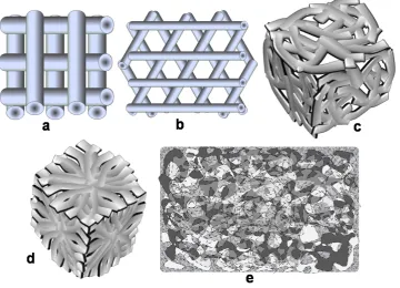
[image:6.595.149.447.80.395.2]Growth of 3D tissues and Specific 3D shape of electrospun scaffolds organs using preferred unit cell geometry [43]
Figure 3. Unit cells for 3D scaffolds: (a) cubic, (b) diamond, (c) gyroid, (d) honeycomb and (e) accordion-like honeycomb.
2.2. Scaffold material development
Many scaffold materials have been exploited recently for a wide range of TE applications. Among the three classes of scaffolding materials, namely, ceramics, polymers and composites only the last two can be easily electrospun into nanofibers, as ceramic materials have not been electrospun thus far without adding an organic functional material or polymer. Table 3 presents a summary of attempts [49–53] to fabricate porous 3D scaffolds for TE applications by electrospinning.
In the previous decade, conventional techniques such as solvent casting, particle leaching, salt fusion, gas foaming,
Figure 4. Different fiber arrangements in 3D scaffolds: (a) 0◦/90◦,(b) 0◦/60◦/120◦, (c) randomly oriented and (d) converging oriented fibers. (e) Surface morphology of a regenerative scaffold.
stem cells (MDSCs) [74], embryonic stem cells (ESCs) [75], adipose tissue-derived stem cells (ADSCs) [76] and dental pulp stem cells (DPSCs) [77] have been extensively explored in the field of connective TE owing to their distinct biological capability to differentiate into osteogenic lineages [78].
3. Progress in bone scaffold development
More than 50 polymers were employed during the last decade [24, 67, 68, 79] for scaffolding hard tissues, more specifically bone tissue. However, no single-phase polymer has shown the desired properties for bone TE applications. Regeneration or reconstruction of bone tissue involves three key factors namely, osteogenic progenitor cells, osteoinductive growth factors, and osteoconductive matrices [53]. Also, the selection of both materials and fabrication techniques is crucial for an ideal scaffold. The bone is such a structural composite material that it has a very high specific strength (i.e. strength-to-weight ratio). The tubular shape of bone can resist a bending force that causes compression on one side and tension on the reverse side.
3.1. Strategy for selection of scaffold materials to improve mechanical properties
Selection of materials for hard TE scaffolds depends on the properties required for real applications. For bone tissue, one of the major criteria is high strength and toughness. The ultimate strength should be such that the scaffold does not fracture before the complete growth of new tissues. The essential mechanical properties of different materials for bone TE scaffolds can be found in [68, 79]. The mechanical
properties of polymers can be improved by several methods as discussed below.
3.1.1. Crystallinity. The mechanical properties of most polymers depend on crystallinity, which generally increases with the number of polar groups in the polymer chain. However, an opposite tendency is observed for some functionalized polymers and attributed to the asymmetric (i.e. atactic or syndiotactic) stereographic position of the pendant functional groups. This asymmetric structure helps to increase the mobility of polymer chains, and therefore reduces the crystallinity and mechanical properties of the polymer. A more detailed explanation of this new finding is given in the original report [80].
3.1.2. Copolymerization. Copolymerization is one of the best techniques to increase the mechanical properties of polymers. The mechanical properties of copolymers can be improved by increasing the size of crystalline domains [81], molecular bond strength [82], tacticity or stereographic position [80], hydrophilicity [83] and so on. In copolymerization, the molecular bond strength is increased by grafting between different polymers, which in turn increases the mechanical properties of bulk copolymer. Polymerization may also affect the mechanical properties. For example, the mechanical strength increases with time and temperature of corona discharge polymerization, whereas ultraviolet irradiation decreases the mechanical strength of polymer materials [82].
Table 3.Materials, techniques and applications of porous 3D TE scaffolds.
Electrospinning as a possible
alternative
Scaffold material Application Technique Technique
Polymers
Porous biodegradable poly Tissue regeneration or reconstruction Emulsion freeze-drying [22] Yes (dl-lactide-co-glycolide)
(PLGA) copolymers
Porous poly(l-lactic acid) (PLLA), Skin tissue scaffolding using ROS Freeze-extraction and freeze- Yes PLGA, chitosan and alginate 17/2.8 osteoblast-like cells (rat gelation [54]
osteosarcoma)
Porous polyethylene glycol Scaffold for cartilage Compression molding and Yes terephthalate/polybutylene TE applications using chondrocytes particle leaching [55]
terephthalate (PEGT/PBT)
Poly(ethylene oxide) and Scaffolds for soft tissues in terms of Stereolithography [50] Yes poly(ethyleneglycol)dimethacrylate elasticity
photopolymerizable hydrogels
Polycaprolactone Bone scaffolds for bone morphogenetic Selective laser sintering [56] Yes protein-7 (BMP-7)-transduced
fibroblasts
Chitosan Electrobiological Electrochemical process [57] Yes
DNA ‘square-U’-based structure Single-strand DNA origami Polymerase chain reaction [9] Yes for biological nanoelectronics
Biodegradable polyurethane (PU) Skin tissue scaffolding using human Melt electrospinning [58] – fetal foreskin fibroblast cells
Ceramics
Porous hydroxyapatite (HAp) Load-bearing bone scaffold Combination of gel casting and No polymer sponge [59]
Biomorphic silicon carbide Bone implants, e.g. load-bearing Biotemplating [60] No ceramics, uncoated or coated prostheses using MG-63 human
with bioactive glass osteoblast-like cells
High-strength HAp Load-bearing bone scaffold Solid-state reaction [5] No
Bioactive, degradable and Bone tissue scaffold using Sol–gel [61] No
cytocompatible bredigite osteoblast-like cells (Ca7MgSi4O16)
Biomorphic HAp Bone tissue scaffold and implant Combination of novel No
biotemplating and sol–gel methods [62]
Nanostructure HAp Low-strength TE including drug Gel casting [63] No
delivery and cell loading Composites
Polyvinyl alcohol (PVA)/HAp Scaffolds for craniofacial and Selective laser sintering [64] Yes joint defects
PLGA/HAp composite and DNA and PLGA/HAp composite Electrospinning [65] – PLGA-dichloromethane-HAp- scaffold for bone TE
DNA/nanoparticles
Chitosan/calcium phosphates TE Membrane diffusion followed by Yes
effective freeze-drying [66]
Polyether etherketone Human trabecular bone TE Unconfined uniaxial compression Yes
(PEEK)/HAp scaffold [67,68]
Thermoplastic PU/ collagen TE scaffold using pig iliac Coaxial electrospinning [69] – endothelial cell (PIEC)
proliferation
Polycaprolactone with 0–50 wt% Scaffold for bone TE Electrospinning [70] – ceramic (20 wt% HAp/ 80 wt%
β-tricalcium)
by increasing their hydrophilicity [84]. Normally, their mechanical properties vary for the dry and hydrated states, whist the last condition is mostly employed in TE scaffolds or implant devices [84]. In 2009, Liet alconducted a tensile test on poly[(R)-3-hydroxybutyrate-co-(R)-3-hydroxyvalerate] (PHBV) and its blends with poly[(R)-3-hydroxybutyrate]-alt-poly(ethylene oxide) (HE) after immersion in phosphate
buffer solution for 24 h at 37◦C. Their results showed that the
a high fraction of fibroblast cells [83]. Moreover, the cell adhesion via adsorbed protein can be improved by surfaces with intermediate wettability. On the other hand, hydrophobic polymers such as polytetrafluoroethylene polypropylene and polyethyleneterapthalate, polystyrene provide a limited support for cell attachment. The poor cell attachment to nonpolar polymers (e.g. polystyrene) is possibly related to the lower degree of crystallinity in amorphous materials or hydrophobic polymers [83]. The different crystallinities of hydrophobic polymers further alter the mechanical properties. However, a technical problem associated with hydrophilic polymers, which is frequently faced when changing the medium during in vitro cell culture experiments, is that smaller hydrophilic electrospun fibers are lifted off the bulk scaffold owing to higher wetting in aqueous medium [40]. This problem can be solved by optimizing the hydrophilicity of polymers using proper surface modifiers.
3.1.4. Surface treatment. A few recent studies have shown that the mechanical properties of the bulk polymers or fibrous polymers can be improved by several surface treatment techniques [80, 86]. The polymer surface is effectively modified with polar groups containing ester (–COOR), ether (–O–), ketone (>CO), epoxy, carboxylic acid (–COOH), hydroxyl (–OH), acetyl (–COCH3), amide
(–CONH2), amine (–NH2) or other moieties by a suitable
surface treatment. The treatment improves the surface activity, polarity, hydrophilicity and mechanical properties of the electrospun polymers. The resulting surface-modified electrospun fiber may be advantageous for TE scaffolds used in connective tissues, including bone, cartilage, ligament and tendon reconstructions or replacements.
3.1.5. Composite fabrication or hybridization. Most of the recent investigations on tissue scaffoldings follow this technique because it is inexpensive and allows easy and precise control of the physical, chemical and biological properties. The organic part of bone tissue, such as protein, gives tensile strength and the inorganic part, calcium and phosphorus salts, provides compressive strength to the bone. The inorganic part of the natural bone is mostly similar to synthetic hydroxyapatite (HAp, [Ca5(PO4)3OH]) in structure
and chemical composition. The structure of bone is a highly organized hierarchy of the inorganic (HAp) and organic (e.g. Type-I collagen) phases [60], and in situ growth of
the network structure and strong hydrogen bonding. The mechanical properties of electrospun composite scaffolds also depend on the solubility of second-phase materials used with the polymer matrix in the body plasma or simulated body fluids. If the materials start to biodegrade or dissolve duringin vivouse, the mechanical properties of the scaffold can deteriorate in an uncontrollable manner. This is often observed in composites of polymers and calcium phosphates. Several types of calcium phosphates have been used in various biomedical fields, and their stability in water under physiological conditions (pH 7.4, at 37◦C)
increases in the following sequence: monocalcium phosphate [MCP, Ca(H2PO4)2·H2O], tetracalcium phosphate [TTCP,
Ca4P2O9], α-tricalcium phosphate [α-TCP, α-Ca3(PO4)2],
dicalcium phosphate dihydrate [DCPD, CaHPO4·2H2O],
dicalcium phosphate [DCP, CaHPO4],octacalcium phosphate
[OCP, Ca8(HPO4)2(PO4)4·5H2O], β-tricalcium phosphate
[β-TCP, β-Ca3(PO4)2], calcium-deficient hydroxyapatite
[CDHAp, Ca9(HPO4)(PO4)5OH], HAp [90]. Therefore,
depending on the mode of application, different ceramics and biopolymers should be combined in composite or hybrid scaffolds.
3.2. Selection of electrospinning technique
Figure 5. Schematic of electrospun fibers with (a) desired properties and required process parameters in aligned morphology and (b) high bead density and randomly oriented (undesired) morphology.
a wide range of fiber diameters, which are limited to microns in the latter technique. Melt electrospinning does not require toxic organic solvents and is ideal for scaled-up processes. However, it requires elevated temperatures for the melt, whereas stable solutions can be electrospun at room temperature. The third variety, coaxial electrospinning, is advantageous in the sense that it produces hollow fibers that can transport nutrients and waste in and out of scaffolds as elsewhere [2]. The morphology or microstructure of such scaffolds is similar to the bone architecture. The crucial electrospinning parameters that can alter the fiber properties are applied voltage, spinneret flow rate, target speed [40], collection distance, target properties, polymer molecular weight and polymer solution properties such as solvent type, concentration, viscosity, conductivity and surface tension [91, 92]. In general, the fiber diameter decreases and the fiber quality increases as the voltage is increased; the fiber diameter also decreases at lower flow rates [40]. Fiber alignment in a particular direction can be improved by increasing the speed of the target in an electrospinning system. The electrostatic forces exerted by interfiber interactions mainly control the motion of fibers ejected from the polymer jet during electrospinning; these forces depend on the external field, collector type and charges. As these forces have no preferential direction, randomly oriented nanofibers are formed on a stationary target. However, more aligned and parallel fibers are deposited on a rotating target or a drum [93]. The bead density in fibers decreases with increasing polymer concentration or viscosity [39], and the uniformity of bead-free fibers can be improved by increasing the ionic conductivity of the source polymer. Conducting polymer solutions carry more electric charge and generate stronger repulsive forces on the polymer jet. Therefore, polymer solutions containing ionic salts form more uniform fibers than pure polymers [94]. The properties and process parameters required for aligned fibers are summarized in figure5.
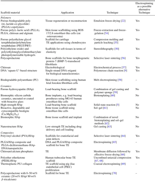
Figure 6. (a) Optimum interfiber space (supporting cell growth) and (b) large interfiber space (hindering cell growth).
3.2.1. Electrospun scaffolds in premade porous scaffolding approach. The research on electrospun scaffolds has recently been invigorated by introducing the premade porous scaffolding approach for skeletal system or connective tissues, including bone repair and/or regeneration. The cell growth property depends on the morphology and fiber orientations in scaffolds [57]. The space between fibers and the position of the fibers in a scaffold should allow the easy growth of cells [95,96] as illustrated in figure6.
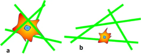
Recently, Dalton et al have obtained a peculiar result (figure7) on coiled electrospun fibers [40]. Fibroblast cells have been found to grow nicely inside and outside coil or ring-like fibers (figures 7(c) and (d)), whereas the cells could not attach properly to randomly oriented, entangled fibers [40] (figure 7(e)). This behavior can be attributed to the amorphous and hydrophobic nature of highly entangled long-chain polymeric fibers. Therefore, better cell attachment and tissue ingrowth are expected for scaffolds made of coiled electrospun fibers compared with randomly oriented, entangled fibers.
[image:10.595.309.546.309.401.2]Figure 7. Redrawn fluorescent images of electrospun fibers (a) with cells grown inside and outside the coil or ring-type fibers and (b) cells outside the randomly oriented entangled structure (adopted from [40] with permission). Schematic of cell attachment to electrospun fibers: (c) cell inside the coil, (d) cell outside the coil and (e) cells outside randomly oriented, long entangled fibers.
[image:11.595.120.481.343.668.2]Kolambkaret al[97] made a formidable effort to introduce electrospun nanofiber mesh tubes (effective pore size
<5µm, porosity 80–90%) as a guide for rat bone regeneration in a segmental bone defect. The nanofibers were made of polycaprolactone and had diameters ranging from 51 to 974 nm, with 82% of the values lying between 50 and 150 nm. The nanofiber mesh combined with peptide-modified alginate hydrogel (i.e., recombinant bone morphogenetic protein-2 (rhBMP-2)) was injected inside the tube for sustaining the growth factor release. A typicalin vivouse of the perforated scaffold or mesh made of electrospun nanofibers, combined with growth factor (rhBMP-2), for bone defects is illustrated in figures 8(B)–(F). Figure 8(G) shows the performance of this model up to 21 days, revealing that most alginate release (98.6%) took place within the first 7 days. This novel electrospun hybrid system allowed complete bony bridging of challenging segmental bone defects in rat.
3.2.2. Electrospun scaffolds in decellularized ECM approach.
Recently, bone regeneration has been attempted with natural materials fabricated into scaffolds, such as demineralized bone as a nanoscale-bone-matrix (NBM) powder, for improving the mechanical and osteoinductive properties [98]. Interactions between cells and ECM are very important for isolating cells and modulating or redirecting their functions [99]. The major advantages of electrospun fibers in the engineered scaffolds are that they provide micro-to nanoscale micro-topography and high porosity similar micro-to the natural ECM [92]. The high surface-to-volume ratio of nanoscale electrospun scaffolds can enhance the cell attachment, drug loading, and mass transfer properties [100]. The porosities resulting from electrospinning help transport nutrients and waste within the decellularized ECM approach. On the other hand, the nonwoven fibrous mats consisting of nanofibers provide a high surface area to interact with and attach to cells [101]. In this context, electrospun fibers of poly(l-lactide) (PLA) with 20% NBM have shown a higher Young’s modulus than pure PLA. These electrospun NBM/PLA composite nanofibers exhibit the properties similar to those of the native collagen-rich mineralized bone matrix and therefore can be used as a temporary substrate for facilitating the isolation and mineralization of bone-forming cells [1].
3.2.3. Electrospun scaffolds in cell-sheets with secreted ECM approach. The uniform aligned electrospun collagen fibers may be an ideal candidate to mimic complex structures in regular connective tissues such as tendons, ligaments and bones. The aligned electrospun fibers will also allow the fibroblasts and collagens to organize in the same orientation. Recently, the cell-sheet approach has been exploited to establish a novel method of tissue reconstruction in bone tissue regenerative medicine [103]. In this approach, there is no need for the scaffold to construct a cell-dense tissue, and it can be transplanted to various damaged tissues without any other treatment (e.g. suturing). The cell sheets can also be controlled with a polymer-coated plunger for transferring and stacking onto other cell sheets. Importantly, the stacking
method allows two cell sheets to contact each other physically, as well as to communicate biologically. The control of cell alignment may be a key factor in the next-generation cell-sheet-based tissue reconstruction technology [103].
3.2.4. Electrospun scaffolds in cell-encapsulated self-assembled hydrogel approach. Several hydrogel materials have been scaffolded for connective TE applications using the cell-encapsulated self-assembled hydrogel approach [104]. The porous hydrogel scaffolds can be used for microcarrier suspension culture of cells and for injection of the cell/microsphere constructs into a tissue defect. The injected porous scaffolds permit infiltration of cells and ingrowth of tissue from the host, and facilitate the regeneration process [105]. A variety of electrospun hydrogels fibers have also been developed for advanced applications [106], and different composites or hybrids of hydrogel materials have been studied for tissue engineering applications [107]. Self-assembling biomolecules (e.g. peptides) have been used forin vitro3D culture of cells,in vivotissue regeneration or repair of bones and optical nerves, as well as drug delivery and other applications [108]. Many important properties (e.g. swelling, mechanical properties, diffusion and degradation) of hydrogel in scaffolds can be tuned by controlling the molecular structure of the hydrogels. The molecular structure depends on both degree and density of crosslinking—the mechanical properties of the hydrogel improve as the crosslinking density increases [36]. However, a higher crosslinking density results in inferior swelling properties and mesh sizes, i.e. the distance between the crosslinks, which influences diffusion through the hydrogel. Thus, while the mechanical strength, which is desirable forin situplacement in articular cartilage, is improved, the swelling and diffusion properties are sacrificed [36]. More research is needed on the cell-encapsulated self-assembled hydrogel approach to develop new strategies in bone tissue regeneration [95,96].
4. Concluding remarks
References
[1] Renner R, Harth W and Simon J C 2009Int. Wound J.6226
[2] Liao I-C and Leong K W 2011Biomaterials321669
[3] Lim K-T, Suh J D, Kim J, Choung P-H and Chung J H 2011 J. Biomed. Mater. Res. B: Appl. Biomater.99B399
[4] Reddi A H 2000Tissue Eng.6351
[5] Pramanik S, Agarwal A K, Rai K N and Garg A 2007Ceram. Int.33419
[6] Liu H, Ge Z, Wang Y, Toh S L, Sutthikhum V and Goh J C H 2007J. Biomed. Mater. Res. B: Appl. Biomater.82129 [7] Hofstetter C P, Holmstrom N A V, Lilja J A, Schweinhardt P,
Hao J, Spenger C, Wiesenfeld-Hallin Z, Kurpad S N, Fris´en J and Olson L 2005Nature. Neurosci.8346
[8] Chen W and Tong Y W 2011Acta Biomater.8540
[9] Pound E, Ashton J R, Becerril H A and Woolley A T 2009 Nano Lett.94302
[10] Butcher J T, Mahler G J and Hockaday L A 2011Adv. Drug Deliv. Rev.63242
[11] Nichols J E, Niles J A and Cortiella J 2009Organogenesis557
[12] Steer D L and Nigam S K 2004Am. J. Physiol. Renal. Physiol.
286F1
[13] Chamuleau R A F M 2009World J. Gastrointest. Surg.121
[14] Petrovic V, Stankovic J and Stefanovic V 2011Sci. World J.
111479
[15] Xu C Y, Inai R and Kotaki M 2004Biomaterials25877
[16] Maemura T, Ogawa K, Shin M, Mochizuki H and Vacanti J P 2004Transpl. Proc.361595
[17] Day R M 2006Therapy1113
[18] Patrick C W Jr. 2004Annu. Rev. Biomed. Eng.6109
[19] Kusuhara H, Isogai N, Enjo M, Otani H, Ikada Y, Jacquet R, Lowder E and Landis W J 2009Wound Repair Regen.
17136
[20] Ge J and Liu J 2007Front. Med. China16
[21] Lee J-L, Bai C-Y, Shen Y-K and Hung J-H 2011IPCBEE (Singapore: IACSIT Press) vol 2 p 62
[22] Whang K, Thomas C H, Healy K E and Nuber G 1995 Polymer36837
[23] Zhong S P, Zhang Y Z and Lim C T 2010WIREs Nanomed. Nanobiotechnol.2510
[24] Spector M, Michno M J, Smarook W H and Kwiatkowski G T 1978J. Biomed. Mater. Res.12665
[25] Agrawal C M and Ray R 2001J. Biomed. Mater. Res.55141
[26] Rainer Aet al2011Ann. Biomed. Eng.391882
[27] Zhang Y, Venugopal J R, El-Turki A, Ramakrishna S, Su B and Lim C T 2008Biomaterials294314
[28] Wnek G E, Carr M E, Simpson D G and Bowlin G L 2002 Nano Lett.3213
[29] Bellamkonda R and Aebischer P 1994Biotechnol. Bioeng.
43543
[30] Riboldi S A, Sampaolesi M, Neuenschwander P, Cossu G and Mantero S 2005Biomaterials264606
J. Biomed. Mater. Res. B: Appl. Biomater.70286
[40] Dalton P D, Joergensen N T, Groll J and Moeller M 2008 Biomed. Mater.3034109
[41] Mikes A G, Bao Y, Coma L G, Ingher D E, Vacanti J P and Langer R 1992J. Biomed. Mater. Res.27183
[42] Cutright D E, Perez D, Beasley J D, Larson W J and Posey W R 1974Oral Surg.37142
[43] Coma L G, Vacanti J P, Vacanti C, Ingber D, Moonev D and Langer R J 1991J. Biomech. Eng.113143
[44] Melchels F P W, Bertoldi K, Gabbrielli R, Velders A H, Feijen J and Grijpma D W 2010Biomaterials316909
[45] Bettinger C J 2009Pure Appl. Chem.812183
[46] Engelmayr G C, Cheng M, Bettinger C J, Borenstein J T, Langer R and Freed L E 2008Nature. Mater.71003
[47] Rumpler M, Woesz A, Dunlop J W C, van Dongen J T and Fratzl P 2008J. R. Soc. Interface51173
[48] Hamid Q, Snyder J, Wang C, Timmer M, Hammer J, Guceri S and Sun W 2011Biofabrication3034109
[49] Nam Y S, Yoon J J and Park T G 2000J. Biomed. Mater. Res. A: Appl. Biomater.531
[50] Dhariwala B, Hunt E and Boland T 2004Tissue Eng.101316 [51] Leukers B, Gulkan H, Irsen S H, Milz S, Tille C, Schieker M
and Seitz H 2005J. Mater. Sci.: Mater. Med.161121
[52] Hollister S J 2005Nature Mater.4518
[53] Schieker M, Seitz H, Drosse I, Seitz S and Mutschler W 2006 Eur. J. Trauma32113
[54] Ho M-H, Kuo P-Y, Hsieh H-J, Hsien T-Y, Hou L-T, Lai J-Y and Wang D-M 2004Biomaterials25129
[55] Malda J, Woodfield T B F, van der Vloodt F, Kooy F K, Martens D E, Tramper J, van Blitterswijk C A and Riesle J 2004Biomaterials255773
[56] Williams J M, Adewunmi A, Schek R M, Flanagan C L, Krebsbach P H, Feinberg S E, Hollister S J and Das S 2005 Biomaterials264817
[57] Twu Y-K, Chang I-T and Ping C-C 2005Carbohydr. Polym.
62113
[58] Karchin A, Simonovsky F I, Ratner B D and Sanders J E 2011 Acta Biomater.73277
[59] Ramay H R and Zhang M 2003Biomaterials243293
[60] de Carlos A, Borrajo J P, Serra J, Gonz´alez P and Le´on B 2006 J. Mater. Sci.: Mater. Med.17523
[61] Wu C, Chang J, Zhai W and Ni S 2007J. Mater. Sci.: Mater. Med.18857
[62] Qian J, Kang Y, Zhang W and Li Z 2008J. Mater. Sci.: Mater. Med.193373
[63] Ghomi H, Fathi M H and Edris H 2011J. Sol–Gel. Sci. Technol.58642
[64] Chua C K, Leong K F, Tan K H, Wiria F H and Chea C M 2004J. Mater. Sci.: Mater. Med.151113
[65] Nie H and Wang C-H 2007J. Control. Release120111
[66] Thanaphat P, Thunyakitpisal P and Tachaboonyakit W 2008 J. Met. Mater. Min.1867
[68] Pramanik S 2011PhD ThesisKanpur, India p 392 [69] Chen R, Huang C, Ke Q, He C, Wang H and Mo X 2010
Colloids. Surf.B79315
[70] Patlolla A, Collins G and Arinzeh T L 2010Acta Biomater.
690
[71] Friedenstein A J 1976Int. Rev. Cytol.47327
[72] Wu M-Y, Chen N, Liu L-K, Yuan H, Li Q-L and Chen S-H 2009Polymers24301
[73] Kim J-Y, Jeon H B, Yang Y S, Oh W and Chang J W 2010 World J. Stem Cells234
[74] Usas A and Huard J 2007Biomaterials285401
[75] Jukes J M, Both S K, Leusink A, Sterk L M T, van Blitterswijk C A and de Boer J 2008Proc. Natl Acad. Sci. USA1056840
[76] Sterodimas A, de Faria J, Nicaretta B and Pitanguy I 2010 J. Plast. Reconstr. Aesthetic Surg.631886
[77] Cordeiro M M, Dong Z, Kaneko T, Zhang Z, Miyazawa M, Shi S, Smith A J and N¨or J E 2008J. Endod.34962
[78] Seong J M, Kim B-C, Park J-H, Kwon I K, Mantalaris A and Hwang Y-S 2010Biomed. Mater.5062001
[79] Pramanik S and Kar K K 2012Development in
Nanocompositesed K K Kar and A Hodzic (Singapore: RPS Publisher) (at press)
[80] Pramanik S and Kar K K 2012J. Appl. Polym. Sci.1231100
[81] Grijpma D W and Pennings A J 1994Macromol. Chem. Phys.
1951649
[82] Lei J and Liao X 2001Eur. Polym. J.37771
[83] Lydon M, Minett T W and Tighe B J 1985Biomaterials6396
[84] Li X, Liu K L, Wang M, Wong S Y, Tjiu W C, He C B, Goh S H and Li J 2009Acta Biomater.52002
[85] Lu H, Nguyen B and Powers J M 2004J. Prosthet. Dent.
92151
[86] Abdolahifard M, Bahrami S H and Malek R M A 2011ISRN Org. Chem.2011265415
[87] Tong H W and Wang M 2011Bioceram. Dev. Appl.1D110107 [88] Kar K K and Pramanik S 2012US Patent20120107612 A1
Published on May 3, 2012 (World Patent. WO 2012/004637 Date: 12 January 2012)
[89] Park H N, Lee J B, Moon H-J, Yang D H and Kwon I K 2011 Nanofibers—Production, Properties and Functional Applicationsed T Lin (Rijeka, Croatia: InTech Publisher) p 327
[90] Horvath L, Smit I, Sikiric M and Filipovic-Vincekovic N 2000 J. Cryst. Growth21991
[91] Kumbar S G, James R, Nukavarapu S P and Laurencin C T 2008Biomed. Mater.3034002
[92] Sill T J and von Recum H A 2008Biomaterials291989
[93] Zhang Q, Chang Z, Zhu M, Mo X and Chen D 2007 Nanotechnology18115611
[94] Kim S J, Lee C K and Kim S I 2005J. Appl. Polym. Sci.
961388
[95] Stevens M M and George J H 2005Science3101135
[96] Mager M D, LaPointe V and Stevens M M 2011Nature. Chem.3582
[97] Kolambkar Y M, Dupont K M, Boerckel J D, Huebsch N, Mooney D J, Hutmacher D W and Guldberg R E 2011 Biomaterials3265
[98] Traphagen S and Yelick P C 2009Regen. Med.4747
[99] Li M, Mondrinos M J, Gandhi M R, Ko F K, Weiss A S and Lelkes P I 2005Biomaterials265999
[100] Flemming R G, Murphy C J, Abrams G A, Goodman S L and Nealey P F 1999Biomaterials20573
[101] Sharma B and Elisseeff J H 2004Ann. Biomed. Eng.32148
[102] Ko E K, Jeong S I, Lee J H and Shin H 2008Macromol. Biosci.81098
[103] Takahashi H, Nakayama M, Shimizu T, Yamato M and Okano T 2011Biomaterials328830
[104] Kisiday J, Jin M, Kurz B, Hung H, Semino C, Zhang S and Grodzinsky A J 2002Proc. Natl. Acad. Sci. USA999996
[105] Chung H J and Park T G 2007Adv. Drug Deliv. Rev.59 249
[106] Burdick J A and Prestwich G D 2011Adv. Mater.23H41
[107] Jha A K, Yang W, Kirn-Safran C B, Farach-Carson M C and Jia X 2009Biomaterials306964

![Figure 7. Redrawn fluorescent images of electrospun fibers (a) with cells grown inside and outside the coil or ring-type fibers and (b) cellsoutside the randomly oriented entangled structure (adopted from [40] with permission)](https://thumb-us.123doks.com/thumbv2/123dok_us/8650672.867602/11.595.120.481.343.668/redrawn-uorescent-electrospun-cellsoutside-oriented-entangled-structure-permission.webp)
