Characterisation of a novel bioactive complex
polysaccharide from a marine invertebrate with
potent anticancer and antimalarial activities
Chrow Ahmed Khurshid
A thesis submitted to The University of Salford Manchester in partial
fulfilment of the requirements for the degree of Doctor of Philosophy
School of Environment & Life Sciences
College of Science & Technology
University of Salford
Salford, UK
i Contents
List of Tables ... v
List of Figures ... vi
Abstract ... viii
Declaration ... x
Acknowledgement ... xi
List of Abbreviations ... xii
Chapter1: Introduction... 1
1.1 General research background ... 2
1.2 Glycosaminoglycans GAGs ... 3
1.2.1 GAG biosynthesis Pathway ... 4
1.2.2 HS and Heparin Chain Polymerization and Modifications ... 6
1.2.3 Heparin and Heparan Sulphate Domains ... 8
1.2.4 Patterns of 2-O- sulphation, 6-O- sulphation and 3-O- sulphation groups in HS and Heparin ... 9
1.2.5 CS/DS Chain Polymerisation and Modifications ... 11
1.3 De - Polymerisations Techniques ... 13
1.3.1 Enzymatic De-Polymerisation of HS/Hep ... 13
1.3.2 Chemical De-Polymerisation of HS/Hep ... 15
1.3.3 Enzymatic De-Polymerisation of CS/DS ... 16
1.4 Heparan sulphate/heparin-protein interactions ... 17
1.4.1 FGF and the FGFR ... 17
1.4.2 The FGF signalling pathways ... 18
1.4.3 HS/FGF/FGFR Complex interactions ... 19
1.7 Marine sulphated glycan ... 21
1.7.1 Structural composition of sulphated polysaccharides (SP) from marine sources ... 22
1.7.2 Potential drug development from marine glycan. ... 26
1.7.3 Therapeutic value of mammalian glycosaminoglycan in cancer ... 28
1.8 Breast cancer ... 29
1.8.1 Causes, Risk Factors, and Prevention of breast cancer ... 29
1.8.2 Epidemiology of breast cancer ... 30
1.8.3 Histology of normal breast vs histology of breast cancer ... 31
1.8.4 Molecular profiling of breast cancer ... 33
1.8.5 Role of metastasis in breast cancer... 35
1.8.6 Treatment options for breast cancer ... 36
1.9 Malaria ... 38
ii
: Materials and Methods ... 43
2.1 Materials ... 44
2.2 Methods ... 44
2.2.1 Method of GAG isolation from soft body of Whelk. ... 44
2.3 Enzyme Preparations ... 45
2.3.1 Preparing of Hep lyase ... 45
2.3.2 Preparation of ABC lyase ... 46
2.4 Enzymatic digestion ... 46
2.4.1 Enzymatic degradation of Heparan Sulphate (GAG-HS) ... 46
2.4.2 Enzymatic degradation of Chondroitin Sulphate (GAG-CS) ... 47
2.5 Standard Preparation of HS and CS ... 47
2.6 Size exclusion analysis ... 47
2.7 Quantitative analysis of Monosaccharides by HPAEC-PAD ... 48
2.8 Ion exchange of intact GAG ... 49
2.9 PD-10 de-salting technique ... 50
2.10 Enzymatic degradation of ion-exchange fractions ... 51
2.10.1 Degradation of HS fractions ... 51
2.10.2 Degradation of CS fractions... 52
2.11 LC/GC-MS Techniques ... 52
2.11.1 Isotopic Aniline tagging for GAG disaccharide analysis by mass spectrometry (GRIL-Glycan Reductive Isotope Labelling) ... 53
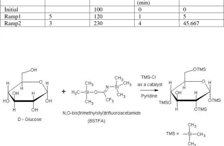
2.11.2 Trimethyl Silyl derivative for GC-MS (TMS) ... 54
2.12 Periodic acid oxidation (H3IO4 or H5IO6) ... 56
2.13 NMR spectrometry ... 57
2.14 Cell culture ... 58
2.14.1 Cell lines studied ... 58
2.14.2 Routine cell culture ... 58
2.14.3 MTT colorimetric Assay ... 60
2.14.4 Statistical approach ... 61
2.14.5 Protein extraction and quantification ... 62
2.15 Label-free protein quantitation proteomics ... 65
2.16 Mammosphere assay ... 66
2.17 General material and methods for in vitro culturing of malaria ... 67
2.17.1 Culture of Plasmodium falciparum ... 67
2.17.2 Drug susceptibility assays ... 69
: Biological activity of whelk-GAG crude extraction as an anticancer and antimalarial therapeutic agent ... 71
iii
3.2 Results ... 74
3.2.1 Cytotoxicity assay on lung carcinoma ... 74
3.2.2 Cytotoxicity assay on two subtypes of positive breast cancer ... 76
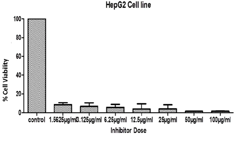
3.2.3 Cytotoxicity assay of intact whelk GAG extracts on hepatoblastoma-derived cell line Hep G2 ... 78
3.2.4 Cytotoxic assay of crude whelk GAG extracts on chronic myelogenous leukaemia ... 79
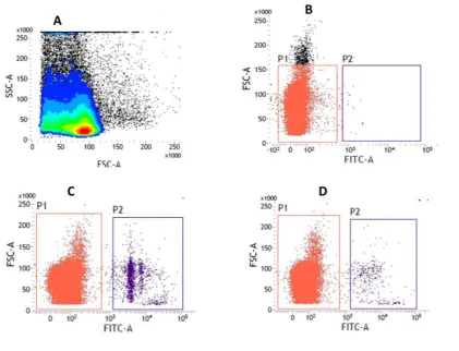
3.2.5 Crude Whelk GAG extracts as an antimalarial therapeutic agent ... 81
3.2.5.1 Blood smear of parasite ... 81
3.2.5.2 Cytotoxic activity of whelk GAG samples toward the malaria parasite using fluorescence-based drug susceptibility assay ... 82
3.3 Discussion ... 86
: Isolation and structural analysis of whelk GAGs ... 92
4.1 Introduction ... 93
4.2 Results ... 94
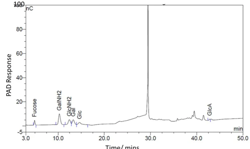
4.2.1 Monosaccharides analysis of intact GAG by HPAEC-PAD ... 94
4.2.2 Enzymatic digestion and Gel filtration chromatography of Intact GAG ... 98
4.2.3 Ion exchange chromatography and PD-10 de-salting techniques ... 103
4.2.4 Quantitative analysis of monosaccharides on different –GAG fractions obtained from DEAE-fragmentation by HPAEC-PAD ... 104
4.2.5 GC-MS spectrum of fraction CK-1 GAG as trimethyl silyl derivative ... 108
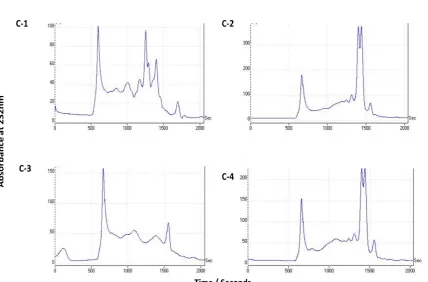
4.2.6 Disaccharide analysis by enzymatic digestions of GAG fractions from ion-exchange chromatography ... 111
4.2.7 Glycan Reductive Isotope Labelling (GRIL) LC-MS analysis of Isotopic Aniline tagging for GAG disaccharide ... 112
4.2.8 Monosaccharides analysis by HPAEC-PAD technique of whelk-GAG sample oxidised by periodic acid. ... 125
4.2.9 NMR spectra analysis ... 128
4.2.10 1D (1H-NMR) and 2D (HSQC-NMR) experiments ... 129
4.3 Discussion ... 133
: The effect of whelk GAG on triple negative breast cancer and elucidation of their mechanisms of action ... 137
5.1 Introduction ... 138
5.2 Results ... 141
5.2.1 Effect of intact GAG on triple negative breast cancer ... 141
5.2.2 Effect of HS/CS-disaccharide observed from digestion of whelk GAG and commercial porcine HS/CS on triple negative breast cancer ... 143
5.2.3 Cytotoxic effect of ion-exchange chromatography fractions of whelk GAG before and after oxidation with periodic acid on triple negative breast cancer cell lines ... 147
iv 5.2.5 Cytotoxic effect of crude whelk GAG extracts on mammosphere formation in breast
cancer ... 154
5.2.6 Label free protein quantification ... 157
5.2.6.1 Quantitative proteomics analysis ... 157
5.2.6.2 Mapping identified proteins to functional clusters in two triple negative breast cancer cell lines ... 159
5.2.6.3 Cell signalling pathway analysis ... 176
5.3 Discussion ... 193
: Conclusion and Future Works ... 200
References ... 204
Appendices ... 217
Appendix I: Recipes for solutions and buffers ... 218
Appendix II: A: LC-MS spectra of aniline-tagged standards and whelk-GAG flow throw (FT) sample fractions. B: MS spectra of the unknown peak in CK1 fraction (CK-1 sample in negative mode direct injection MS), Standard CS in negative mode direct injection MS, Standard GlcA in negative mode direct injection MS, and Reagent blank run on MS ... 224
Appendix III: Monosaccharide quantification... 241
Appendix IV: Monosaccharide analysis of the flow through fraction of the DEAE ion exchange column. ... 243
v List of Tables
Table 2-1: The gradient settings used for monosaccharides. ... 49
Table 2-2: GCMS conditions setting. ... 56
Table 2-3: List of cell lines studied, classification, immune profile and complete growth media. ... 58
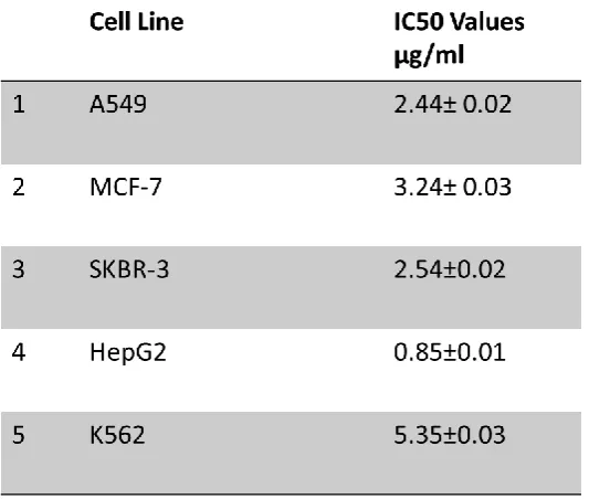
Table 3-1: IC50 Values represented in (µg/ml) of different cancer cell lines over 5 day’s incubation with crude whelk GAGs. ... 81
Table 4-1: Monosaccharide quantification in µg/ml of whelk crude sample. ... 97
Table 4-2: Quantification of monosaccharide in three fractions of ionic exchange chromatography. ... 108
Table 4-3: HS and CS Disaccharides codes. ... 113
Table 4-4: Disaccharide quantities of fractions CK1, CK2 and CK3 in percentage mole. ... 117
Table 4-5: Type of sulphating in HS and CS derivatives of each whelk-GAG fractions. ... 119
Table 5-1: IC50 values of whelk GAG fragments libraries from Hep lyase and ABC lyase on TNBC cell lines. ... 144
Table 5-2: Adjusted functional annotation clustering of significant proteins from DAVID online analysis of label--free proteomic data from the MDA-MB-468 cell line... 169
Table 5-3: Adjusted functional annotation clustering of significant proteins from DAVID online analysis of label-free proteomic data from MDA-MB-231 cell line. ... 172
Table 5-4: Pathways enriched in MDA-MB-468 from PANTHER online analysis. ... 176
Table 5-5: Pathways enriched in MDA-MD-231 from PANTHER online analysis. ... 177
Table 5-6: Pathways enriched from REACTOME in MDA-MB-468 cell line. ... 180
vi List of Figures
Figure 1-1: The biosynthesis of GAG chain. ... 6
Figure 1-2: Typical HS and Heparin Chain Polymerization and Modifications. ... 8
Figure 1-3: Typical organisation of heparin vs heparan sulphate domains. ... 9
Figure 1-4: Biosynthesis enzymes and 2-O- sulphation, 6-O- sulphation and 3-O- sulphation patterns found in HS/Hep. ... 11
Figure 1-5: CS/DS Chain Polymerisations and Modifications. ... 13
Figure 1-6: Heparinase (I, II, III) lyase action on each fractions substrate. ... 15
Figure 1-7: Chondroitin (ABC) lyase action on GAG. ... 16
Figure 1-8: HS/FGF/FGFR Complex interactions. ... 21
Figure 1-9: types of sulphated polysaccharide from marine organisms. ... 23
Figure 1-10: Breast anatomy. A: Histology of normal breast. B: Histology of breast cancer (Howard M. Reisner 2015). ... 32
Figure 1-11: Metastasis in breast cancer. ... 36
Figure 1-12: The life cycle of malaria. ... 40
Figure 2-1: Whelk GAG extraction process. ... 45
Figure 2-2: Size exclusion analysis of GAG samples and STDs. ... 48
Figure 2-3: Ion exchange chromatography and de-salting technique of intact GAG. ... 50
Figure 2-4: Sugars reduction with sodium borohydride following conversion of polyols to polyacetate esters, then formation of TMS ethers of sugars. ... 56
Figure 2-5: MTT assay, plate dose pattern. ... 61
Figure 3-1: Cytotoxic effects of different concentrations of whelk GAG crude extracts on pulmonary adenocarcinoma A549 cells. ... 75
Figure 3-2: Cytotoxic activity of whelk GAG extracts on two subtypes of breast cancer. Panel A: MCF-7. Panel B: SKBR3. ... 77
Figure 3-3: Cytotoxic activity of whelk GAG extracts on HepG2 cell line. ... 79
Figure 3-4: Cytotoxic activity of crude whelk GAG extracts on K562 cell line. ... 80
Figure 3-5: Thin blood smear for untreated and treated infected blood. ... 82
Figure 3-6: IC50 values of crude whelk GAG extracts on two mononuclear parasite stages. ... 84
Figure 3-7: Comparison of uninfected blood (negative control), infected blood (positive control) and sample treated with crude whelk GAG extracts. ... 85
Figure 4-1: Monosaccharide composition analysis. ... 96
Figure 4-2: Gel filtration chromatography of intact GAG, digested GAG with HEP lyase and commercial HS. ... 101
Figure 4-3: Gel filtration chromatography of digested GAG with ABC lyase and commercial CS. ... 103
Figure 4-4: The monosaccharide analysis form HPAEC-PAD system for ionic exchange chromatography fractions. ... 106
Figure 4-5: Composition analysis of CK1- GAG fraction and standards by GC-MS. ... 111
Figure 4-6: Disaccharide yield from whelk-GAG sample using GRIL-LC-MS. ... 115
Figure 4-7: The interaction of amino group in the aniline tag with the reducing sugar form. ... 120
Figure 4-8: The separation technique of Nanosep Centrifugal Devices with Omega Membrane. ... 121
Figure 4-9: Monosaccharide analysis of retained fraction of the mixture in all sample fractions. .... 124
Figure 4-10: Monosaccharide composition analysis by HPAEC-PAD of whelk-GAG sample fractions oxidised with periodic acid. ... 127
Figure 4-11: Brucker NMR spectrometer at 1D and 2D, frequency of 800MHz at 70 °C and 600MHz at 37 °C. ... 132
Figure 5-1: Inhibition activity of intact whelk GAG sample on negative breast cancer cells. ... 142
vii Figure 5-3: Inhibition activity of whelk GAG fractions (CK1, CK2, and CK3) from anion-exchange comparing with the biological activity of fractions oxidised with periodic acid on triple negative breast cancer cells. ... 149 Figure 5-4: Anti-proliferation activity of whelk GAG fractions CK1, CK2 and CK3 before and after enzymatic degradation on triple negative breast cancer. ... 153 Figure 5-5: Mammosphere formation in three breast cancer cell lines. ... 156 Figure 5-6: Principal Component Analysis PCA of Triple Negative Breast Cancer TNBC cells proteome at protein level. ... 159 Figure 5-7: Volcano plot images were produced using R, for each analysis, for corrected p-values. 162 Figure 5-8: Function classification from PANTHER online tool analysis of differential proteins expressed in the MDA-MB-468 cell line upon GAG treatment... 164 Figure 5-9: Function classification from PANTHER online tool analysis of differential proteins expressed in the MDA-MB-231 cell line upon GAG treatment... 166 Figure 5-10: The integrin cell surface interactions pathway. ... 192
viii
Abstract
Considerable excitement has been generated by the discovery of active compounds from
marine organisms. These compounds have a remarkable role in cancer treatment, with fewer
side effects on general health. Sulphated glycosaminoglycan (GAGs) are present in all
animals, vertebrate and invertebrate. They are of proven economic importance, not only in
the food industry but also in the pharmaceutical field.
The present study identifies the key structural differences between the GAGs isolated from
whelk and mammalian GAG and investigates their biological activity in relation to cancer
and malaria. Glycans from marine sources are unique in terms of their structure and function.
They work as an alternative natural source to provide effective treatment for several types of
cancer, especially triple negative breast cancer (TNBC), which is the most aggressive type of
cancer with fewer available treatment options. In addition, marine GAGs have a proven
influence on malaria, although their mechanisms of action are not fully understood.
Several methods have been employed to achieve the aims of this study. The structure of
whelk GAG was determined using several analytical techniques such as gel filtration
chromatography, ion exchange chromatography, mass spectrometry and nuclei magnetic
resonance spectroscopy. Enzymatic depolymerisation was also used to generate a library of
fragments; each was then evaluated for its biological activity on cancer growth.
Anti-proliferation activity on several cancer cells, including pulmonary adenocarcinoma A459, the
MCF-7 ER-positive cell line, overexpression of protein HER2- SKBR3 cell line, the
hepatoblastoma-derived cell line (Hep G2), chronic myelogenous leukaemia (K562), TNBC
and mammosphere formation in breast cancer subtypes, and on malaria has been investigated
in vitro using drug sustainable assays. Focusing on TNBC, the label-free quantitative
proteomic approach has been used to gain overall insight into the mechanisms of action of
ix
The results from whelk GAG analysis suggest a complex fine structure with a high sulphation
levels that is clearly distinct from mammalian GAG. Few impurities were detected within the
whelk GAG structure, which exhibits enzymatic resistance; this generated structurally
indeterminate fragments. These resistant fragments still have significant biological activity
against cancer growth. In vitro assays demonstrated significant inhibition activity of whelk
GAG toward all types of cancer cells, mammosphere formation from breast cancer cells and
malaria.
The mechanisms of action by which whelk GAG inhibits the growth of two TNBC cell lines
appear to involve influencing the cell integrin signalling cascade, extracellular organisation
pathways including the regulation of fibroblast growth factor FGF signalling and fibroblast
growth factor receptors FGFR, cell adhesion and reduced glycolysis metabolism. In addition,
whelk GAG affected the cell mitosis pathways by downregulating DNA replication proteins.
To our knowledge, this is the first study to identify significant structural differences between
whelk GAGs and mammalian GAGs, which helps to explain the structural-functional
relationship of marine glycans in inhibiting cancer cell growth. It also examines the
biological role of GAGs isolated from whelk as anti-proliferation agents toward a wide
variety of cancer cell lines and mammosphere formation in breast cancer. Finally, this study
x
Declaration
This thesis is submitted under the University of Salford requirements for the award of a PhD
degree by research. I certify that this thesis has not been submitted for any other degree in
any other university of higher learning, is not being submitted as part of candidature for any
such degree, and does not contain any material, which has been accepted as part of the
requirements for any such degree.
I clarify that the thesis does not contain any material previously published or written by
another person except where due acknowledgment is made. Throughout the PhD study, some
of the contents and findings have been published in conference posters only prior to the
submission of the thesis.
xi
Acknowledgement
First and foremost, I would like to express my sincere gratitude to my supervisor Dr David
Pye for all his help and continuous support for this research. Without his help, I would not be
in the position that I am today. Special thanks for Dr. Biswa Choudhury, for his patience,
encouragement and advice. We had few helpful discussions and he gave me very valuable
points. I would like to express my gratitude to Dr. Niroshini and her PhD student Muna
Abobaker for their collaboration at the parasitology labs.
I would like to thank all my colleagues at the biomedical and cancer research department for
their kind help. Special thanks for the amazing lab teams at University of California San
Diego, University of Manchester, NMR facility at MIB and University of Liverpool for their
help throughout this project. Special thanks for University of Kirkuk and Ministry of Higher
Education and Scientific Research for their funding throughout this project.
I would like to dedicate this thesis to my wonderful parents, sisters and my husband, who
have supported me throughout my life in each, and every aspect.
xii
List of Abbreviations
∆UA 4, 5 unsaturated Uronic Acid
2OSTs Uronosyl+2+O+sulphotransferase enzymes 3OSTs Glucosaminyl 3+O+sulphotransferase enzymes 6OSTs Glucosaminyl 6+O+sulphotransferase enzymes ABC lyase Chondroitin sulphate enzymes
AT III Antithrombin III BSA Bovine serum albumin
CML Chronic Myelogenous Leukaemia CS Chondroitin sulphate
CSC Cancer stem cells CTC Circulating tumour cells
DAVID Database for Annotation, Visualization and Integrated Discovery DBA Dibutylamine
DCIS Ductal in situ carcinomas DEAE Diethylaminoethyl
DMEM Dulbecco's Modified Eagle's medium DMSO Dimethyl sulphoxide
DNA Deoxyribonucleic acid dp Degrees of polymerisation DS Dermatan sulphate
ECM Extra cellular matrix ECM Extracellular matrix
EDTA Ethylenediaminetetraacetic acid EGF Endothelial Growth Factor EGFR Epidermal growth factor receptor EI Electron impact
ER Estrogen receptors EXT Exostoses
EXTL Exostoses like
FASP Ilter-aided sample preparation FBS Fetal Bovine Serum
FDR False discovery rate
FGFRs Fibroblast Growth Factor receptors (IIIc isoform unless stated otherwise) FGFs Fibroblast Growth Factors
Fuc Fucan
GAG Glycosaminoglycan Gal Galactose
GalNAc N+acetyl galactosamine GalT+I Galactosamine transferase I GalT+II Galactosamine transferase II GalT+III Galactosamine transferase III GL Gel filtration
Glc Glucosamine
GlcNAc N+acetyl glucosamine GlcNS Glucosamine N+sulphate
GlcNS (6S, 3S) Glucosamine N+sulphate, 6+sulphate, 3+sulphate GlcNS(6S) Glucosamine N+sulphate, 6+sulphate
GlcUA Glucuronic acid
xiii GS Gas chromatography
HA Hyaluronic acid
HBGF Heparan sulphate binding Growth Factor Hep Heparin
HEPES 4-(2-Hydroxyethyl) piperazine-1-ethanesulfonic acid HER2 Epidermal growth factor receptor
HexA Hexose amine
HexUA Hexuronic acid (either IdoUA or GlcUA). HGF Hepatocytes Growth Factor
HPAEC-PAD High-Performance Anion-Exchange Chromatography- pulsed amperometric detection
HPLC High performance liquid chromatography HS Heparan sulphate
HS Heparan Sulphate
HSPG Heparan Sulphate Proteoglycan IDC Invasive ductal carcinoma IdoUA Iduronic acid
IdoUA(2S) 2+O+sulphated Iduronic acid ILC Invasive lobular carcinoma KS Keratin sulphate
LCIS lobular in situ carcinomas LMWH Low molecular weight heparin MS Mass spectrometry
NA+domain N+acetylated domain
NDST Glucosaminyl N-deacetylase/N-sulfotransferase NMR Nuclear magnetic resonance spectroscopy NS/NA+domain N+sulphated/N+acetylated alternating domain NSCLC Non-small-cell lung carcinoma
OST O+sulphotransferase
PANTHER Protein ANalysis THrough Evolutionary Relationships PAPS 3’+phosphoadenosine 5’+phosphosulphate
PARP Poly (ADP-ribose) polymerase PBS Phosphate buffered saline PCA Principal component analysis PG Proteoglycan
PR Progesterone receptor
PTM Post-translational modifications RBC Red blood cells
RPMI Roswell Park Memorial Institute Medium SCLC Small-cell lung carcinoma
SDS Sodium dodecyl sulphate Ser Serine residue
ST Serine/threonine rich domains TFA Trifluoroacetic acid
TKIs Kinase inhibitors TMS Trimethylsilyl
TNBC Triple negative breast cancer Tyr Tyrosine
UDPsugar Uridine diphosphate UV Ultra violet
VEGF Vascular endothelial growth factor Xyl Xylose
1
2 1.1 General research background
Marine biology is an exceptionally rich source of many biological compounds and exogenous
sulphated glycans. They have important values in the pharmaceutical industry. Interest has
significantly increased in this field with many reports of compounds that have divers
biochemical structure (Liu & Rein, 2010). Compositional diversity is predominant in marine
organisms, almost each class revealing individually unique available compounds.
Marine species have been the source of a wide variety of metabolites with interesting
biological effects, interest has increased significantly in this field which has led to
considering as one of the natural and alternative sources of compounds with vital biological
functions worthy of further in depth investigation (Nur Hanim Zainudin, 2014).
Some marine natural products have derived drugs which are registered and published
either in the EU or in the US while more are under clinical and pre-clinically investigation.
Over 14,000 natural compounds have been developed from marine organisms recently and
hundreds of patents illustrating the activities of marine natural products have been
submitted, most of their remarkable activity have been on cancer (MarinLit, 2003; Proksch
et al., 2002).
Marine-derived bio active molecules include polyunsaturated compounds with sulphated
glycosaminoglycan as a part of their structure and they have been shown anticancer,
anti-inflammatory, antioxidant and antimicrobial activities in vitro (Schmitz, 1994). Sulphated
glycosaminoglycan is present in all animals, vertebrate and invertebrate (Yamada et al.,
2011). The polymeric form of sugars and amino sugars from marine organisms, known as
glycosaminoglycans (GAGs), are of proven importance not only in the food industry but also
in the pharmaceutical field. Interest in GAG has increased in response to their remarkable
biological activity as co-receptors for a variety of growth factors, cytokines, chemokines, and
3
Many patent applications disclose GAG isolated from various marine species for
anticoagulant and antithrombotic activity exclusively, while other patents present antituomur
activity such as GAG extracted from cartilaginous fish. Anti-inflammatory properties
reported from GAG isolated from Perna canaliculis due to sulphated hexosamine of GAG
derived. Heparan sulphate glycosaminoglycans express the highest structural variability of
their GAGs from other forms of sulphated glycosaminoglycan such as keratan sulphate (KS)
(Newman et al., 2000). Due to their pharmaceutical properties, various structures of heparan
sulphate glycosaminoglycan have been extensively studied.
Cancer is one of the major causes of mortality worldwide; it is defined as abnormal cells
dividing in an uncontrolled manner, which eventually could spread into other tissues. There
are many types of cancer, this diversity in cancer types and the fact that they are initiated or
generated from human body’ own cells make it the most complicated disease to treat. There
are several treatment options for cancer, mainly involve a combination of three approaches,
surgery, radiotherapy and chemotherapy. All of these three options have severe side effects
on the patient, especially chemotherapy. For example, doxorubicin is a useful chemotherapy
agent, it used to treat several cancer types but after surviving cancer, the patient in serious
danger of develop life- threatening conditions such as cardiomyopathy, nephropathy or
congestive heart failure (Sharkey et al., 2013).
1.2 Glycosaminoglycans GAGs
GAGs are found covalently attached to proteins, giving conjugates known as proteoglycans.
GAGs are a linear glycan chain composed of a repetitive structure of disaccharide units of
uronic acid and glucosamine. Initially it thought that GAGs only exist in mammals; however,
they found in wide variety of organisms such as fish, insects, fungi, yeast, and bacteria. It is a
4
GAGs found on the cell surface and the extracellular spaces in many tissues, especially
connective tissues. Such as that found in cartilage and the walls of blood vessel. Connective
tissues contain large amounts of GAGs, which also contains collagen and elastin fibres
embedded in a gel-like matrix called the ground substance. There are many types of GAG
families: hyaluronic acid (HA), chondroitin sulphate (CS), dermatan sulphate (DS), keratan
sulphate (KS), heparin (Hep), and perhaps the most widely studied is heparan sulphate (HS).
The main differences between the family members are the position and configuration of their
glyosidic linkages, amount and location of their sulphate substituents and monosaccharide
composition. Some of these species have very simple structure, for instance; hyaluronic acid,
whereas the others such as CS, DS, Hep and HS contain extensive blocks of modified
disaccharide units, with complex sulphation patterns which appear to be under tight
biosynthetic control (Mathews, 1974).
1.2.1 GAG biosynthesis Pathway
GAG biosynthesis occurs through three steps; chain initiation, chain polymerization and
polymer modification. It is generally believed that the polymerization takes place by stepwise
addition of monosaccharide units to non- reducing polysaccharide chain ends. Those chains
have various properties, which fixed at the initiation phase of polymerization and tightly
controlled by glycosyltransferases, sulphotransferases and an epimerase enzyme in the lumen
of the Golgi apparatus. The expression level of these enzymes during chain initiation is
responsible for the display of high structural complexity within GAG chain later (Wei et al.,
1993). The position and arrangement of glycosides linkages that join the monomer sugar
residues and determine, in part, the type of GAG synthesised are specified by the local
configuration of four groups, sulphate, hydroxyl, carboxyl and acetyl within the
5
Before the completion of GAG chain elongation, the polymerization products are extensively
modified to produce sulphate sequences within the polysaccharide using 3 -phosphoadenyl-
phosphorsulphate (PAPS) as a sulphate donor. The GAG chain contains uronic acid (either
L-iduronic acid or D- glucouronic acid) and hexosamine (represented in D-glucosamine or D-
galactosamine) residues respectively. The type of monosaccharide building block, the
position of the glycosidic linkages and the location of sulphate groups on the monosaccharide
rings will determine the subspecies of GAG synthesised.
The GAGs chain initiates with the protein core attached to tetrasaccharide contains GAG (n)–
GlcA–Gal–Gal–Xyl–Ser sequence. The chain initiates by adding an O-serine residue using
glycosyltransferases (Kim et al., 2001). Glycosyltransferases such as XylT-1 responsible for adding next residue Xyl from UDP-Xyl to the hydroxyl group of the serine core proteins in
the endoplasmic reticulum. Then two D-galactose monosaccharides are transferred to the Xyl
residue, this occurs by the activity of two types of galactosyltransferases GalT-I and GalT-II
(Carey, 1997). The GlcAT-I enzyme completes the biosynthesis of the linkage trisaccharide
by addition of a D-glucuronic acid residue from the UDP - GlcA transferase I (Pedersen et al.,
2000; Perrimon & Bernfield, 2000; Prydz & Dalen, 2000).
Two extensive modifications are carrying on GAG chain after it has been synthesised. First,
phosphorylation of the Xyl residue at position 2, which appears to happen in both CS and HS.
The second modification takes place on Gal residues by sulfation on either one or both Gal
residues at the 4 or 6 position. This step is found only in CS and DS, thus this might be
important for the selective assembly of CS/DS rather than HS/heparin as the last two never
shows any kind of modification in Gal residues onto the linkage region tetrasaccharide
6 Figure 1-1: The biosynthesis of GAG chain.
Modified from (Moehler et al., 2013)
1.2.2 HS and Heparin Chain Polymerization and Modifications
Chain polymerisation continues by adding hexosamine residues to the linkage-region
tetrasaccharide core. The sugar backbone of HS/Heparin is initiated by adding GlcA and
α-GlcNAc by α-α-GlcNAc transferase I (α-GlcNAcT-I) and α-α-GlcNAc transferase II (α-GlcNAcT-II)
enzymes respectively, these enzymes are also known as EXT1 and EXT2. In this step, the
chain will be classified and growing as HS/Hep chain (Fritz et al., 1994; Kim et al., 2001;
Sugahara & Kitagawa, 2000).
The addition of these residues results in the final length of the Hep/HS chain which could be
typically between 50 and 200 disaccharide units (Duncan et al., 2001; Turnbull et al., 2001).
The next step is an extensive modification of the chain by enzymes, which yields specific
sequences of sulphated and unsulphated regions within the Hep/HS chain. Glucosaminyl
N-deacetylase/N-sulfotransferase (NDST) is a golgi enzyme that initiates modification steps by
7
Phosphoadenosine 5-phosphosulphate PAPS is considered as the main donor for all sulphate
groups (Kakuta et al., 1999).
Recently, it was found that The NDST enzyme is active one both N-deacetyl and N-sulphate
residues, in vitro studies have characterised four isoforms of NDST enzymes in which each
enzyme’s isoform carrying the same reaction, with regards to the chemical context. NDST
isoforms 1 and 2 appear to be remarkably important in HS modification due to overlapping
issuing of two of these isoforms among others which might be produced by different genes
(Pikas et al., 2000).
The next step in chain modification is epimerisation by GlcA C5 epimerase, this enzyme
selectively epimerise GlcA to IdoA (Pinhal et al., 2001). The reaction was found to be freely
reversible in a solubilised enzyme system in the presence of 3H
2O trihydrate which is
basically 3 molecules of water, and the O-sulphate group on the C2 position of the IdoUA
residue which both seem to promote this reaction (Lindahl et al., 1998; Pinhal et al., 2001).
The action of epimerase is to increase the flexibility of the GAG, thus the IdoUA units are
considered as highly flexible units across the chain (Salmivirta et al., 1996).
HS is further modified in the golgi, heparan sulphate O-sulfotransferases (HS-2/3/6OST)
selectively add sulphate groups to IdoA or GlcA residues at the 2-O position, and the 3-O or
6-O positions of GlcNS or GlcNAc (Nakato & Kimata, 2002).Ultimately, the final structure
of the polysaccharide chain is determined by the action of glucosaminyl N-deacetylase/N
-sulphotransferase (the NDST’s) golgi enzymes (Ringvall et al., 2000).
In specific cells or tissues, further chain modification occurs by the action of 6-O sulfatases
(sulfs) which selectively remove the 6-O sulfation (Ai et al., 2003).
Despite the complicated modifications among HS chain, on average only 40-50% of HS
disaccharides are sulphated, this giving HS great structural diversity leading to increase the
8
activities, such as anticoagulation when HS binding to antithrombin protein, also promote
angiogenesis by HS binding to FGF (Lamanna et al., 2006; Robinson et al., 2006).
Figure 1-2: Typical HS and Heparin Chain Polymerization and Modifications.
Modified from (Esko et al., 2009)
1.2.3 Heparin and Heparan Sulphate Domains
The structural features between Hep/HS has been identified; the disaccharides units that
configure the heparin chain are extensively modified unlike HS chain. In the HS biosynthesis
and modification, selected GlcNAc residues likely undergo the N-deacetylation /N-sulfation
modification with no further modification. This known as an incomplete biosynthetic
modification pathway. Thus, HS chain appears as blocks of unmodified sequences (NAc-
domains), separated by highly modified residues or (S- domains).
However, in heparin the chain essentially contains S-domain interrupted by NAc domains.
During the modification reactions, three types of domains are likely to be generated. First,
9
amounts in the predominant disaccharide unit in HS. The second type is adjacent N-sulphated
regions with 2-O- and 6-O-sulphated in both IdoA and GlcA residues in (NS domains).
Finally alternating regions of N-sulphated and N-acetylated disaccharides domains appear in
HS chains (Mulloy et al., 2011; Robinson et al., 2006).
Figure 1-3: Typical organisation of heparin vs heparan sulphate domains.
Modified from (Chavaroche et al., 2013)
1.2.4 Patterns of 2-O- sulphation, 6-O- sulphation and 3-O- sulphation groups in HS and Heparin
Once the initiated synthesis and modification pathways in Hep/HS chain biosynthesis is
completed, further modifications will occur to add additional complexity to its structure and
function. Three types of HS sulphotransferases are responsible for the modification of
HS/Hep domains. Heparan sulphate 2-O-Sulphotransferase (HS2ST) is one of these
evaluative enzymes, and has a key role in forming 2-sulphated L-iduronic acid (IdoUA (2S)),
which widely exists in HS chains. The mechanism of action is to catalyse the transfer of
sulphate groups from PAPS to the C2 site on the L-iduronic acid (IdoUA) (Kobayashi et al.,
10
As this enzyme acts only on IdoUA units, it mainly modifies the S-domain sections of the
GAG chain. It may also play a critical role in HS degradation especially within IdoUA (2S) –
GlcNS (6S) domains (Habuchi et al., 2000; Kobayashi et al., 1997).
The second enzyme is heparan sulphate 6-O-sulphotransferase (HS6ST). This enzyme
catalyses the process of adding a sulphate group to the C6 position of a glucosamine using
PAPS as a sulphate donor. There are three isoforms of this enzyme, HS6ST-1, HS6ST-2 and
HS6ST-3. The first enzyme can transfer sulphate groups to GlcNS sites of IdoUA – GlcNS
disaccharide units (Sedita et al., 2004).
The second isoform transfers sulphate groups to a wide variety of substrates. More sulphate
groups may transfer to GlcUA – GlcNS domain, and less to IdoUA – GlcNS domain. Finally,
the third isoform HS6ST-3 is thought to act on both domains, IdoUA – GlcNS and GlcUA –
GlcNS (Habuchi et al., 2000; Sedita et al., 2004). In other words, HS6ST only act or transfer
O-sulphate to GlcNS.
During the biosynthesis of the HS chain, the final and important modification occurs on
carbon C3 of N- sulphated GlcN residues, this is the substrate that 3-O-sulphotransferase
enzyme (3-OST-1) acts on. This enzyme is considered most important, as it is thought to be
engaged in creating the antithrombin III (AT III) binding site within HS chains, thus
converting non-anticoagulant HS into anticoagulant HS as the residue is critical for the
formation of the antithrombin binding region the 3-O-sulphated GlcNS (3S) units (Liu et al.,
1999). 3-O-sulphation has been shown to be rare among other sulphations of glucosamine
during the modification process. There are three different isoforms of this enzyme, 3-OST-1,
3-OST-2, 3-OST-3a and 3-OST-3b. Each of these isoforms is expressed in different tissues.
3-OST-1 is found in the brain, kidney and the heart, while 3-OST-2 is highly represented in
brain tissue, the third isoform is found within a wide variety of different tissues (Habuchi et
11
transfers to the GlcNS residue regardless of the proceeding UA unit hence the domain is
GlcUA – GlcNS or IdoUA(2S) – GlcNS, whilst the 3-OST-3a and b enzymes act on specific
sequences limited by IdoUA(2S) – GlcNS domains (Liu et al., 1996).
Figure 1-4: Biosynthesis enzymes and 2-O- sulphation, 6-O- sulphation and 3-O- sulphation patterns found in HS/Hep.
1.2.5 CS/DS Chain Polymerisation and Modifications
Biosynthesis of CS/DS seems to be similar to that in HS/Hep chain synthesis. The CS/DS
biosynthesis enzymes are localised in trans-golgi complex, which is different from golgi
network part in the lower end of the trans-golgi matrix where HS/hep biosynthesis enzymes
are confirmed in golgi apparatus (Prydz & Dalen, 2000).
The diverging step in chain polymerisation begins after the formation of the linker
tetrasaccharide section completed. The enzyme EXTL2, which also known as GalNAcT1 is
[image:25.595.81.536.159.468.2]12
acts by adding GalNAc to the chain, to produce CS/DS chains. EXTL2 is the enzyme that
adds GlcNAc residue to HS chains. There are two distinct enzymes for the synthesis of the
CS/DS backbone known as GlcUA transferase II and GalNAc transferase II, the action of
these enzymes will lead produce chains with repeating 4- GlcUA β1-3 GalNAc units
(Sugahara et al., 2003).
After the biosynthesis, the DS/CS chain undergoes a significant modification process,
including the epimerization of GlcUA to IdoUA. The outcome of the C5 epimerase enzyme
leads to the chain classified as DS. The main differences between CS and DS is that CS only
has a glucuronic acid unit, while DS has both D-glucuronic acid and L-Iduronic acid units
(Prydz & Dalen, 2000).
The final chain modifications are by sulphotransferase enzymes action, the chain eventually
undergoes 2-O, 6-O-sulphation and 4-O-sulphation. The Chondroitin-6-sulphotransferase is
known as C6ST, the action of this enzyme produces GlcUA – GalNAc (6S) domains from
GlcUA – GalNAc by catalysing the conveyance of sulphate groups to the 6-O site of GalNAc.
Unlike Chondroitin-4-sulphotransferase that shift the sulphate groups to the 4-O site of
GalNAc residues, thus producing in GlcUA – GalNAc (4S) structure. Uronosyl 2-
O-sulphotransferase is found to only be active on DS chains via the shifting of a sulphate group
13 Figure 1-5: CS/DS Chain Polymerisations and Modifications.
Modified from (Maeda, 2015)
1.3 De - Polymerisations Techniques
There are two known methods for de-polymerisation of GAG chains, each one could be used
separately or combined and yield different compounds. A method for determining the
sequence type of GAG at the level of disaccharide provides key information for their
structure-function relationships. Disaccharide is obtained by depolymerisation of the
polysaccharide chains via enzymes and/or chemical degradation.
1.3.1 Enzymatic De-Polymerisation of HS/Hep
The enzymes generally used for the enzymatic depolymerisation process have varying
degrees of specificity for HS/Hep chains and are found in the soil bacterium, Flavobacterium
heparinum. These enzymes act especially on the structure uronic acid-HexA releasing
disaccharide or oligosaccharide compounds, with approximate fragment lengths of 10-20
14
unsaturated uronic acid at the non-reducing terminal (Galliher et al., 1981). This will create
an unsaturated double bond between C4 and C5 on the non-reducing end of the chain, which
is known as unsaturated uronic acid (ΔUA). This has UV sensitivity, which can detected by
wave length 232 nm, this allows enzyme digestion process to be detected (Linker, 1979).
There are three different isoforms of heparanase known as lyases I, II and III, each of these
lyases has a very high substrate specificity making them useful tools when comparing
isolated GAGs from different cells or tissue types, also they play an important role in
producing specific types of oligosaccharide fragments which vary in sulphate pattern, chain
structure and length. Heparanase I (hep I) typically acts on the sequence IdoUA (2S) -
GlcNS-(6S), thus it will release disulphate disaccharides and as a result releases the modified
NS regions. The enzyme also cleaves the antithrombin III binding pentasaccharide domain in
the heparin molecule. Heparanase II (hep II) acts on the sequences IdoUA-2S
GlcNS/NA-3S/6S and GlcUA -GlcNS/NA-GlcNS/NA-3S/6S. This is why Heparanase II is considered the least
specific of the heparainase enzymes and able to cleave a wide range of substrates in HS/Hep
chains. Heparanase III (hep III) acts on the sequence GlcUA –GlcNAc6S (Lindahl et al.,
15 Figure 1-6: Heparinase (I, II, III) lyase action on each fractions substrate.
http://www.sigmaaldrich.com
1.3.2 Chemical De-Polymerisation of HS/Hep
Nitrous acid is involved in the chemical method used for generating disaccharide units from
polysaccharide chains. Nitrous acid acts very selectively and quantitatively on the GlcNS
sequence, and is also able to convert GlcNS, GlcNS (6S), GlcNS (3S) and GlcNS (3,6S) units
into 2, and 5- anhydro D-mannose (Man). A specific range of pH is important to nitrous acid
substrate cleavage sites, high pH (3-4) or a low pH (1.5) can be used (Rabenstein, 2002). Low
pH nitrous acid-catalyzed deamination, which cleaves chains at N-sulphated GlcN residues
while high pH nitrous acid-catalyzed deamination will not act on GluNS domains, instead the
cleavage occurs at a small number of N-unsubstituted glucosamine residues GluN that exist
16 1.3.3 Enzymatic De-Polymerisation of CS/DS
Chondroitin sulphate ABC lyase is the enzyme used to de-polymerise chondroitin sulphate. It
is purified first from Proteus Vulgaris with two isoforms, known as an endoeliminase,
capable of depolymerizing chondroitin sulphate to produce, as products, a mixture of
∆4-unsaturated tetra- and disaccharides and an exoeliminase acting on chondroitin sulphate tetra-
and hexasaccharides to yield the respective disaccharides. It acts on β1, 4-galactosaminidic
bonds between N-acetylgalactosamine and either D-glucuronic acid or L-iduronic acid in CS
or DS and yield oligosaccharide mixtures of unsaturated tetra- and disaccharides (Yamagata
et al., 1968). The ABC lyase has a variety of applications, it has been used in the
quantification of CS and DS from a crude mixture (Yamagata et al., 1968), also it has an
important role in structural analysis of the carbohydrate moiety of proteoglycans studies
(Sugahara et al., 1992). A complete depolymerisation of CS to unsaturated disaccharides
yields from the combined activity of “endoeliminase (chondroitin sulphate ABC endolyase)”
and exoeliminase (chondroitin sulphate ABC exolyase) (Hamai et al., 1997).
Figure 1-7: Chondroitin (ABC) lyase action on GAG.
17 1.4 Heparan sulphate/heparin-protein interactions
Consensus sequences exist on the surface of some proteins with positively charged amino
acids that are able to interact strongly with negative-charged groups on the HS chains. The
interactions occurs within the binding regions in specific HS binding proteins (Goupille et al.,
1997).
The type of interaction is principally an ionic interaction involving negatively charged
groups in HS chain specifically sulphate and the carboxylates, which exists in all GAG chains.
So far, hundreds of proteins including in particular antithrombin III and the FGF family have
been shown to have significant interactions with HS chains. In particular with NS domains,
these domains provide multiple binding sites for protein ligands (Kreuger et al., 2006). Their
affinity for HS oligosaccharides basically correlates with the overall degree of sulfation
(Jemth et al., 2002). Heparin oligosaccharides were generally the most efficient complex
promoters, whereas less sulphated HS species were less efficient (Jastrebova et al., 2006).
More selective interactions would require either sequences containing rare components or
precise spacing of two or more sulphated domains N-sulphated/acetylated/sulphated (SAS)
(Kreuger et al., 2006).
1.4.1 FGF and the FGFR
Fibroblast growth factors are known as signalling molecules, which have a capacity to
promote angiogenesis by stimulating the proliferation of endothelial cells and can act directly
on tumour cells by increasing their growth. They are considered as one of the principle
angiogenic factors. Many members of the FGF family have the ability to bind to HS GAGs
and to FGFR receptors. These receptors bind to members of the fibroblast growth factor
18
Such interactions have been shown to be an absolute prerequisite for FGF mediated
biological activity. There are a great number of fibroblast growth factor family members
(FGF’s) in humans and mice, all of them are similar in terms of structure, but FGF1 is the
only one which is able to bind to all FGFR’s (Rieckmann et al., 2008; Wu et al., 2003).
FGF1 and FGF2 are known as essential angiogenic factors more than vascular endothelial
growth factor (VEGF) or platelet-derived growth factor (PDGF) (Cao et al., 2003).
These FGFs bind the HSPGs chain with different affinities. There are two types of FGFR
receptors on the surface of cells, each binding to FGF within different affinities, the low
affinity cell surface receptors are known as HSPG’s co-receptors, and the high affinity termed
as tyrosine kinase receptors (FGFR’s). Five distinct mammalian genes are encoding tyrosine
kinase receptors. Each of these FGFR is expressed in variety of different tissues, for instance,
FGFR1 is found mainly in connective tissue, FGFR2 mostly exists in bone, FGFR3
contributes to the structure of cartilage, and finally FGFR4 identified in muscle. There are
two types of each aforementioned FGFR, unspliced receptors or binding receptors, which are
divided into many variants known as A, B, and C. Their function secreted as receptor such as
FGFR- type A or supply binding ligand requirements for interactions such as FGFR- 3c and
FGFR -3b types. Some of the FGFRs binding to FGF1 and FGF2 equally such as FGFR- 2c,
while FGFR-3 binds to FGF1 with approximately 1000 fold higher affinity than FGF2
(Goodger et al., 2008; Rieckmann et al., 2008).
1.4.2 The FGF signalling pathways
Signal transduction from FGF requires the interaction between the growth factors and their
specific FGFR receptors including HSPG as a co-receptor, forming a complex that usually
exists on most of cell surfaces. The interaction between FGF and HS chains occur with many
19
Many factors can influence HS activation of FGFs including chain length andthe presence of
specific saccharide components, for e.g., HS oligosaccharides fractions of (dp6-dp12) have
different capability to activate bFGF in a mitogenic assay and this was correlated with their
length and disaccharide composition (Pye et al., 1998).
Another factor correlates to the charge distribution, which is mainly depend on sulphated
patterns. For e.g. the N- and 6-O-sulfation apparently sufficed to satisfy the requirement for
HS in important VEGF and FGF signalling events (Kreuger et al., 2006).
HSPGs are considered as co-receptors as they have secondary role in FGFR’s signal
transduction (Goodger et al., 2008). Cellular response initially starts from cell surfaces where
a particular type of FGF binding to their cell surface receptors, occurs the strength of the
signalling from this interaction will mainly depend on the environment of the cell including
the number of receptors available and the activated period of these receptors. All this will
lead to receptor dimerization followed by tyrosine auto phosphorylation of the receptor and
eventually activation of the appropriate signalling cascades (Pye et al., 2000).
1.4.3 HS/FGF/FGFR Complex interactions
Highly sulphated residues or NS domains within HS are important in FGF activation as they
have the ability to bring together FGFs with their tyrosine kinase receptors, they also increase
the stability of FGF-FGFRs interaction (Goodger et al., 2008; Kreuger et al., 2006). The
ternary complex of FGF1: HS: FGFR1 is highly stable and loses its complex structure only
when heated to 100 °C, while both of FGF1: HS and HS: FGFR1 binary complexes loss their
binding structure when heated to 60 °C (Wu et al., 2003).
The second crucial function of the complex interaction between HSPG and FGF-FGFRs is
the ability to prevent the proteolysis of FGF proteins (Ornitz, 2000), and to protect both
20
known to occur in different ratio and ways. The FGF: HS: FGFR interactions happen in three
modes known as Trans, cis and mix (contains both cis and Trans interactions). Multiple
models generated from each of these modes (Wu et al., 2003). In these complexes both
ligand and receptor might interact with HSPGs, or only the ligand might interact with
HS/Hep chain (Ornitz, 2000; Pellegrini et al., 2000). For example, in Trans model, the HS
derived hexasaccharide (dp6) is the smallest sequences that might interact with two FGF1’s
on each side of the isoform interaction. In the cis oriented model, HSPGs of (dp12) units is
required and the minimum size required for the mix cis/trans to form 2:2 FGF1:FGFR2
complex is a HSPGs of dp16 (Ornitz, 2000).
There are two well-known structural models for this complex that have been confirmed by
X-ray crystallography, which are the “symmetrical complex model” and the “asymmetrical
complex model”. The symmetrical complex is more stable among the other complexes this
due to extensive protein- protein contact (Goodger et al., 2008). This model involves a 2:2:2
complex, two HS dp10 chains facilitate the complex and were confirmed by the X-ray crystal
structure (Schlessinger et al., 2000). The asymmetrical complex contains only a single chain
of HS and is able to join the FGF and FGFR’s together in a 2:2:1 ratio such as FGFR2 IIIc:
FGF1: hep decasaccharide seen by Pellegrini et al., (2000). The size of the HS chain and the
amount of sulphation determines which HS chain will pick by the FGFR to create the
21 Figure 1-8: HS/FGF/FGFR Complex interactions.
Modified from (Gallo et al., 2015)
1.7 Marine sulphated glycan
Marine organisms have been shown to be one of the most extensive and diverse sources of
many bioactive compounds. During the last 50 years, over 14,000 different natural products
from marine organisms or species have been reported (MarinLit, 2003).
The first isolation of sulphated glycans from a marine organism was by Killing (1913), as he
reported a structure of sulphated fucan from marine brown algae (Phaeophyta). Different
types of sulphated GAGs have been purified from microalgae such as brown, green, and red
algae, and other invertebrate species such as echinoderms (sea cucumber and sea urchins)
Sulphated GAGs are found in other living phyla and have been confirmed in many other
classes of invertebrates such as Porifera, Actinopterygii fish (Zierer & Mourão, 2000), also
in commercially relevant species such as sharks, skate, codfish, and salmon. Most of these
sulphated GAG’s have shown biological activites in different aspects such as anti-pathogenic,
22
Other species have been found with a rich content of sulphated GAG in their composition,
such as S. hermanni and S. vastus. Other species of sea cucumbers known as ‘GAMAT are
considered as a source of traditional medicines, due to containing high bioactive substances
(Hawa et al., 1999).
1.7.1 Structural composition of sulphated polysaccharides (SP) from marine sources
Sulphated polysaccharide is a significant component in marine organisms’ structures that
have been extensively studied to date. Two types have been identified so far, sulphated fucan
and sulphated galactans. Essentially, the first type occurs in two forms, heterogeneous
non-repetitive sulphated fucans from algae and homogeneous, non-repetitive sulphated fucans of
echinoderm. While heterogeneous sulphated galactans have been extracted from red algae,
green algal species, some species of invertebrates and marine angiosperm polysaccharide
(Pomin & Mourão, 2008). Many factors affect the composition of this polysaccharide; first,
different species express different structures of sulphated polysaccharides. Second factor is
the method of extraction
The third factor is the season or the environment of conditions, such as the crude fucan that is
extracted monthly from makonbu, Laminaria japonica Areschoug, cultured from April to
October at a southern site of the Hokkaido bay (Japan). The crude fucan tends to increase
gradually from April to September, and rise markedly in October (Honya et al., 1999;
23 Figure 1-9: types of sulphated polysaccharide from marine organisms.
Heterogeneous sulphated fucans are more abundant in marine brown algae (Phaeophyta).
Early study by Percival & Ross, (1950), reported the structure of these sulphated fucan,
which mainly contain 1→2 glycosidic linkages and sulfation at the 4-position. Recently,
another structure has been confirmed for brown algae’s sulphated fucan which contains equal
amounts of alternating units of 2,3-disulphated, 4-linked and 2-sulphated, 3-linked
α-L-fucopyranosyl units and has been confirmed by NMR analysis. The heterogeneous nature of
these structures is thought to be a result of occurrence of branches of non-sulphated fucose
residues. However, a unique structure is reported from other brown algae species with NMR
studies suggesting the occurrence of O-acetylation in very high amounts (Chevolot et al.,
1999). The structure of a polysaccharide from brown algae is known as the most complex
marine polysaccharide due to branching chains, diverse glyosidic linkages, acetylation,
methylation and random sulphation pattern.
Suphated polysaccharide
from marine organisms
Sulphated fucan
Homogeneous, repetitive sulphated fucan
Heterogeneous non repetitve sulphated fucan
Sulphated galactan
24
Homogeneous, repetitive sulphated fucans have been reported in marine invertebrates
especially in sea urchins and sea cucumbers (Echinodermata, Holothuroidea) with simple
repetitive structures composed of unbranched chain of α-L-fucose either [α (1→3) or α
(1→4)] type of glycosidic linkages and 4-O and/or 2-O sulfation positions (Pomin & Mourão,
2008).
Heterogeneous sulphated galactans are the other main type of suphated glycan and are found
principally in marine red algae. The main reason for heterogeneity of the glycans isolated
from red algae is the sulphate distribution along the chain. There are two types of red algae
termed as Carrageenans and Agarans both express an homogeneous galactan backbone unlike
other red algae species (Bixler, 1994; Knutsen et al., 1994; Lahaye, 2001; van de Velde et al.,
2004).
Both of these red algae have glycans with a non-branched (linear) chain comprised of
alternating 3-linked β-D-galactopyranose and 4-linked α-D/ α-L-galactopyranose residues.
They have a variety of industrial applications due to their simple structure, they also have a
unique ability to create strong aqueous gels. The physicochemical properties of these glycans
are affected by variation in the backbone structure such as modified α-D-galactopyranoses in
carrageenans to α-L-galactopyranoses in agaran (Lahaye, 2001). Sulphated galactans are also
found in green algal species like Codium (Bilan et al., 2007; Farias et al., 2008). The chain
here is without repetitive units of disaccharide and instead has a complicated sulphated
pattern in positions 4 and/or 6 –sulfation unlike red algae. Chain complexity is higher in
green algae than red algae but is considered as simpler structure in comparison to brown
algae (Pomin, 2009). Sulphated galactans have also been reported in some species of
invertebrates in ascidians (Urochordata, Ascidiacea), and in two species of sea urchins. The
25
In general, the location of these sulphated glycans, either sulphated fucan or galactan, is
determined within the body wall or extracellular matrix, this matches the amount of
glycosaminoglycan found in the extracellular matrix of mammalian connective tissue. Marine
sulphated fucans and galactans are more sulphated than CS and DS in vertebrate GAGs.
Marine sulphated glycans are found with extremely high molecular weights that can be >1
million Da such as the mass of sulphated fucans that found in sea urchin egg jellies for the
glycan fraction, as this sulphated glycan has high molecular weight itself without any core
protein interaction, while the GAG molecular mass in mammals could reach ∼15 and ∼60
kDa, or approximately >100 kDa in case of covalent attach to core protein (Pomin & Mourão,
2008).
The presence of sulphated GAGs in some invertebrates is now well-documented. Different
classes of invertebrates have shown that CS/DS, HS-like, and/or heparin-like compounds are
present in many species (Medeiros et al., 2000). studies have also shown that heparin is
present in several species of molluscs. A compound from the clam Mercenaria mercenaria
exhibits several structural similarities to heparin (Jordan & Marcum, 1986). Heparins with
high anticoagulant activity have been isolated from the molluscs Anomalocardia brasiliana,
TiVela mactroides (Dietrich et al., 1985; Pejler et al., 1987) and Tapes phylippinarum
(Cesaretti et al., 2004). CS, DS, and acharan sulphate have also been isolated and
characterized from different families of molluscs (Cassaro & Dietrich, 1977; Kim et al.,
1996).
Whereas HSs and CSs are ubiquitous components of most tissues, heparin has shown a very
peculiar distribution in mammalian and other vertebrate tissues, as well as invertebrates
(Medeiros et al., 2000). Furthermore, a large variation of the concentration of heparin among
species is evident, with the non-mammalian vertebrate tissues showing considerably lower
26
echinoderma, and cnidaria, with the anticoagulant activity varying according to the species
analysed (Nader et al., 2004).
1.7.2 Potential drug development from marine glycan.
Marine sulphated polysaccharides can express their activity on mammal’s biological system
and provide pharmacological activity with different diseases, such as antithrombotic,
anti-inflammatory, antiangiogenic, and antimetastatic. Sulphated fucan from brown and sulphated
galactans from red and green algae are well known for their activity as anticoagulants as they
interact with both antithrombin and heparin cofactor II (Berteau & Mulloy, 2003; Pereira et
al., 2005). However, the complex, heterogeneous structures of these polysaccharides has
limited the possibility of identifying specific sequences required for their activity. Many
researches, have worked in this field in order to confirm the structure of these
polysaccharides and the relationships related to their activity. Chemical oversulphation or
desulphation of the intact chain from these algae has been reported (Haroun-Bouhedja et al.,
2000; Soeda et al., 1993). The results suggest that the activity increases with increasing
sulphate regions and decreases when the crude or intact pattern of sulphation is reduced. The
same role is applied to the molecular weight, the activity decreases with smaller molecular
size of oligosaccharide or polysaccharide (Soeda et al., 1993).
In order to identify the structure-function relationships of these polysaccharides, it was
important to identify clearly oligosaccharides type, glycosidic bonds between the residues,
and sulphate positions which are required for blood coagulation protein interaction to occur
that exhibit the anticoagulant process. The effect of the type of saccharide residue, whether
this was fucose or galactose on modifying anticoagulant activity has been reported by Pereira
et al., (2002). Comparisons were made between two different sulphated polysaccharides, the
27
their ability to modify the activity from active to inactive sequence model. The position of the
glycosidic linkage in this polysaccharide affected their activity; this was shown by comparing
two models of sulphated galactans, the inactive 3-sulphated, 4-linked and the active
2-sulphated, 3-linked α-L-galactans. The effect of the sulphation pattern is important in the
structure-function relationship; increasing the sulphation content only ∼1.8-fold caused
extensive increases in their anticoagulant activity of nearly 38-fold. This was linked to the
occurrence of two, 4-di-sulphated of 3-linked fucans from 2-sulphated, 3-linked
α-L-fucan (Mourão, 2004).
Sulphated fucans have been shown to have angiogenesis inhibition activity. According to
Koyanagi et al., (2003), some fucoidans have been extracted from Fucus vesiculosus which
interfere with the binding of VEGF with their receptors. The same interaction between these
molecules and FGFs with their respective receptors is also reported which causes
angiogenesis inhibition as a consequence (Soeda et al., 2000).
Moreover, apoptosis and cell death due to autophagy in tumour cells was reported as a
biological activity with some active compounds within sulphated fucans from brown algae.
Sulphated heparin, fucoidan and Carrageenan lambda are reported to significantly inhibit
activity toward lung adenocarcinoma in rat. This appears to be due to an interference with the
passaging process in the tumour cells, which induces early apoptosis and leads to a
remarkable loss of tumour cells from the lung after one hour, followed by cell death (Coombe
et al., 1987). Most of the bioactive compounds in algae are available commercially as an
intact or crude compound containing sulphated polysaccharide but also contaminated with
non-glycan compounds. Designing low molecular weight glycans with known sequences of
saccharide remains unachieved as yet, due to the structural complexity of polysaccharide