2018 International Conference on Computer Science and Software Engineering (CSSE 2018) ISBN: 978-1-60595-555-1
Extraction of Tongue Crack Based on Gray
Level and Texture
Feifei Chen, Chunming Xia, Jiayun Sui, Yiqin Wang
and Qian Peng
1
ABSTRACT
Tongue diagnosis is one of the most important content of Traditional Chinese Medicine (TCM). While the tongue crack is an important index of tongue diagnosis, there are just a few existing researches of tongue crack extraction and the results of them are unsatisfactory. This paper proposes a new algorithm for tongue crack extraction based on crack’s grayscale and texture in the tongue image. The method analyses and handles the tongue image’s luminance component in HSV color space. Firstly, we use the morphological opening closing reconstruction to eliminate small texture and noise of tongue images. Then we use Bot-hat transform and Otsu adaptive threshold segmentation to extract tongue cracks. Finally, we use correlation and entropy of grayscale symbiotic matrix to screen extracted tongue cracks and realize automatic extraction of tongue cracks. The result has shown that the method can extract tongue cracks with high accuracy and lay a good foundation for subsequent analysis of the crack.
INTRODUCTION
Tongue diagnosis is a clinical diagnostic method which is to know the physiological function and pathological changes of the body by observing and analyzing various changes in the tongue. It is one of main contents of the TCM
Feifei Chen1, Chunming Xia1, Jiayun Sui1, Yiqin Wang2 and Qian Peng2
1
inspection. Traditional tongue diagnosis is judged by doctor's experience. The diagnosis result is easy to be influenced by the knowledge level of doctors, diagnostic skills and light conditions, which makes the objectification, quantification and standardization of tongue diagnosis difficult to achieve and restricts its development in modern times. It is an inevitable trend for the development of tongue diagnosis to realize the objectification, quantification and standardization of tongue diagnosis by modern means [1, 2].
The tongue crack is an important index of tongue diagnosis. The definition of tongue crake in TCM is different depths and shapes of cracks in the tongue. Modern studies suggest that shallow tongue cracks are mainly due to atrophy of tongue mucosa which makes the upper lingual cortex lose its normal structure. Some nipples become flattened and fused. Some of them atrophy and break. Deep tongue cracks are severe atrophic tongue pathological changes.
Tongue crack is a kind of special surface texture in the tongue image which is the most changeful. It can be viewed as curve structure on the surface of tongue, so we can use the method of line detection which is roughly divided into 3 categories: contour-based, center line-based and area-based [3-5].Yang got line response image using tongue image gray-level and color information as well as the pixel distant gradient and extracted tongue cracks from line response image by hysteresis threshold algorithm [6].Liu proposed an improved line width detector which can avoid the effect of undesirable width and uneven illumination [7]. There is also extraction algorithm using region characteristics such as color feature of cracks. Chen used color features in L*a*b and regional division and merger to divide the tongue image into several regions, then extract cracks by adaptive threshold [8]. Yang used kernel false color transformation to increase the image contrast, then calculated gradient image for the G component, finally used the lag threshold method to get crack images [9]. The resent researches’ results are difficult to apply in the subsequent analysis of tongue cracks directly. This study aims to improve accuracy of crack extraction by combining the characteristics of the crack’s color and texture.
The experimental samples are typical cracked tongue images provided by Shanghai University of Traditional Chinese Medicine. And use manual segmentation method to extract tongue body. All the samples were marked by Shanghai University of Traditional Chinese Medicine experts for the crack areas which are used as the true value targets.
METHODS
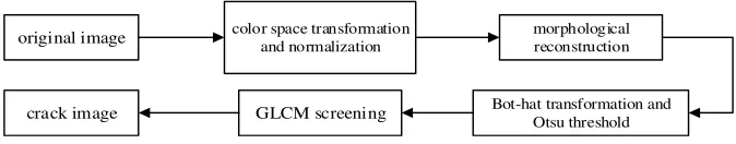
color space transformation and normalization
morphological reconstruction
Bot-hat transformation and Otsu threshold
GLCM screening original image
[image:3.612.125.462.85.151.2]crack image
Figure 1. Flow chart of crack extraction algorithm.
Color Space Transform
This paper firstly analyzes and selects the color space of samples. Since RGB is proposed for hardware, while HSV is conforming to the visual characteristics of human eyes [10,11], so the first step is converting RGB space to HSV space as equations (1-3) [12].
0 , max min
60 0 , max ,
max min
60 360 , max ,
max min
60 120 , max
max min
60 240 , max
max min if
g b
if b g b
g b
h if r g b
b r if g r g if b = − × + = ≥ − −
= × + = < − − × + = − − × + = − (1)
0 , max 0
max min min
1 ,
max max if s otherwise = = − = − (2) max
v= (3)
where,
(
r g b, ,)
is the red, green and blue values of a pixel color,(
h s v, ,)
is the hue, saturation and intensity values of a pixel color, and their value is a real number between 0 and 1. max is equivalent to the maximum value in R, G, B, respectively. min is equivalent to the minimum value in R, G, B, respectively.The next step is selecting the human eyes most sensitive component, luminance component (V) in HSV, for subsequent analysis, and normalizing the value by equation (4) to make different samples comparable.
where, V is the gray value of V component, Vnor is the normalized gray value of
V component, Vmax is the maximum value and Vmin is the minimum value.
Mathematical Morphological Opening and Closing Reconstruction
There are many tiny structures and textures and noise in tongue images which cause some interference on crack extraction. This paper uses mathematical morphology reconstruction(MMR) to filter them and achieves good results[13,14]. MMR is a part of image transformation set and is a very important content in mathematical morphology image processing. It is often applied in image segmentation field. MMR based on the characteristics of the labeled image (Marker) processes the mask image (Mask). In binary images, one image, I (Mask), contains another image, J (Marker), and the binary image’s reconstruction is extracting connected domain from the I marked by the J.
The process can be expanded from a binary image to a grayscale image by line expansion. The definition of gray MMR based on line expansion is described as equation (5).
( )
1
( ) n ( )
I I
n
D J δ J
≥
= ∨ (5)
where, ( )n( )
I J
δ is n times grayscale line expansion operations of the J in the I. ∨
means to seek maximum point by point.
Its dual operation, the operation of corrosion reconstruction is as equation (6):
( )
1
( ) n ( )
I I
n
E J ε J
≥
= ∧ (6)
where, ( )
( )
n
I J
ε is n times grayscale line corrosion operations of the J in the I.
∧ means to seek minimum point by point.
If take the gray image, f , as a mask image and fΘA as the marker image, the
morphological opening reconstruction operation is defined as equation (7).
( )
(
)
(
)
,
rec
f
O f A =D fΘA (7)
where, A is the structural element, Θ is gray corrosion operation.
If take f ⊕A as the marker image, the morphological closing reconstruction
operation is defined as equation (8).
( )
(
)
(
)
,
rec
f
C f A =E f ⊕A (8)
where, ⊕ is grayscale expansion operation.
Combining the two forms, called opening and closing reconstruction, is defined as equation (9).
(rec)
(
,)
(rec)(
(rec)(
,)
,)
OC f A =C O f A A (9)
Opening and closing reconstruction is a nonlinear filter based on mathematical morphology. After the opening and closing reconstruction, details and noise in the image are suppressed and local extremum is eliminated and then form extreme value regions. It can be seen from the pictures that small texture and noise are completely eliminated and crack paths are more smooth.
Extraction Based on Bot-hat Transform and GLCM
The noise and other effects of the reconstructed image are well eliminated and the cracks are well preserved. In this paper, the Bot-hat transformation and a single threshold is used to extract cracks. The Bot-hat transformation is a morphological filter, also called the trough detector, which has same characteristics of high pass filters. The Bot-hat transformation is the difference of the gray levels between the result of closed operation image and the original image [15]. Choose a structure element. The Bot-hat transformation is defined as equation (10):
( ) ( )
hat
B f = f A⋅ − f (10)
where, f A⋅ is morphological close operation.
Therefore, it can detect high frequency troughs, such as image cracks, and enhance details of cracks in the image. The reconstructed image is carried out to reduce the complexity of crack extraction by the Bot-hat transformation.
After the Bot-hat transformation, the image mainly contains troughs. Meanwhile, the contrast of the whole image is reduced. After processing, the cracks can be easily extracted by adaptive threshold method.
In this paper, Otsu method is used for image segment, which is defined as below [16].
If the gray range of the image is 1 ~m, the gray value of the pixel is ni. The
total gray value is as equation (11):
1 =
m i i
N n
=
∑
(11)The probability of each gray value is as equation (12):
i i
n P
N
Use t to divide it into two groups, that is Co={1 ~ }t and C1={t+1 ~m}. 0
ω and ω1 is the probability of the corresponding two groups. µ0 and µ1 is the average of the two groups. The generation probability of each group is as follows:
The generation probability of Co is as equation (13):
0 1 t i i P ω =
=
∑
(13)The generation probability of C1 is as equation (14):
1 1 m i i t P ω = +
=
∑
(14)The average of Co is as equation (15):
0 1 0 t i i iP µ ω =
=
∑
(15)The average of C1 is as equation (16):
1 1 1 m i i t iP µ ω = +
=
∑
(16)Then The threshold of Otsu method is defined as equation (17):
2 2
1 1
( )t o ( o )
σ =ω ω µ −µ (17)
When calculating the max value of 2
( )t
σ by changing t from 1 ~m, the value
of is the best threshold.
Filter the segmented image by morphological opening and closing operation to optimize the result. Because different diseases manifested on the tongue, such as ecchymosis, yellow fur and so on, the region which has similar gray value as crack region is need to be extracted too. Due to the texture feature of the crack region has obvious differences, this paper uses gray level co-occurrence matrix(GLCM) to extract texture features and optimize the results by the texture features[17].
The GLCM was proposed by Haralick et al [18]. It is the second order statistical feature of image texture. The GLCM is obtained by statistical correlation between two pixels of a distance and direction in a gray image, which is defined as equation (18):
ˆ ( , | , ) ˆ ( , | , ) P i j d P i j d
S
θ
θ = (18)
Suppose image gray level as N, P as GLCM, then P is the matrix of N×N,
between the two pixels in this statistical process , and the values of the two pixels are probabilities in this grayscale map.
The secondary characteristics of the matrix are obtained by statistical calculation of GLCM, which is used to represent the texture features of the image. Correlation and entropy characteristics of the GLCM are used as criterions to
screen the crack regions. Calculate correlation, C d( , )θ , and entropy, ENT, of the
matrix Pˆ, which is defined as equation (19-20):
1 2 2 2
, 1 2
ˆ ( , | , ) ( , )
i j
P i j d
C d θ θ µ µ
σ σ
−
=
∑
(19),
ˆ( , | , ) log( ( , | , ))ˆ
i j
ENT = −
∑
P i j d θ P i j d θ (20)where, the gray level is 64 and each gray level is extracted from four directions at angles of 0°, 45°, 90° and 135°, respectively. The distance is 5. The mean value in different directions of correlation and entropy is respectively used to represent the texture feature of the crack.
Algorithm Steps
The steps of crack extraction are as follows:
Step 1 Choose 6x6 pixels disk structure element, se , and perform
morphological corrosion operation on the normalized V component image, I to
get the corrosion image Ie.
Step 2 Takes I as the mask image, Ie as the marked image and perform
morphological expand operation on Ie to get the open reconstructed image Iorec .
Step 3 Continue to take the structure element, se, and performs morphological
expansion operation on Iorec to get the expansion image Id.
Step 4 Take Iorec as the mask image, Id as the marked image and perform
morphological corrosion operation to get the opening and closing reconstruction image, Iocrec.
Step 5 Take 12 sizes disk structure element and perform Bot-hat transform on
ocrec
I to get the image, Jbot.
Step 6 Use Otsu threshold to segment Jbot and get the cracks image. Preform
morphological opening and closing filter on the cracks image to get the image C.
Step 7 Divide image C into separate regions Cip according to the connected
domain and calculate the circumscribed rectangle of each area. Use The rectangle
Step 8 Calculate the correlation average value and the entropy average
value(d=5,θ=0, 45,90,135) of GLCM according to each criterion image, Cip, and
use the equation (21) to judge.
( 0.6, 0.7)
i
i
Cor Ent
C
C
∅ < >
=
(21)
Performance Index
The accuracy of the extraction results is calculated by the overlap degree of the segmented areas as the evaluation criterion. The specific definition is as equation (22).
100%
auto manu auto manu
S S
SA
S S
∩
= ×
∪ (22)
At the same time, divide the crack tongue image into three different extraction difficulties, i.e., simple, common and difficult, by using the visible index VI and the depth index DI of the crack. VI and DI are defined as equations (23-24):
( )
( )
crackle area VI
common area
= (23)
( )
( )
crackle averagegray DI
common averagegray
= (24)
The simple group contains images with VI>0.03 and DI<0.60; the difficult group contains images with VI<0.01 and the DI>0.80; and the rest belong to the ordinary group.
RESULTS
The result of opening and closing reconstruction is shown in Figure 2. Many tiny structures and textures and noise are eliminated.
Every step of the crack extraction process is shown as Figure 3.
(a) (b) (c)
Figure 2. Morphological opening and closing reconstruction[(a)original image; (b)the opening reconstruction result; (c)the opening and closing reconstruction result].
(a)origin image (b) reconstructed image (c) Bot-hat transformJbot
(d)Otsu method C (e)Crack criterion image Cip (f)result
Figure 3. Crack extraction process.
TABLE I. STATISTICAL RESULT.
Group Evaluating Indicator
SA Avg VI Avg DI
Simple 95.54±0.83% 0.0375 0.5501
Normal 93.13±1.31% 0.0212 0.6971
Difficult 90.77±2.92% 0.0168 0.8359
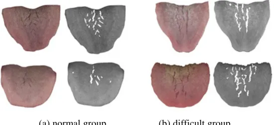
[image:9.612.130.457.258.673.2](a) normal group (b) difficult group
Figure 4. Results of normal group and difficult group.
Some of the results are shown in Figure 4-5.
It is seen from table I and Figure 4 that the extraction effect of the simple group and the normal group are good. Those cracks are obvious. The extraction effect of the difficult group is relatively poorer than the simple group and the normal group. The main reasons are as follows:
When tiny cracks and coarse cracks both presented in the tongue images, the coarse cracks are extracted successfully but the tiny cracks are easily ignored. Besides, Different diseases are manifested in the tongue images. Some of them are similar with cracks and are extracted too.
Nevertheless, the extraction accuracy of the difficult group is above 90%. Combined with the results of all the groups, the crack extraction algorithm achieves the satisfaction level and lay a good foundation for the future analysis of tongue cracks.
CONCLUSION
This paper proposes a new algorithm to extract cracks from tongue images based on characteristics of color and texture, which has a good extraction accuracy.
and the grayscale of crack. Then, these two indexes were used to classify the difficulty of the crack samples. For simple group and normal group, this algorithm had high accuracy, while for difficult group, the accuracy was also above 90%. As a result, the algorithm for extraction of tongue cracks lay a good basis for the future analysis of tongue cracks. All manuscripts must be in English, also the table and figure texts, otherwise we cannot publish your paper. Please keep a second copy of your manuscript in your office.
REFERENCE
1. Pang B, Zhang D, Li N, et al. Computerized tongue diagnosis based on Bayesian networks[J]. IEEE Transactions on Biomedical Engineering, 2004, 51(10):1803-1810. 2. Ren Z, Liu GD, Huang Z. Development of Spectrometer for Tongue Diagnosis Based on
Plane Holography Concave Grating[J]. Spectroscopy & Spectral Analysis, 2013, 33(9):2586.
3. Fernandes L A F, Oliveira M M. Real-time line detection through an improved Hough transform voting scheme[J]. Pattern Recognition, 2008, 41(1):299-314.
4. Yao X, Guo L, Zhao T. Power Line Detection Based on Region Growing and Ridge-Based Line Detector[J]. Lecture Notes in Electrical Engineering, 2013, 255:431-437.
5. Qin H, Huang Z, Zhao Y, et al. New MLBP-Otsu method and its application in tongue crack image segmentation[J]. Computer Engineering & Applications, 2014.
6. Yang Z H, Li N M. Detection of Tongue Crack Based on Distant Gradient and Prior Knowledge[J]. International Journal of Image & Graphics, 2010, 10(02):100-377.
7. Liu L L, Zhang D. Extracting Tongue Cracks Using the Wide Line Detector[C]// International Conference on Medical Biometrics. Springer-Verlag, 2008:49-56.
8. Chen X F, Cui-Hua L I, Xiao-Feng D U. Detection of Tongue's Crack Based on Adaptive Threshold[J]. Computer Technology & Development, 2009.
9. Yang Z, Li N. Detection of Tongue Crack Based on Distant Gradient and Prior Knowledge[J]. International Journal of Image & Graphics, 2010, 10(02):273-288.
10. Yoon I, Kim S, Kim D, et al. Adaptive defogging with color correction in the HSV color space for consumer surveillance system[J]. IEEE Transactions on Consumer Electronics, 2012, 58(1):111-116.
11. Huang K Q, Wang Q, Wu Z Y. Multi-scale color image enhancement algorithm based on color space and human visual system (HVS)[J]. Acta Electronica Sinica, 2004, 32(4):673-676.
12. Trambadia S, Mayatra H. Food detection on plate based on the HSV color model[C]// Online International Conference on Green Engineering and Technologies. IEEE, 2017:1-6. 13. Serra J P. Image Analysis and Mathematical Morphology[J]. Biometrics, 1982, 39(2):536. 14. Zhou N, Cui Y. Skeletonization and Reconstruction via Mathematical Morphology[J].
Journal of Image & Graphics, 1997.
15. Bai X, Zhou F, Xue B. Multi scale multi structuring element top-hat transform for linear feature detection[J]. 2012:1920-1923.
16. Vala M H J, Baxi A. A review on Otsu image segmentation algorithm[J]. International Journal of Advanced Research in Computer Engineering & Technology, 2013, 2(2):387– 389.
![Figure 2. Morphological opening and closing reconstruction[(a)original image; (b)the opening reconstruction result; (c)the opening and closing reconstruction result]](https://thumb-us.123doks.com/thumbv2/123dok_us/271713.1027503/9.612.130.457.258.673/figure-morphological-reconstruction-original-reconstruction-opening-closing-reconstruction.webp)