0095-1137/95/$04.0010
Copyrightq1995, American Society for Microbiology
Outer Surface Protein C Gene Sequence Analysis of
Borrelia burgdorferi Sensu Lato Isolates
from Japan
MASAHITO FUKUNAGA*
ANDAKIKO HAMASE
Faculty of Pharmacy and Pharmaceutical Sciences, Fukuyama University, Fukuyama 729-02, Japan
Received 20 March 1995/Returned for modification 18 May 1995/Accepted 20 June 1995
The nucleotide sequences of the outer surface protein C gene (
ospC
) from
Borrelia burgdorferi
sensu lato
isolates representing six different restriction fragment length polymorphism (RFLP) ribotype groups were
determined, and the deduced amino acid sequences were aligned in comparison with the previously published
OspC protein sequences. The sequence similarity analysis revealed the high sequence variability of OspC
protein, and the degree of amino acid similarity ranged from 53.8 to 100% among 25 isolates. It has been
reported that the representatives belonging to the three species of
B. burgdorferi
sensu lato showed a
species-specific amino acid sequence motif at positions 23 to 35 (B. Wilske, S. Jauris-Heipke, R. Lobentanzer, I. Pradel,
V. Preac-Mursic, D. Ro
¨ssler, E. Soutschek, and R. C. Johnson, J. Clin. Microbiol. 33:103–109, 1995). Alignment
with the OspC sequences of RFLP ribotype group IV, V, and VI isolates revealed that a sequence motif of all
the isolates was quite similar to that of
Borrelia garinii
. A phylogenetic analysis based on OspC protein
sequences also showed that most of the Japanese isolates were closely related to the species
B. garinii
. The
RFLP ribotype group IV species is predominant among clinical isolates of Lyme disease patients, reservoir
rodents, and adult ticks in Japan. Although the isolates differed from type strains of the three delineated
genospecies in genetic and immunological characteristics, it is likely that the spirochetes diverged within the
species level. Therefore, the representatives of ribotype groups IV, V, and VI appear to have evolved within
B.
garinii
and to have adapted to an Asiatic habitat, and there appeared to be a sufficient ecological pressure to
allow bacterial species level development.
Three species of the genus Borrelia, Borrelia burgdorferi
sensu stricto, Borrelia garinii, and Borrelia afzelii, associated
with Lyme disease have been recognized to date, and
repre-sentatives of the three species were isolated from patients,
reservoir rodents, and relatively limited species of ixodid ticks
(1, 3, 4, 13, 32, 35). B. burgdorferi sensu stricto is geographically
restricted to North America and Europe, and B. garinii and B.
afzelii are widely distributed in Eurasia (2, 3, 13, 17). The
spirochetes are maintained in locations where the disease is
endemic by vector-reservoir transmission cycles involving the
Ixodes ricinus species complex ticks, North American Ixodes
scapularis and Ixodes pacificus, European Ixodes ricinus, and
East European and Asiatic Ixodes persulcatus (13, 21).
Spirochetes were isolated from ticks, rodents, birds, and
Lyme disease patients in Hokkaido, the northern island of
Japan where Lyme disease is endemic (16, 22, 26). We have
demonstrated the usefulness of the restriction fragment length
polymorphism (RFLP) ribotyping system based on the
borre-lial 23S-5S rRNA gene repetition for assessing the genetic
diversity of B. burgdorferi sensu lato (7, 9, 24). This grouping
was in full agreement with the three-species classification of
Lyme disease agents. In Japan, no isolates of B. burgdorferi
sensu stricto have been identified with our RFLP ribotyping
system and some of the isolates belonged to ribotype groups II
(B. garinii) and III (B. afzelii) (24, 25). However, most of the
Japanese isolates (about 70%) from I. persulcatus, reservoir
rodents, and Lyme disease patients bearing erythema migrans
showed a unique RFLP ribotype pattern, distinct from those
assigned to date to three delineated species (24). These
iso-lates were tentatively classified as members of group IV
(un-known species assignation), and the rest were classified as
members of groups V and VI (unknown species assignation).
Outer surface protein C gene (ospC) of B. burgdorferi sensu
lato has been cloned and mapped to a 26-kb circular plasmid
(19, 30). The ospC was present in all B. burgdorferi sensu lato
isolates, and nucleotide sequence analysis of the gene revealed
significant diversity within the species (20, 31, 34, 36). Despite
the variability among ospC sequences, alignment of deduced
amino acid sequences showed a sequence motif specific for the
species (34). Sequence analysis of ospC and predicted amino
acid alignment is, therefore, thought to be a useful tool for
species identification of borrelial isolates. Thus, the present
study was undertaken to identify the unknown species
tenta-tively classified as members of RFLP ribotype groups IV, V,
and VI. These Japanese isolates were determined to be closely
related to the species B. garinii by the ospC nucleotide
se-quence analysis.
MATERIALS AND METHODS
Bacterial strains and culture conditions.The designation, origins, and RFLP
ribotype groups of B. burgdorferi sensu lato isolates used in this study are shown in Table 1. The identities of all isolates have been established, and the isolates have been classified by the RFLP ribotyping system described previously (9, 23). The spirochetes were cultivated in BSKII medium at 318C as described previ-ously (22). Escherichia coli JM109 was used for transformation with a recombi-nant vector plasmid. E. coli transformant cells were grown at 378C in TYP medium (16 g of Bacto tryptone, 16 g of Bacto yeast extract, 5 g of NaCl, and 2.5 g of K2PO4per liter) which contained 50mg of ampicillin per ml.
DNA extraction, PCR amplification, and cloning of theospCgene.Spirochete
cells at late exponential phase were harvested by centrifugation at 15,0003g for
30 min at 258C and washed twice with saline-EDTA (0.15 M NaCl, 0.1 M EDTA, pH 8.0) at 48C. The cells were then collected by centrifugation at 10,0003g for
30 min at 48C and used for DNA preparation. Total DNA was extracted as
* Corresponding author. Mailing address: Department of Molecular Microbiology, Faculty of Pharmacy and Pharmaceutical Sciences, Fukuyama University, Gakuencho 1-1, Fukuyama 729-02, Japan. Phone: 81 849 36 2111. Fax: 81 849 36 2024.
2415
on May 15, 2020 by guest
http://jcm.asm.org/
previously described (11) and used for PCR amplification. ospC-specific primers (forward primer, 59-TAA TGA AAA AGA ATA CAT TAA GTG-39; reverse primer, 59-TTA AGG TTT TTT TGG ACT TTC TGC-39) were designated from the published ospC nucleotide sequence from the German strain, B. afzelii PKo (5). PCR was carried out as previously described (9). PCR-amplified product was then deproteinized with phenol extraction and precipitated with polyethylene glycol 6000. The DNA solution (200ml) was mixed with 120ml of 20% polyeth-ylene glycol solution (molecular weight, 8,000 to 10,000) and chilled in ice for 1 h. DNA fragments were precipitated by centrifugation, washed with 900ml of 70% ethanol, and dried under reduced pressure. Finally, a DNA sample was dissolved in 50ml of TE buffer (10 mM Tris-HCl, 1 mM EDTA, pH 8.0) and used for further experiments. Vector ligation was performed by using the pGEM-T vector system (Promega Biotech., Madison, Wis.) according to the manufactur-er’s instructions. The DNA fragments amplified by PCR were ligated into pGEM5Zf vector and introduced into E. coli JM109. Competent cells of JM109 (100ml; Takara Shuzo Co. Ltd., Kyoto, Japan) were mixed with DNA (10 to 20 ng) and incubated on ice for 30 min. The mixture was heated for 45 s at 428C and cooled on ice for 1 min. Bacterial cells were then incubated with 1 ml of TYP broth for 1 h. Bacterial colonies harboring recombinant plasmid DNA were selected on the TYP plates with 50mg of ampicillin per ml. Five independent clones for each PCR product were established and used for nucleotide sequence determination.
Preparation of single-stranded DNA.Overnight culture of E. coli JM109 cells
harboring recombinant DNA was inoculated into 10 ml of TYP broth and shaken at 378C. After incubation for about 1 h, 50ml of M13KO7 helper phage (about 53109PFU) was added at a bacterial-cell density ranging from 53107to 13
108per ml, and incubation was continued for about 15 h. Bacterial cells were
removed by centrifugation, and the cleared supernatant was treated with 5mg each of DNase and RNase per ml at 378C for 30 min. The phage particles were then precipitated with polyethylene glycol 6000, collected, and washed by cen-trifugation, and phage DNA was extracted and purified as described previously (6).
DNA sequencing.DNA sequencing was performed by the dideoxy chain
ter-mination method, using an autoread sequencing kit in an ALFred sequencer (Pharmacia Biotech. Japan, Osaka, Japan). Sequence alignments were per-formed and a similarity matrix and a phylogenetic tree were constructed on a Macintosh personal computer with the DNASTAR program (DNASTAR, Inc., Madison, Wis.) and the CLUSTAL V software package (12).
Nucleotide sequence accession numbers.The ospC nucleotide sequences of
the 17 B. burgdorferi sensu lato isolates have been assigned DNA Data Bank of Japan accession numbers as follows (strains are in parentheses): D49497 (B31T
), D49501 (HT10), D49502 (HT25), D49379 (VS461T
), D49503 (HT61), D49498 (20047T
), D49499 (HT17), D49500 (JEM4), D49377 (HT57), D49378 (HT64), D49504 (HT22), D49505 (JEM1), D49506 (JEM2), D49381 (HT37), D49507 (HT19), D49508 (JEM3), and D49509 (HT55). The previously published acces-sion numbers of ospC sequences of various strains (5, 31, 34) are as follows: B.
afzelii PKo, X62162; B. burgdorferi 297, U08284; B. garinii T25, X69592; B. garinii
TN, X69593; B. garinii PBi, X69595; B. garinii DK6, X73626; B. burgdorferi DK7, X73625; B. afzelii DK26, X73624.
RESULTS AND DISCUSSION
The nucleotide sequences of ospC were determined for the
14 Japanese isolates and the type strains of the three B.
burg-dorferi sensu lato species (strains B31, 20047, and VS461). A
nucleotide mismatch was observed when sequenced clones
were aligned. This discrepancy was thought to have resulted
from a Taq enzyme synthesis error. Since this PCR
amplifica-tion error was detected once per about 2,000 bases under our
experimental conditions, four independent clones for each
PCR product were sequenced. These sequences have been
submitted to the DNA Data Bank of Japan database, and their
accession numbers are listed in Materials and Methods. We
compared the ospC sequence of B. burgdorferi B31
Twith the
previously published sequence of the strain. One base pair
difference in the ospC sequence was seen at position 25:
Jauris-Heipke et al. (14) have reported a G in this position, whereas
in our sequence there was an A. Two base pair substitutions
(positions 433 and 588) and three base pair deficiencies (TTC,
between positions 431 and 432) were observed when compared
with the ospC sequence reported by Theisen et al. (31).
Over the region sequenced, both terminal ends of the gene
appeared to be conserved and the nucleotide differences or
deficiencies were observed in the internal part of the gene
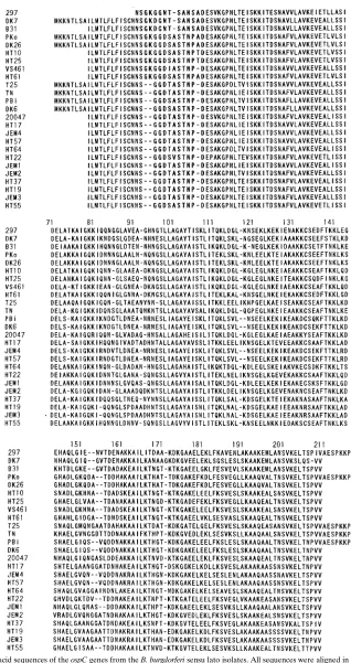
(data not shown). Figure 1 shows the predicted amino acid
sequences of the Japanese isolates, aligned and compared with
the previously published OspC sequences. Sequence
compar-ison of the OspC proteins from the 25 isolates revealed
signif-icant variability of OspC. Recently, Wilske et al. (34) have
studied OspC polymorphism and found the species-specific
motif of the OspC protein sequence at the N-terminal end
(from amino acid positions 23 to 35). The B. afzelii-specific
sequence motif was well conserved in all Japanese RFLP
ri-botype group III isolates, HT10, HT25, and HT61. One gap at
amino acid position 34 was observed in the type strain of B.
afzelii, strain VS461. In comparison with B. afzelii sequences, B.
garinii sequences have two gaps (two amino acid residues at
positions 23 and 24 and one amino acid residue at position 34)
and were significantly different from those of B. afzelii. This B.
[image:2.612.57.554.83.285.2]garinii-specific motif was highly conserved among B. garinii
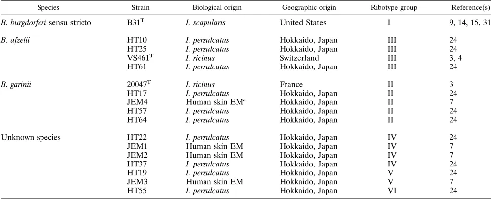
TABLE 1. B. burgdorferi sensu lato strains characterized by outer surface protein gene sequence analysisSpecies Strain Biological origin Geographic origin Ribotype group Reference(s)
B. burgdorferi sensu stricto B31T I. scapularis United States I 9, 14, 15, 31
B. afzelii HT10 I. persulcatus Hokkaido, Japan III 24
HT25 I. persulcatus Hokkaido, Japan III 24
VS461T I. ricinus Switzerland III 3, 4
HT61 I. persulcatus Hokkaido, Japan III 24
B. garinii 20047T I. ricinus France II 3
HT17 I. persulcatus Hokkaido, Japan II 24
JEM4 Human skin EMa Hokkaido, Japan II 7
HT57 I. persulcatus Hokkaido, Japan II 24
HT64 I. persulcatus Hokkaido, Japan II 24
Unknown species HT22 I. persulcatus Hokkaido, Japan IV 24
JEM1 Human skin EM Hokkaido, Japan IV 7
JEM2 Human skin EM Hokkaido, Japan IV 7
HT37 I. persulcatus Hokkaido, Japan IV 24
HT19 I. persulcatus Hokkaido, Japan V 24
JEM3 Human skin EM Hokkaido, Japan V 7
HT55 I. persulcatus Hokkaido, Japan VI 24
aEM, erythema migrans.
on May 15, 2020 by guest
http://jcm.asm.org/
FIG. 1. Deduced amino acid sequences of the ospC genes from the B. burgdorferi sensu lato isolates. All sequences were aligned in comparison with the previously published OspC sequences. The amino acids corresponding to the PCR primers are not shown. Dashes indicates gaps. 297, DK7, and B31T, B. burgdorferi sensu stricto;
PKo, DK26, HT10, HT25, VS461Tand HT61, B. afzelii; T25, TN, PBi, DK6, 20047T, HT17, JEM4, HT57, and HT64, B. garinii; HT22, JEM1, JEM2, HT37, HT19,
JEM3, and HT55, unknown species.
on May 15, 2020 by guest
http://jcm.asm.org/
TABLE 2. Amino acid similarity between OspC sequences from B. burgdorferi sensu lato isolates a Isolate % Amino acid similarity with OspC sequence from isolate: DK7 B31 T PKo DK26 HT10 HT25 VS461 T HT61 T25 TN PBi DK6 20047 T HT17 JEM4 HT57 HT64 HT22 JEM1 JEM2 HT37 HT19 JEM3 HT55 297 68.1 70.7 66.0 62.3 64.4 62.8 62.3 65.4 62.3 58.1 61.8 58.6 60.7 54.5 58.1 59.2 56.0 59.2 64.9 56.5 59.2 58.1 58.1 61.3 DK7 72.8 64.0 64.0 62.2 61.2 66.5 62.9 60.1 62.6 70.0 71.5 64.4 53.8 66.7 67.7 56.9 56.9 66.0 60.2 62.6 59.0 59.0 62.4 B31 T 69.7 69.7 72.8 70.8 72.7 71.3 64.1 64.1 64.6 65.1 63.4 58.5 64.6 65.1 65.1 65.6 68.6 62.1 64.1 58.5 58.5 68.6 PKo 100.0 77.0 79.6 70.1 78.2 69.7 68.2 69.6 69.0 64.9 60.4 65.6 66.1 67.7 72.8 73.2 66.3 66.7 65.6 65.6 75.8 DK26 77.0 79.6 70.1 78.2 68.3 66.8 66.8 69.0 64.9 60.4 65.6 66.1 67.7 72.8 73.2 66.3 66.7 65.6 65.6 75.8 HT10 85.2 91.8 81.1 71.4 66.3 64.8 65.3 73.2 63.3 63.0 64.1 72.8 71.8 70.1 67.9 66.7 67.2 67.2 71.1 HT25 77.8 76.5 67.3 67.9 64.3 63.8 71.6 56.6 60.9 62.0 70.3 68.2 69.6 63.8 65.1 64.1 64.1 74.7 VS461 T 76.8 72.2 70.1 68.6 69.1 74.2 67.0 67.7 68.8 72.7 69.1 71.1 71.1 70.1 69.6 69.6 69.1 HT61 65.5 65.0 64.0 64.5 66.5 63.5 63.5 64.1 69.2 72.3 73.7 65.8 68.2 65.6 65.6 77.8 T25 68.7 71.0 70.5 74.2 73.6 67.7 68.2 72.8 71.8 69.6 76.0 74.9 72.8 72.8 67.5 TN 68.6 68.0 71.1 69.5 67.2 68.2 69.7 62.1 73.2 77.0 71.3 70.8 70.8 70.6 PBi 99.5 69.1 64.5 88.5 89.6 65.1 65.6 71.1 68.4 67.7 69.7 69.7 69.1 DK6 69.6 64.5 89.1 90.1 65.1 65.6 71.1 68.9 67.7 69.7 69.7 69.6 20047 T 68.0 64.6 65.6 75.3 64.9 71.6 71.6 71.1 74.7 74.7 69.6 HT17 68.2 69.3 64.6 67.7 66.0 74.5 73.3 79.5 79.5 63.4 JEM4 99.0 65.1 64.6 73.4 66.7 68.2 71.9 71.9 67.2 HT57 65.6 65.1 74.0 67.7 68.8 72.9 72.9 67.7 HT64 63.6 73.2 65.6 69.7 69.7 69.7 68.6 HT22 72.2 71.8 71.3 67.7 67.7 74.7 JEM1 68.6 71.6 69.6 69.6 75.8 JEM2 73.8 75.9 75.9 68.0 HT37 76.4 76.4 71.1 HT19 100.0 67.0 JEM3 67.0 aStrains: 297, DK7, and B31 T, B. burgdorferi sensu stricto; PKo, DK26, HT10, HT25, VS461 T, and HT61, B. afzelii ; T25, TN, PBi, DK6, 20047 T, HT17, JEM4, HT57, and HT64, B. garinii ; HT22, JEM1, JEM2, HT37, HT19, JEM3 and HT55, unknown species.
on May 15, 2020 by guest
http://jcm.asm.org/
isolates from Japan and also in the representatives of RFLP
ribotype groups IV, V, and VI.
A sequence similarity matrix based on OspC amino acid
sequences was constructed, and representative results are
shown in Table 2. The similarity matrix showed the high
vari-ability of the OspC protein, and the degree of sequence
simi-larity ranged from 53.8 to 100% for all of the isolates
exam-ined. The lowest degree of similarity is shown between
Japanese HT17 and Danish DK7, and lower similarity values
were observed for B. garinii isolates when compared with B.
burgdorferi sensu stricto strains. A great OspC variability may
suggest that the OspC protein plays an important role in
eva-sion of the immune system of the vertebrate reservoir host.
Sequences for PKo and DK26 were identical, as were those for
JEM3 and HT19, and the sequence of PBi differs by only one
amino acid residue from the DK6 sequence. Two amino acid
substitutions between the JEM4 and the HT57 sequences were
observed. Only 10 amino acid substitutions and two gaps
oc-curred between the type strain VS461 sequence and that of the
Japanese HT10 isolate, although the two isolates had different
geographical origins. The higher degree of conservation of
OspC sequences within the isolates (even in the strains isolated
from different geographic regions) suggests that immune
se-lection of OspC might be occurring in the vertebrate host.
A phylogenetic tree derived from the similarity matrix was
constructed and is shown in Fig. 2. B. garinii and B. afzelii
isolates are deeply branched from B. burgdorferi sensu stricto
B31, DK7, and 297. When a bootstrap analysis of the 25
aligned sequences was performed, the clustering of respective
B. garinii and B. afzelii isolates occurred in 905 of 1,000 trees.
Members of each of the species B. garinii and B. afzelii formed
a phylogenetic cluster, and the tree showed that this cluster is
divided into several subgroups. One of the subgroups is
com-posed of the HT55 isolate and B. afzelii HT61. The other
subgroups are closely related with their species. The
phyloge-netic tree revealed that the Japanese isolates classified as
members of ribotype groups IV, V, and VI are all included in
these subgroups. The phylogenetic tree also indicated that B.
garinii isolates have a wide variety of OspC protein sequences.
A significant diversity has been observed by using monoclonal
antibodies against OspC, and there is a greater heterogeneity
in OspC sequences of B. garinii isolates (14, 31, 36). In a
previously published report (31), three OspC sequences were
in complete agreement with the sequences of the three
geno-species of B. burgdorferi sensu lato. The same phylogenetic
analysis was carried out on the basis of ospC nucleotide
se-quences. In the case of DNA sequences used instead of the
deduced amino acid sequences, the similarity matrix showed a
greater degree of sequence similarity, ranging from 65.6 to
100%. A phylogenetic tree derived from the similarity matrix,
however, yielded results quite similar to those obtained with
the tree constructed using deduced amino acid sequences.
Lyme disease is endemic in the temperate latitudes of North
America, Europe, and Asia (13, 33). The etiological agent of
Lyme disease was first isolated in the United States and named
B. burgdorferi (15). Representatives of three species were
fur-ther recognized among strains isolated from patients, rodents,
and ticks. The North American isolates were thought to be
closely related (3, 29). In contrast, considerable divergence in
genetic and immunological properties among the European
isolates was observed (32, 35). Lyme disease spirochetes
ob-tained in Japan also have varied genetic and immunological
determinants (8, 10, 26, 27). We have employed PCR
amplifi-cation based on the species-specific sequence of the 16S rRNA
gene to identify borrelial isolates to the species level according
to recently established nomenclature (18, 28). Most of the
Japanese isolates amplified the appropriately sized fragment,
when the species-specific primer sets were used. However,
some of the isolates of ribotype groups IV, V, and VI were not
amplified by any of the 16S rRNA gene-specific primers (data
not shown). Experiments using DNA-DNA hybridization
be-tween the representatives of B. garinii and a few of the isolates
belonging to ribotype group IV gave borderline results, and the
DNA relatedness was 65 to 85% with the membrane filter
hybridization method (unpublished results).
There are some differences between the spirochetes isolated
from Europe and those isolated from the northern regions of
Japan. Nevertheless, alignment of the OspC sequences of
Jap-anese isolates revealed that the species-specific domain was
conserved in all of the isolates of ribotype groups IV, V, and
VI. Despite differences in some traits, it is likely that the
spirochetes of unknown species diverged within the species
level of B. garinii.
ACKNOWLEDGMENTS
We deeply thank Guy Baranton of the Institut Pasteur for his helpful discussion and comments. This work would not have been possible without the help, supplied strains, and stimulating discussion of Mi-noru Nakao and Kenji Miyamoto of the Asahikawa Medical College. We also thank Osamu Matsushita of Kagawa Medical College for his help with computer work to construct the phylogenetic tree and Ya-suyo Koreki for her technical help.
This work was supported in part by a Grant-in-Aid for Scientific Research (nos. 06044191 and 06640912) from the Ministry of Educa-tion, Science, and Culture of Japan.
REFERENCES
1. Adam, T., G. S. Gassman, C. Rasiah, and U. B. Go¨bel.1991. Phenotypic and genotypic analysis of B. burgdorferi isolates from various sources. Infect. Immun. 59:2579–2585.
[image:5.612.58.296.73.305.2]2. Anderson, J. F. 1989. Epizootiology of Borrelia in Ixodes tick vectors and reservoir hosts. Rev. Infect. Dis. 11(Suppl. 6):s1451–s1459.
FIG. 2. Phylogenetic tree based on OspC protein sequences for B. burgdorferi sensu lato isolates. 905/1,000 represents the estimated confidence levels, as de-termined by a bootstrap analysis. Bar, 10% differences between sequences, as determined by measuring the lengths of the horizontal lines connecting two isolates.
on May 15, 2020 by guest
http://jcm.asm.org/
3. Baranton, G., D. Postic, I. S. Girons, P. Boerlin, J. C. Piffaretti, M. Assous,
and P. A. D. Grimont.1992. Delineation of Borrelia burgdorferi sensu stricto,
Borrelia garinii sp. nov., and group VS461 associated with Lyme borreliosis.
Int. J. Syst. Bacteriol. 42:378–383.
4. Canica, M. M., F. Nato, L. du Merle, J. C. Mazie, G. Baranton, and D.
Postic.1993. Monoclonal antibodies for identification of Borrelia afzelii sp.
nov. associated with late cutaneous manifestations of Lyme borreliosis. Scand. J. Infect. Dis. 25:441–448.
5. Fuchs, R., S. Jauris, F. Lottspeich, V. Preac-Mursic, B. Wilske, and E.
Soutschek.1992. Molecular analysis and expression of a Borrelia burgdorferi
gene encoding a 22kDa protein (pC) in Escherichia coli. Mol. Microbiol.
6:503–509.
6. Fukunaga, M., and I. Mifuchi. 1989. Unique organization of Leptospira
interrogans rRNA genes. J. Bacteriol. 171:5763–5767.
7. Fukunaga, M., M. Sohnaka, M. Nakao, and K. Miyamoto. 1993. Evaluation of genetic divergence of borrelial isolates from Lyme disease patients in Hokkaido, Japan, by rRNA gene probes. J. Clin. Microbiol. 31:2044–2048. 8. Fukunaga, M., M. Sohnaka, Y. Takahashi, M. Nakao, and K. Miyamoto. 1993. Antigenic and genetic characterization of Borrelia species isolated from
Ixodes persulcatus in Hokkaido, Japan. J. Clin. Microbiol. 31:1388–1391.
9. Fukunaga, M., M. Sohnaka, and Y. Yanagihara. 1993. Analysis of Borrelia species associated with Lyme disease by rRNA gene restriction fragment length polymorphism. J. Gen. Microbiol. 139:1141–1146.
10. Fukunaga, M., and Y. Takahashi. 1994. Pulsed field gene electrophoresis analysis of Borrelia burgdorferi sensu lato isolated in Japan and taxonomic implications with Lyme disease spirochetes. Microbiol. Immunol. 38:747– 751.
11. Fukunaga, M., Y. Yanagihara, and M. Sohnaka. 1992. The 23S/5S ribosomal RNA genes (rrl/rrf) are separate from the 16S ribosomal RNA gene (rrs) in
Borrelia burgdorferi, the aetiological agent of Lyme disease. J. Gen.
Micro-biol. 138:871–877.
12. Higgines, D. G., A. J. Bleasby, and R. Fuchs. 1992. CLUSTAL V: improved software for multiple sequence alignment. Comput. Appl. Biosci. 8:189–191. 13. Jaenson, T. G. T. 1991. The epidemiology of Lyme borreliosis. Parasitol.
Today 7:39–45.
14. Jauris-Heipke, S., R. Fuchs, M. Motz, V. Preac-Mursic, E. Schwab, E.
Soutschek, G. Will, and B. Wilske.1993. Genetic heterogeneity of the genes
coding for the outer surface protein C (OspC) and the flagellin of Borrelia
burgdorferi. Med. Microbiol. Immunol. 182:37–50.
15. Johnson, R. C., F. W. Hyde, G. P. Schmid, and D. J. Brenner. 1984. Borrelia
burgdorferi sp. nov.: etiological agent of Lyme disease. Int. J. Syst. Bacteriol.
34:496–497.
16. Kubo, N., Y. Arashima, M. Yoshida, M. Kawabata, S. Nishinarita, T.
Hayata, S. Sawada, T. Horie, M. Nakao, K. Miyamoto, and K. Kawano.1992.
Questionnaire surveys of cases of tick bite and Lyme borreliosis in hunters in Hokkaido with reference to detection of anti-Borrelia burgdorferi antibody. Intern. Med. 31:1163–1168.
17. Lane, R. S., J. Piesman, and W. Burgdorfer. 1991. Lyme borreliosis: relation of its causative agent to its vectors and hosts in North America and Europe. Annu. Rev. Entomol. 36:587–609.
18. Marconi, R. T., and C. F. Garon. 1992. Development of polymerase chain reaction primer sets for diagnosis of Lyme disease and for species-specific identification of Lyme disease isolates by 16S rRNA signature nucleotide analysis. J. Clin. Microbiol. 30:2830–2834.
19. Marconi, R. T., D. S. Samuels, and C. F. Garon. 1993. Transcriptional analyses and mapping of the ospC gene in Lyme disease spirochetes. J. Bacteriol. 175:926–932.
20. Marconi, R. T., D. S. Samuels, T. G. Schwan, and C. F. Garon. 1993.
Identification of a protein in several Borrelia species which is related to OspC of the Lyme disease spirochetes. J. Clin. Microbiol. 31:2577–2583. 21. Matuschka, F. R., O. Fischer, M. Heiler, D. Richter, and A. Spielman. 1992.
Capacity of European animals as reservoir hosts for the Lyme disease spi-rochete. J. Infect. Dis. 165:479–483.
22. Miyamoto, K., M. Nakao, N. Sato, and M. Mori. 1991. Isolation of Lyme disease spirochetes from an ixodid tick in Hokkaido, Japan. Acta Trop.
49:65–68.
23. Nakao, M., and K. Miyamoto. 1993. Reservoir competence of the wood mouse, Apodemus speciosus ainu, for the Lyme disease spirochete, Borrelia
burgdorferi, in Hokkaido, Japan. Jpn. J. Sanit. Zool. 44:69–84.
24. Nakao, M., K. Miyamoto, and M. Fukunaga. 1994. Lyme disease spirochetes in Japan: enzootic transmission cycles in birds, rodents, and Ixodes
persulca-tus ticks. J. Infect. Dis. 170:878–882.
25. Nakao, M., K. Miyamoto, M. Fukunaga, Y. Hashimoto, and H. Takahashi. 1994. Comparative studies on Borrelia afzelii isolated from a patient of Lyme disease, Ixodes persulcatus ticks, and Apodemus speciosus rodents in Japan. Microbiol. Immunol. 38:413–420.
26. Nakao, M., K. Miyamoto, N. Kawaguchi, Y. Hashimoto, and H. Iizuka. 1992. Comparison of Borrelia burgdorferi isolated from humans and ixodid ticks in Hokkaido, Japan. Microbiol. Immunol. 36:1189–1193.
27. Nakao, M., K. Miyamoto, K. Uchikawa, and H. Fujita. 1992. Characteriza-tion of Borrelia burgdorferi isolated from Ixodes persulcatus and Ixodes ovatus ticks in Japan. Am. J. Trop. Med. Hyg. 47:505–511.
28. Park, K. H., W. H. Chang, and T. G. Schwan. 1993. Identification and characterization of Lyme disease spirochetes, Borrelia burgdorferi sensu lato, isolated in Korea. J. Clin. Microbiol. 31:1831–1837.
29. Postic, D., M. V. Assous, P. A. D. Grimont, and G. Baranton. 1994. Diversity of Borrelia burgdorferi sensu lato evidenced by restriction fragment length polymorphism of rrf (5S)-rrl (23S) intergenic spacer amplicons. Int. J. Syst. Bacteriol. 44:743–752.
30. Sˇadzˇiene, A., B. Wilske, M. S. Ferdows, and A. G. Barbour. 1993. The cryptic
ospC gene of Borrelia burgdorferi B31 is located on a circular plasmid. Infect.
Immun. 61:2192–2195.
31. Theisen, M., B. Frederiksen, A. M. Lebech, J. Vuust, and K. Hansen. 1993. Polymorphism in ospC gene of Borrelia burgdorferi and immunoreactivity of OspC protein: implications for taxonomy and for use of OspC protein as a diagnostic antigen. J. Clin. Microbiol. 31:2570–2576.
32. Wallich, R., C. Helmes, U. E. Schaible, Y. Lobet, S. E. Moter, M. D. Kramer,
and M. M. Simon.1992. Evaluation of genetic divergence among Borrelia
burgdorferi isolates by use of OspA, fla, HSP60, and HSP70 gene probes.
Infect. Immun. 60:4856–4866.
33. Weber, K., and H. W. Pfister. 1992. History of Lyme borreliosis in Europe, p. 1–20. In K. Weber and W. Burgdorfer (ed.), Aspects of Lyme borreliosis. Springer-Verlag, Berlin.
34. Wilske, B., S. Jauris-Heipke, R. Lobentanzer, I. Pradel, V. Preac-Mursic, D.
Ro¨ssler, E. Soutschek, and R. C. Johnson.1995. Phenotypic analysis of outer
surface protein C (OspC) of Borrelia burgdorferi sensu lato by monoclonal antibodies: relationship to genospecies and OspA serotype. J. Clin. Micro-biol. 33:103–109.
35. Wilske, B., V. Preac-Mursic, U. B. Go¨bel, B. Graf, S. Jauris, E. Soutschek, E.
Schwab, and G. Zumstein.1993. An OspA serotyping system for Borrelia
burgdorferi based on reactivity with monoclonal antibodies and OspA
se-quence analysis. J. Clin. Microbiol. 31:340–350.
36. Wilske, B., V. Preac-Mursic, S. Jauris, A. Hofmann, I. Pradel, E. Soutschek,
E. Schwab, G. Will, and G. Wanner.1993. Immunological and molecular
polymorphism of OspC, an immunodominant major outer surface protein of
Borrelia burgdorferi. Infect. Immun. 61:2182–2191.