S T U D Y P R O T O C O L
Open Access
Gait training assisted by multi-channel
functional electrical stimulation early after
stroke: study protocol for a randomized
controlled trial
Maijke van Bloemendaal
1,2*, Sicco A. Bus
2, Charlotte E. de Boer
1, Frans Nollet
2, Alexander C. H. Geurts
3and Anita Beelen
1,2Abstract
Background:Many stroke survivors suffer from paresis of lower limb muscles, resulting in compensatory gait patterns characterised by asymmetries in spatial and temporal parameters and reduced walking capacity. Functional electrical stimulation has been used to improve walking capacity, but evidence is mostly limited to the orthotic effects of peroneal functional electrical stimulation in the chronic phase after stroke. The aim of this study is to investigate the therapeutic effects of up to 10 weeks of multi-channel functional electrical stimulation (MFES)-assisted gait training on the restoration of spatiotemporal gait symmetry and walking capacity in subacute stroke patients.
Methods:In a proof-of-principle study with a randomised controlled design, 40 adult patients with walking deficits who are admitted for inpatient rehabilitation within 31 days since the onset of stroke are randomised to either MFES-assisted gait training or conventional gait training. Gait training is delivered in 30-minute sessions each workday for up to 10 weeks. The step length symmetry ratio is the primary outcome. Blinded assessors conduct outcome assessments at baseline, every 2 weeks during the intervention period, immediately post intervention and at 3-month follow-up.
Discussion: This study aims to provide preliminary evidence for the feasibility and effectiveness of MFES-assisted gait rehabilitation early after stroke. Results will inform the design of a larger multi-centre trial. Trial registration: This trial is registered at the Netherlands Trial Register (number NTR4762, registered 28 August 2014) Keywords:Stroke, Lower Limb, Functional Electrical Stimulation, Gait, Training, Spatiotemporal Parameters
Background
Regaining independent gait is considered one of the pri-mary goals in stroke rehabilitation [1–3]. In the early phase after stroke, the musculature of the affected side is often paretic or even paralytic. As a consequence, com-pensatory gait patterns characterised by asymmetries in spatial and temporal parameters may arise that tend to be persistent, even in patients who show substantial res-toration of paretic leg motor control, perhaps due to
mechanisms related to‘learned non-use’as has been de-scribed for the upper extremity [4]. These compensatory gait patterns are less energy-efficient and may negatively affect balance control leading to an increased risk of falls and injury as well as to limitations in functional mobility [5–8]. Furthermore, they may cause secondary complica-tions, such as muscle shortening and joint deformation [6]. Restoration of gait symmetry can be accomplished by motor relearning and neuroplasticity, for which highly in-tensive, repetitive and task-specific training is essential in the early rehabilitation phase after stroke [9, 10]. The use of functional electrical stimulation (FES) timed to the gait cycle in the early phase after stroke may improve gait sym-metry by enhancing neuroplasticity, preventing secondary
* Correspondence:[email protected]
1Merem Rehabilitation Centre De Trappenberg, Huizen, The Netherlands 2Department of Rehabilitation, Academic Medical Centre, Amsterdam, The
Netherlands
Full list of author information is available at the end of the article
complications, and by supporting the acquisition of an ad-equate compensatory strategy. Although the orthotic ef-fects of peroneal FES (PFES) have been established, the therapeutic effect of PFES in the subacute phase has been scarcely investigated [11–19]. Furthermore, PFES assists the ankle dorsiflexion movement only during the swing phase and early stance phase of gait and does not support the more proximal movements of the lower limb. Several studies have shown that strength and range of motion of the knee flexors and extensors are associated with gait performance [20–22]. Thus, multi-channel FES (MFES) of the distal and proximal parts of the lower limb may be more effective in normalising the gait pattern by compen-sating for thigh and dorsiflexor muscle weakness. There is preliminary evidence of a positive therapeutic effect of MFES in early stroke rehabilitation on balance control and mobility [23–25]. However, it remains unclear whether MFES is effective for the restoration of gait sym-metry. Furthermore, it remains unclear whether it is feas-ible to implement MFES in functional gait training including pre-gait activities. Due to the limited evidence of MFES-assisted gait training during early stroke rehabili-tation we designed a proof-of-principle study. The aim of this study is to examine the feasibility and preliminary efficacy of MFES-assisted gait training on gait symmetry and walking capacity in patients in the subacute phase after stroke during their inpatient rehabilitation. We hypothesise that MFES-assisted gait training for maximally 10 weeks in the early phase after stroke is feasible and improves the step length symmetry compared to conventional gait training. In this paper we describe the protocol of our study according to the SPIRIT guidelines (Additional file 1).
Methods
Design
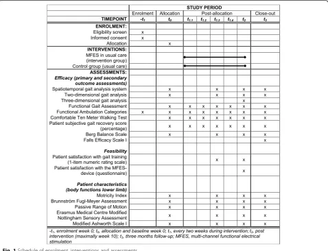
A prospective, assessor-blinded, single-centre, proof-of-principle study with a randomised controlled two-armed parallel design is being conducted. Forty participants with gait impairments in the subacute phase after stroke who are referred for inpatient rehabilitation are randomly assigned in a 1:1 ratio to either an intervention group, receiving MFES-assisted gait training, or a control group, receiving gait training as usual. The intervention period lasts 10 weeks or until discharge from inpatient rehabilita-tion, whichever is sooner. Outcomes will be assessed every 2 weeks during the 10-week intervention period as well as after a 3-month follow-up period (Fig. 1).
Ethics
The study protocol has been approved by the Medical Ethics Committee (MEC) of the Academic Medical Centre Amsterdam (protocol number NL50002.018.14). Any changes to the study protocol or study procedures will be reviewed and approved by the MEC and
communicated to relevant parties. A Dutch rehabilita-tion centre (Merem Rehabilitarehabilita-tion Centre De Trappen-berg in Huizen) granted approval to include and train participants. The study has been registered at the Netherlands Trial Register (number NTR4762, registered 28 August 2014). Additional file 2 provides an overview of the trial registration data.
Participants
Participants are recruited at the rehabilitation centre. All stroke survivors admitted for inpatient rehabilitation are screened for eligibility by their physiatrist. Inclusion teria are: (1) a clinical diagnosis of stroke (diagnostic cri-teria according to the World Health Organization definition) [26]; (2) within 31 days since stroke onset; (3) age between 18 and 80 years; (4) indication for gait training (according to the treating physiatrist); (5) suffi-cient capacity to stand between parallel bars with or without physical assistance and able to walk with aids and physical assistance from one physical therapist (Functional Ambulation Categories [FAC] score≥1); and (6) passive range of motion (PROM) upon ankle dorsi-flexion ≥0° with full knee extension. Exclusion criteria are: (1) subarachnoid haemorrhage or stroke in the cere-bellum or brain stem; (2) severe spasticity of the knee or ankle flexors or extensors (i.e., Modified Ashworth Scale [MAS]≥3); (3) pre-existing lower limb deficits or any other medical co-morbidities that might significantly interfere with gait (indicated by a self-reported max-imum walking distance <300 meter or walking duration <6 minutes walking pre stroke); (4) severe cognitive problems or aphasia leading to severely impaired com-prehension of test instructions; (5) medical conditions that might lead to inability to comply with the study protocol (e.g., congestive heart failure, chemotherapy, uncontrolled epilepsy, pregnancy, depression or psych-otic disorder, etc.); (6) demand-type cardiac pacemaker, defibrillator or electrical implant; (7) metallic implant at the affected lower limb; or (8) present or suspected can-cerous lesion at the affected lower limb. Potentially eli-gible participants receive verbal and detailed written information (see Additional file 3) about the study and are invited to participate. In case of willingness to par-ticipate, an intake assessment is performed by a re-searcher who explains the purpose and procedures of the study and asks for informed consent. The following demographics are recorded for each participant: gender, date of birth, body length, body mass, type of stroke, lo-cation of stroke (left, right or both), hemiplegic side (left or right), date of stroke, neglect (tactile and visual present or not), relevant co-morbidities, medication and FAC. Furthermore, the following sensorimotor character-istics of both lower limbs are recorded for each partici-pant: Motricity Index (muscle strength) [27], Brunnström
Fugl-Meyer Assessment (motor selectivity) [28], and spe-cific parts of the Erasmus Medical Centre Modified Not-tingham Sensory Assessment (tactile and proprioceptive sensation) [29], MAS (muscle tone) [30, 31] and passive range of motion at the hip, knee and ankle (PROM). Strat-egies for patient retention include sending newsletters, ac-commodating their schedules when planning follow-up visits, sending reminders of upcoming visits, and provid-ing transport support.
Randomisation and blinding
Concealed randomisation and allocation is effectuated by an assigned researcher (AB), who is not involved in any patient contact, using a computerised randomisa-tion system. Randomisarandomisa-tion takes place stratified by functional walking capacity (dependent gait [FAC 1–2] versus independent gait [FAC 3–5]). Outcome asses-sors are kept blinded to allocation of the participants during all assessments. Participants are instructed not to reveal their group allocation or therapy content to the assessors. Data will be analysed by an independent
statistician. Randomisation will be concealed to the primary researcher until data analysis has been completed.
Interventions Control group
Participants in the control group will receive regular gait training by a physical therapist and/or movement therapist depending on their needs. Typically, per week, three to eight 30-minute sessions of gait-oriented physical therapy are given on five working days for 6 to 12 weeks. This ‘usual care’ may include individual gait training, gait training in groups, fitness training, sports, and hydrotherapy. Walking aids, orthoses, orthopaedic shoes and medication may all be used, but not lower limb FES. Participants will not be restricted in their activities. Therapists are instructed to document characteristics of the gait training (duration, frequency and content) for each participant in weekly logs.
[image:3.595.59.538.88.454.2]Intervention group
Participants in the intervention group receive the same amount of gait-oriented physical therapy, but gait train-ing is assisted by MFES. Per week, MFES is delivered during one 30-minute session on five working days up to 10 weeks. Physical therapists and movement thera-pists specifically trained in the use of MFES carry out the gait training. They are instructed to document char-acteristics of the gait training (duration, frequency, con-tent and intensity of MFES) for each participant in weekly logs. During an initial adaptation period of 4 days, the duration of MFES is gradually increased from 15 mi-nutes (day 1) to 30 mimi-nutes (day 4). Thereafter, partici-pants receive 30-minute session of MFES-assisted gait training on each workday.
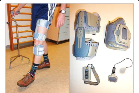
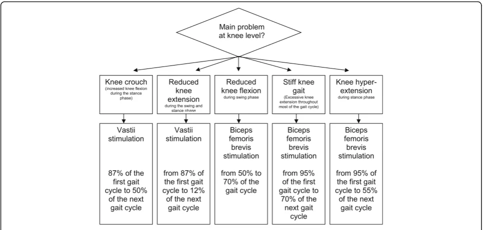
Multi-channel functional electrical stimulation device The MFES device used in this study (NESS L300™Plus, Bioness, Valencia, CA, USA; CE 0473) delivers electrical pulses during gait to muscles in the affected leg to pro-mote ankle dorsiflexion in combination with knee flexion or extension. The device consists of two cuffs (lower leg and thigh), a foot switch, and a wireless con-trol unit that activates the system by radio frequency sig-nals (Fig. 2). In each cuff two cotton electrodes and a stimulation unit are embedded. The electrodes of the lower leg cuff are located over the common peroneal nerve and the tibialis anterior muscle to elicit ankle dorsiflexion. The electrodes of the thigh cuff are posi-tioned over the vastus medialis muscle to promote knee extension or over the biceps femoris brevis muscle to promote knee flexion. With this configuration, either paretic muscles can be stimulated or spastic muscles antagonised. Figure 3 illustrates some examples of posi-tioning of the thigh cuff and timing of the upper leg stimulation expressed as percentage of the gait cycle [32]. Authorised clinicians are specially trained to fit and
set the MFES device. They fit the device at baseline and evaluate the settings of the device every two-and-a-half weeks. A force-sensitive resistor in the foot switch de-tects the force under the foot. A dynamic gait-tracking algorithm is used to detect whether the foot is on the ground (e.g., initial contact) or in the air (e.g., heel off ) by analysing the foot pressure. Average stance and swing phases are calculated by the system and data is transmit-ted by radio signals to the stimulation unit allowing for the synchronisation of the stimulation in accordance with the timing of gait events (gait mode). During the fitting process, the clinician sets the stimulation parame-ters (intensity level, phase duration, pulse rate, waveform and maximum duration of stimulation, ramp up, exten-sion and ramp down) for the gait mode with a hand-held computer (personal digital assistant; PDA). The peroneal stimulation starts at‘heel off’and terminates at ‘heel contact’. Stimulation can be extended beyond heel contact to control the first rocker. The thigh stimulation – biceps femoris brevis or vastus muscles – can start and end once or twice at any segment in the gait cycle, which is determined by the clinician. Participants who cannot walk without personal assistance receive MFES treatment in the NESS L300™ Plus clinician mode (pre-gait and balance training) and (pre-gait mode ((pre-gait training) during individual physical therapy. The clinician mode is used to manually start and stop stimulation in the thigh and lower leg unit simultaneously. The clinician mode uses the stimulation parameters set for gait mode.
Outcome measures Primary outcome
The primary outcome to determine efficacy of MFES-assisted gait training is the step length symmetry ratio. Step length during comfortable gait is measured with a spatiotemporal gait analysis system (SGAS) using a lat-erally placed camera (Panasonic HC-V550 High Defin-ition camera 50 Hertz; Panasonic, Osaka, Japan) and discrete linear transform matrix software designed for this study. Participants walk at comfortable walking speed along a 10-metre walkway until three valid gait trials are collected in which each foot lands within the 1300-millimetre-wide video frame for both sides. The primary condition is walking without shoes and orthosis with minimal use of walking aids. The symmetry ratio is calculated as the difference between the step length of the affected and non-affected leg divided by the mean of the step length of both legs.
Secondary outcomes
The SGAS is also used to examine other spatiotemporal parameters (step length, stride length, cadence, stance time symmetry ratio, double support time, and swing/ stance time symmetry ratio) for two conditions (walking
Fig. 2The functional electrical stimulation device including two cuffs, a foot switch, and a control unit
[image:4.595.57.290.546.702.2]with and without shoes and orthoses). Furthermore, sa-gittal and frontal plane video (Basler Scout GigE; Basler AG, Ahrensburg, Germany), electromyography (Mobita and Porti 7 8bt; TMSi, Oldenzaal, the Netherlands) and force plate recordings (OR6-7; AMTI, Watertown, MA, USA) are used to collect kinematic, electromyographic and kinetic data, respectively. One valid gait trial is col-lected for different conditions (walking with and without shoes, orthoses and walking aids). In addition, at the end of the intervention period, a full three-dimensional gait analysis is performed with an 8-camera VICON MX1.3 motion capture system operating at a sample rate of 100 Hertz (VICON, Oxford, United Kingdom) with two force plates in series recording at 1000 Hertz (OR6-7; AMTI, Watertown, MA, USA) positioned along a 12-metre walkway. Three valid gait trials are collected to register gait width and other kinematic and kinetic pa-rameters that cannot be determined with the SGAS. Walking capacity is assessed with the Functional Gait Assessment (FGA), the FAC and the 10-Meter Walk Test (10MWT), all validated measurement instruments in the stroke population [33]. The FGA is a 10-item test to assess functional gait activities. The FAC is an instru-ment for categorising gait (in)dependency from ‘no ability to walk or with the help of two or more persons’(FAC 0) to the‘ability to walk independently’(FAC 5). The 10MWT assesses comfortable and maximum walking speed. In this study, only comfortable walking speed will be recorded. Walking capacity is also assessed by a subjective walking capacity recovery score. During each visit the participant is asked to score his or her recovery of walking capacity since the onset of stroke by giving a percentage between 0 % (‘no
recovery’) and 100 % (‘full recovery’). Balance control is assessed with the Berg Balance Scale [34–36] and fear of falling with the Falls Efficacy Scale I (FES-I) [37].
Feasibility
Feasibility of the intervention is evaluated on the basis of compliance with the MFES-assisted gait training and pa-tient satisfaction with this type of training using the MFES device. The following criteria are used: (1) MFES-assisted gait training took place during ≥75 % of all therapy ses-sions; and (2) patient satisfaction with MFES-assisted gait training was ≥7 on a numeric rating scale from 0 (‘most unsatisfied’) to 10 (‘most satisfied’) assessed at the end of the intervention period. Patient satisfaction with the MFES device is evaluated by a questionnaire designed for this study.
Sample size
Due to lack of data on effect size, sample size is based on the feasibility of recruitment in one centre with an ap-proximate yearly admission rate of 80 stroke survivors. Using an inclusion period of 3 years and estimating that 25 % of the patients are eligible and willing to participate, the sample size is set at 40 participants (20 in each group).
Data management and statistical analysis
Data entry takes place by digital and paper case report forms. Personal information of the participants is treated confidentially. Every participant receives an identification number. This number is used on all forms so that no names or other personal information have to be used. Data is saved in a locked cabinet in a locked office and
[image:5.595.56.540.87.317.2]stored digitally in a trial master file for the duration of 15 years. Data quality is guaranteed by random checks of the research database and range checks for data values.
Descriptive statistics
Patient characteristics will be described using means, standard deviations, medians, and interquartile ranges (dependent on whether data is normally distributed or not) and percentages. Group comparisons at baseline will be performed with Student’sttests, Mann–Whitney
Utests andχ2tests where appropriate.
Primary and secondary analysis
Primary efficacy analysis will be performed on an intention-to-treat basis. In addition, per protocol ana-lyses will be performed. A linear mixed model for re-peated measures will be used to analyse differences in the primary outcome and secondary outcomes. A squared time variable will be included to test for a curvi-linear recovery curve. The interaction of time by inter-vention (MFES versus control) assesses whether the slopes of the recovery curves differ between groups. In these analyses both the intercept and the time variable are included as random effects. Group comparisons at the end of the intervention period for the three-dimensional gait analysis parameters and FES-I will be performed with Student’st tests. To assess feasibility of the intervention, the proportion of participants in the intervention group who are compliant with the gait training and who scored ≥7 on the numeric rating scale will be determined. Patient satisfaction with the MFES device will be described. In all analyses, statistical uncer-tainty will be expressed by means of 95 % confidence in-tervals. Significance will be set atp< 0.05.
Monitoring and quality assurance
Internal monitoring of the conduct of the study is per-formed once a year by researchers of the Merem Re-habilitation Centre De Trappenberg and the Academic Medical Centre Amsterdam. The completeness, accur-acy, consistency, and procedures are checked according to the monitoring plan. Adverse events (AEs) of the in-dividual participants are reported in the period from signing informed consent (introduction meeting) until the last follow-up meeting. All AEs reported spontan-eously by the participant or observed by the primary re-searcher or staff are recorded. All AEs are followed until they have abated or a stable situation has been reached. Depending on the event, follow-up may require add-itional tests or medical procedures as indicated, and/or referral to the general physician or a medical specialist. Serious AEs (SAEs) are reported up to the end of study. The sponsor reports the SAEs to the MEC within 15 days after the sponsor has first knowledge of the SAE. SAEs
that result in death or appear to be life threatening are reported expedited, i.e. not later than 7 days after the primary researcher has obtained first knowledge of the adverse event. The primary researcher reports the pro-gress of the trial once a year to the MEC.
Dissemination policy
Trial results are communicated to participants, health-care professionals, the public, and other relevant groups via newsletters and (inter)national, peer-reviewed jour-nals (Medline database). The results will be presented at relevant (inter)national conferences in rehabilitation and neurology. Furthermore, results will be published on websites of patient societies.
Discussion
The aim of this study is to evaluate the therapeutic ef-fects of up to 10 weeks of daily MFES-assisted gait train-ing on spatiotemporal parameters, walktrain-ing capacity, and motor recovery early after stroke. We hypothesise that stroke survivors will benefit from the therapeutic effect of MFES-assisted gait training by larger improvements on spatiotemporal parameters compared to conventional gait training. These data will inform the design of a suffi-ciently powered (multi-centre) randomised controlled trial. The strength of our study is that we investigate the effects of MFES during functional gait activities. Two out of three studies investigating MFES in the early phase after stroke applied MFES with the patient in a supine position [24, 25]. Moreover, the stimulation pe-riods in the three studies regarding this topic were only 3–4 weeks [23–25]. There is no evidence for the mini-mum intensity of MFES required to enhance recovery of walking capacity in stroke survivors. Different treatment doses of electrical stimulation have been studied in the past from 15 minutes up to all day long and from once to more sessions a day. The three studies investigating MFES in the early phase after stroke applied MFES for 30–45 minutes and found positive effects on several out-comes [23–25]. In our study, MFES will be applied each workday for minimally 15 minutes to maximally 30 mi-nutes to aim for a feasible protocol in early stroke re-habilitation. Findings from this study will provide insight into the initial effects of MFES-assisted gait training on regaining gait symmetry and several other outcomes in early stroke rehabilitation. The collection of detailed data will generate new knowledge regarding early use of MFES to promote motor and gait recovery in the early phase after stroke. If this study confirms the feasibility and initial efficacy of MFES-assisted gait training, a lar-ger study would be warranted to further determine the effectiveness of this intervention.
Trial status
At the time of manuscript submission, the enrolment of participants was ongoing at Merem Rehabilitation Centre De Trappenberg, Huizen, the Netherlands.
Additional files
Additional file 1:SPIRIT checklist. (DOC 127 kb)
Additional file 2:Trial registration data. (DOCX 19 kb)
Additional file 3:Dutch information letter and informed consent. (PDF 284 kb)
Abbreviations
10MWT:10-Meter Walk Test; AEs: adverse events; FAC: Functional Ambulation Categories; FES: functional electrical stimulation; FES-I: Falls Efficacy Scale I; FGA: Functional Gait Assessment; MAS: Modified Ashworth Scale; MEC: Medical Ethics Committee; MFES: multi-channel functional electrical stimulation; PDA: personal digital assistant; PFES: peroneal functional electrical stimulation; PROM: passive range of motion; SAEs: serious adverse events; SGAS: spatiotemporal gait analysis system
Acknowledgements
The authors are grateful to Merem Rehabilitation Centre De Trappenberg for the support given to the study.
Funding
The purchase of the MFES devices was covered by a grant from the Dutch foundation‘Stichting GKZ’, Huizen. No further external funding has been provided.
Availability of data and materials
Not applicable.
Authors’contributions
MB conceived the idea for the study, contributed to the study design and the methodology of the study and was principally responsible for the drafting of the manuscript. SB contributed to the study design and the methodology of the study and helped to draft the manuscript. CB helped conceive the idea for the study, contributed to the study design and the methodology of the study and assisted in editing the final manuscript. FN contributed to the study design and the methodology of the study and assisted in editing the final manuscript. AG contributed to the study design and the methodology of the study and helped to draft the manuscript. AB conceived the idea for the study, contributed to the study design and the methodology of the study and was principally responsible for the drafting of the manuscript. All authors read and approved the final manuscript.
Authors’information
Maijke van Bloemendaal (MB): Physical therapist, clinical health scientist and PhD candidate.
Sicco Bus (SB): Human movement scientist, senior researcher and head human performance laboratory.
Charlotte E. de Boer (CB): Physiatrist neurorehabilitation.
Frans Nollet (FN): Physiatrist, chair department of Rehabilitation at the Academic Medical Centre Amsterdam.
Alexander C.H. Geurts (AG): Physiatrist neurorehabilitation, clinical epidemiologist, chair department of Rehabilitation at Radboud University Medical Centre.
Anita Beelen (AB): Human movement scientist, senior researcher department of Rehabilitation at the Academic Medical Centre Amsterdam and manager Research at Merem.
Competing interests
The authors declare that they have no competing interests.
Consent for publication
[image:7.595.304.537.302.738.2]Written informed consent was obtained from one person for publication of Fig. 2 in this manuscript. The consent form is held by the authors and is available for review by the Editor-in-Chief.
Ethics approval and consent to participate
The study protocol has been approved by the Medical Ethics Committee of the Academic Medical Centre Amsterdam (protocol number
NL50002.018.14). Patients sign informed consent before participation in the study.
Author details
1Merem Rehabilitation Centre De Trappenberg, Huizen, The Netherlands. 2
Department of Rehabilitation, Academic Medical Centre, Amsterdam, The Netherlands.3Department of Rehabilitation, Donders Centre for
Neuroscience, Radboud University Medical Centre, Nijmegen, The Netherlands.
Received: 1 June 2016 Accepted: 10 September 2016
References
1. Maeda A, Yuasa T, Nakamura K, Higuchi S, Motohashi Y. Physical performance tests after stroke: reliability and validity. Am J Phys Med Rehabil. 2000;79(6):519–25.
2. Langhammer B, Stanghelle JK, Lindmark B. Exercise and health-related quality of life during the first year following acute stroke. A randomized controlled trial. Brain Inj. 2008;22(2):135–45.
3. Wandel A, Jorgensen HS, Nakayama H, Raaschou HO, Olsen TS. Prediction of walking function in stroke patients with initial lower extremity paralysis: the Copenhagen Stroke Study. Arch Phys Med Rehabil. 2000;81(6):736–8. 4. Taub E. Somatosensory deafferentation research with monkeys: implications
for rehabilitation medicine. In: Ince LP, editor. Behavioral Psychology in Rehabilitation Medicine: Clinical Applications. New York: Williams & Wilkins; 1980. p. 371–401.
5. Lewek MD, Bradley CE, Wutzke CJ, Zinder SM. The relationship between spatiotemporal gait asymmetry and balance in individuals with chronic stroke. J Appl Biomech. 2014;30(1):31–6.
6. Chaitow L, DeLany JW. Clinical applications of neuromuscular techniques. Volume 2: the lower body. London: Churchill Livingstone; 2002. 7. Awad LN, Palmer JA, Pohlig RT, Binder-Macleod SA, Reisman DS. Walking
speed and step length asymmetry modify the energy cost of walking after stroke. Neurorehabil Neural Repair. 2015;29(5):416–23.
8. Franceschini M, Rampello A, Agosti M, Massucci M, Bovolenta F, Sale P. Walking performance: correlation between energy cost of walking and walking participation. New statistical approach concerning outcome measurement. PLoS One. 2013;8(2):e56669.
9. Jørgensen HS, Nakayama H, Raaschou HO, Olsen TS. Recovery of walking function in stroke patients: the Copenhagen Stroke Study. Arch Phys Med Rehabil. 1995;76(1):27–32.
10. Veerbeek JM, van Wegen E, van Peppen R, van der Wees PJ, Hendriks E, Rietberg M, et al. What is the evidence for physical therapy poststroke? A systematic review and meta-analysis. PLoS One. 2014;9(2):e87987. 11. Dunning K, Black K, Harrison A, McBride K, Israel S. Neuroprosthesis peroneal
functional electrical stimulation in the acute inpatient rehabilitation setting: a case series. Phys Ther. 2009;89(5):499–506.
12. Wilkinson IA, Burridge J, Strike P, Taylor P. A randomised controlled trial of integrated electrical stimulation and physiotherapy to improve mobility for people less than 6 months post stroke. Disabil Rehabil Assist Technol. 2015; 10(6):468-74.
13. Sabut SK, Sikdar C, Kumar R, Mahadevappa M. Improvement of gait & muscle strength with functional electrical stimulation in sub-acute & chronic stroke patients. Conf Proc IEEE Eng Med Biol Soc. 2011;2011:2085–8. 14. Morone G, Fusco A, Di Capua P, Coiro P, Pratesi L. Walking training with
foot drop stimulator controlled by a tilt sensor to improve walking outcomes: a randomized controlled pilot study in patients with stroke in subacute phase. Stroke Res Treat. 2012;2012:523564.
satisfaction, walking speed and physical activity level. J Rehabil Med. 2010;42:117–21.
16. Ring H, Treger I, Gruendlinger L, Hausdorff JM. Neuroprosthesis for footdrop compared with an ankle-foot orthosis: effects on postural control during walking. J Stroke Cerebrovasc Dis. 2009;18(1):41–7.
17. Van Swigchem R, Van Duijnhoven HJ, Den Boer J, Geurts AC, Weerdesteyn V. Effect of peroneal electrical stimulation versus an ankle-foot orthosis on obstacle avoidance ability in people with stroke-related foot drop. Phys Ther. 2012;92(3):398–406.
18. Sheffler LR, Hennessey MT, Naples GG, Chae J. Peroneal nerve stimulation versus an ankle foot orthosis for correction of footdrop in stroke: impact on functional ambulation. Neurorehabil Neural Repair. 2006;20(3):355–60. 19. Kluding PM, Dunning K, O’Dell MW, Wu SS, Ginosian J, Feld J, et al. Foot
drop stimulation versus ankle foot orthosis after stroke: 30-week outcomes. Stroke. 2013;44(6):1660–9.
20. Flansbjer U, Downham D, Lexell J. Knee muscle strength, gait performance, and perceived participation after stroke. Arch Phys Med Rehabil. 2006;87(7):974–80.
21. Bowden MG, Balasubramanian CK, Neptune RR, Kautz SA. Anterior-posterior ground reaction forces as a measure of paretic leg contribution in hemiparetic walking. Stroke. 2006;37(3):872–6.
22. Balasubramanian CK, Bowden MG, Neptune RR, Kautz SA. Relationship between step length asymmetry and walking performance in subjects with chronic hemiparesis. Arch Phys Med Rehabil. 2007;88(1):43–9.
23. Kojovic J, Djuric-Jovicic M, Dosen S, Popovic MB, Popovic DB. Sensor-driven four-channel stimulation of paretic leg: functional electrical walking therapy. J Neurosci Methods. 2009;181(1):100–5.
24. Tan Z, Liu H, Yan T, Jin D, He X, Zheng X, et al. The effectiveness of functional electrical stimulation based on a normal gait pattern on subjects with early stroke: a randomized controlled trial. Biomed Res Int. 2014;2014:545408. 25. Yan T, Hui-Chan CW, Li LS. Functional electrical stimulation improves motor
recovery of the lower extremity and walking ability of subjects with first acute stroke: a randomized placebo-controlled trial. Stroke. 2005;36(1):80–5. 26. Stroke - 1989. Recommendations on stroke prevention, diagnosis, and
therapy. Report of the WHO Task Force on Stroke and other Cerebrovascular Disorders. Stroke. 1989;20(10):1407–31.
27. Collin C, Wade D. Assessing motor impairment after stroke: a pilot reliability study. J Neurol Neurosurg Psychiatry. 1990;53(7):576–9.
28. Gladstone DJ, Danells CJ, Black SE. The fugl-meyer assessment of motor recovery after stroke: a critical review of its measurement properties. Neurorehabil Neural Repair. 2002;16(3):232–40.
29. Connell LA, Tyson SF. Measures of sensation in neurological conditions: a systematic review. Clin Rehabil. 2012;26(1):68–80.
30. Gregson JM, Leathley MJ, Moore AP, Smith TL, Sharma AK, Watkins CL. Reliability of measurement of muscle tone and muscle power in stroke patients. Age Ageing. 2000;29(3):223–8.
31. Brashear A, Zafonte R, Corcoran M, Galvez-Jimenez N, Gracies JM, Gordon MF, et al. Inter- and intrarater reliability of the Ashworth Scale and the Disability Assessment Scale in patients with upper-limb poststroke spasticity. Arch Phys Med Rehabil. 2002;83(10):1349–54.
32. Perry J. Gait Analysis: Normal and Pathological Function. 1st ed. Thorofare: SLACK Incorporated; 1992.
33. Van Bloemendaal M, Van de Water AT, Van de Port IG. Walking tests for stroke survivors: a systematic review of their measurement properties. Disabil Rehabil. 2012;34(26):2207–21.
34. Berg K, Wood-Dauphinee S, Williams JI. The Balance Scale: reliability assessment with elderly residents and patients with an acute stroke. Scand J Rehabil Med. 1995;27(1):27–36.
35. Mao HF, Hsueh IP, Tang PF, Sheu CF, Hsieh CL. Analysis and comparison of the psychometric properties of three balance measures for stroke patients. Stroke. 2002;33(4):1022–7.
36. Liaw LJ, Hsieh CL, Lo SK, Chen HM, Lee S, Lin JH. The relative and absolute reliability of two balance performance measures in chronic stroke patients. Disabil Rehabil. 2008;30(9):656–61.
37. Yardley L, Beyer N, Hauer K, Kempen G, Piot-Ziegler C, Todd C. Development and initial validation of the Falls Efficacy Scale-International (FES-I). Age Ageing. 2005;34(6):614–9.
• We accept pre-submission inquiries
• Our selector tool helps you to find the most relevant journal
• We provide round the clock customer support
• Convenient online submission
• Thorough peer review
• Inclusion in PubMed and all major indexing services
• Maximum visibility for your research
Submit your manuscript at www.biomedcentral.com/submit