Restoration of synaptic function in sight for
degenerative retinal disease
Timm Schubert, Bernd Wissinger
J Clin Invest.
2015;
125(7)
:2572-2575.
https://doi.org/10.1172/JCI82577
.
Synaptic disorganization is a prominent feature of many neurological diseases of the CNS,
including Parkinson’s disease, intellectual development disorders, and autism. Although
synaptic plasticity is critical for learning and memory, it is unclear whether this innate
property helps restore synaptic function in disease once the primary cause of disease is
abrogated. An answer to this question may come from a recent investigation in X-linked
retinoschisis, a currently untreatable retinopathy. In this issue of the
JCI
, Ou, Vijayasarathy,
and colleagues showed progressive disorganization of key functional elements of the
synapse between photoreceptors and ON-bipolar cells in a retinoschisin-deficient mouse
model. Moreover, they demonstrated that adeno-associated virus–mediated
(AAV-mediated) delivery of the retinoschisin gene restores structure and function to the
photoreceptor to ON–bipolar cell synapse in mouse models, even in adults at advanced
stages of the disease. The results of this study hold promise that AAV-based supplemental
gene therapy will benefit patients with X-linked retinoschisis in a forthcoming clinical trial.
Commentary
Find the latest version:
Restoration of synaptic function in sight
for degenerative retinal disease
Timm Schubert1 and Bernd Wissinger2
1Werner Reichardt Centre for Integrative Neuroscience (CIN) and Institute for Ophthalmic Research, and 2Molecular Genetics Laboratory, Institute for Ophthalmic Research,
University of Tübingen, Tübingen, Germany.
X-linked retinoschisis: a
disease of synapse dysfunction
X-linked retinoschisis (XLRS) is a reti-nal degenerative disease leading to early visual loss. Morphologically, the disease is characterized by splitting through reti-nal layers and formation of intraretireti-nal fluid–filled cystic cavities that are most prominent in the foveomacular region. XLRS is caused by mutations in the ret-inoschisin-encoding gene (RS1), which is mainly expressed in photoreceptors in the mature retina. Retinoschisin is a secreted protein that adopts an octameric structure and is a prominent component of the extracellular matrix (ECM) of both the photoreceptor inner segment and the retinal outer plexiform layer, where it may act as a cell-adhesion molecule. Although some interaction partners have been iden-tified, the precise function of retinoschisin
is largely unknown. A hallmark of XLRS is a reduction of the b-wave amplitude in light-evoked electroretinography (ERG) responses, which points to a principal defect in light-dependent signal transmis-sion from photoreceptors to depolarizing ON–bipolar cells (1). In this issue, Ou, Vijayasarathy, and colleagues used Rs1–/–
knockout mouse model to study XLRS-associated alterations of the rod photore-ceptor–bipolar cell synapse in detail (2).
In the retina, substantial synaptic remodeling has been described at the very first synapse of the visual system in several mouse degeneration models. In particular, the postsynaptic side of bipo-lar cells has been studied in great detail. Various degeneration-induced synaptic alterations have been observed, includ-ing sproutinclud-ing of bipolar cell dendrites (3), downregulation of the metabotropic
glu-tamate receptor (mGluR6)(4), alteration of downstream signaling cascade proteins (G proteins, nyctalopin, and transient receptor potential melastatin subfamily M member 1 [TRPM1]) (5), and compen-satory expression of ionotropic glutamate receptors (6) and glutamate transport-ers (7). Interestingly, photoreceptor loss can affect glutamate receptor expression within a time span of a few hours, indicat-ing a high degree of plasticity in the outer degenerative retina (8).
Murine XLRS model provides
insight into pathosynaptic
phenotypes
The extent and type of pathosynaptic alter-ations in retinal disease strongly depend on how degeneration affects the photore-ceptor. For example, when rod photorecep-tors vanish — as is the case in rd1 and rd10 mouse models — rod bipolar cells retract their dendrites, but express alternative somatic glutamate receptors to integrate glutamatergic input from remnant cones (4, 6, 7). Conversely, if only glutamate release from the presynapse is reduced, but the rod itself is maintained, ectopic synapses between rods and rod bipolar cells are formed and mGluR6 distribution is strongly altered (reviewed in ref. 9). In this issue of the JCI, Ou, Vijayasarathy, and colleagues report a subtle pathosynap-tic phenotype in a mouse model for XLRS. Using Rs1–/– mice, these authors present
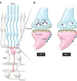
evidence that proteins downstream of the intracellular signaling cascade are dis-lodged from postsynaptic bipolar cell den-drites; however, the glutamate-binding receptor mGluR6 itself remains at this site (Figure 1). Notably, compared with other murine retinal degeneration models, pre-synaptic structures were maintained, but the calcium levels in the presynaptic ter-minals were seemingly reduced in Rs1–/–
mice. Moreover, expression of the glu-tamate transporter vGluT1 was reduced in the outer retina of Rs1–/–, pointing to a
Related Article: p. 2891
Conflict of interest: The authors have declared that no conflict of interest exists. Reference information: J Clin Invest. 2015;125(7):2572–2575. doi:10.1172/JCI82577.
ers) remain unclear, which in theory would allow the glutamate concentration in the synaptic cleft to be decreased or increased. A recently developed, genetically encoded glutamate-sensing indicator (14) has been successfully expressed in retinal cells to measure glutamate in the synaptic cleft; however, this sensor has not been used to evaluate glutamate in murine models of degeneration. Adeno-associated virus– mediated (AAV-mediated) expression of such a bioindicator in bipolar cells to moni-tor glutamate release from phomoni-torecepmoni-tors has the potential to substantially advance our understanding of how altered gluta-matergic drive from degenerating photo-receptors affects postsynaptic structures. As there is a high degree of plasticity at the first synapse of the mouse visual system in degeneration mouse models, a finding that presumably translates to patients, therapeutic intervention to rescue func-tion of this important synapse may be fea-sible in the future.
Gene therapy restores visual
acuity in adult
retinoschisin-deficient mice
Currently, there is no effective treatment for patients with XLRS. Topical admin-istration of carbonic anhydrase inhibi-tors ameliorates prominent morphologi-cal distortions, such as central macular thickness and the size of macular cysts; however, improvement of visual acuity upon treatment is controversial (15, 16). In contrast, in the Rs1–/– mouse model for
XLRS, supplemental AAV vector–mediat-mGluR6 and TRPM1 are differently
regu-lated. In this view, the distinct pathosynap-tic patterns of the mGluR6-signaling cas-cade proteins observed in different mouse retinal degeneration models suggest that the postsynaptic macromolecular organi-zation in bipolar cells depends on highly complex protein expression, trafficking, and sorting processes. However, a much more complete picture of the composition and regulation of the photoreceptor–bipo-lar cell synapse needs to be established to fully understand the pathosynaptic phe-notype in Rs1–/– animals. For example,
fur-ther ultrastructural studies are required to evaluate fine-scale morphological altera-tions, similar to what has been performed at the photoreceptor synapse to study the ECM protein pikachurin (13). Additionally, it is not clear how glutamate release from photoreceptors is changed in regard to degenerative downstream processes. For example, in the Rs1–/– retina, the calcium
concentration in photoreceptor axon ter-minals is lower, but the expression levels of proteins that remove glutamate from the synaptic cleft (e.g., glutamate transport-strong alteration of the glutamate release
at the photoreceptor–bipolar cell synapse. When synaptic transmission is reduced, postsynaptic neurons normally respond in one of two possible ways. They either increase postsynaptic receptor density in response to the reduced level of transmit-ter to adjust the synaptic efficiency or they reduce the number of postsynaptic recep-tors to strengthen other connections. The compensatory pathway chosen very much depends on the specificity of the synapse and the overall activity level (reviewed in ref. 10). Surprisingly, the decreased calcium level in the Rs1–/– photoreceptor
[image:3.585.46.370.50.391.2]terminals and the expected downregula-tion of glutamate release did not directly alter expression or localization of the glutamate-binding mGluR6 receptor on the dendrite tips; however, the calcium decrease reduced expression of down-stream signaling molecules, including the cation channel TRPM1, subsequently lead-ing to homeostatic hyperpolarization of the bipolar cells. Unlike earlier studies (11, 12), the study by Ou, Vijayasarathy, and col-leagues suggests that expression levels of
Figure 1. Functional remodeling of the rod photoreceptor–to–rod bipolar cell synapse in the retinoschisin-deficient retina. (A) This
schematic shows the synaptic connection between rod photoreceptors (blue, rods) and rod bipolar cells (red, RBC) in the outer retina. For simplicity, all other cell types are shown in gray. (B) Magnified synapse illustrates the
differ-ences in organization of the rod-to-rod bipolar cell synapse between WT and synaptically remodeled Rs1–/– mutant retinae (right). In the
1. Molday RS, Kellner U, Weber BH. X-linked juvenile retinoschisis: clinical diagnosis, genetic analysis, and molecular mechanisms. Prog Retin
Eye Res. 2012;31(3):195–212.
2. Ou J, et al. Synaptic pathology and therapeutic repair in adult retinoschisis mouse by AAV-RS1 transfer. J Clin Invest. 2015;215(7):2891–2903. 3. Chang B, et al. The nob2 mouse, a null muta-tion in Cacna1f: anatomical and funcmuta-tional abnormalities in the outer retina and their con-sequences on ganglion cell visual responses.
Vis Neurosci. 2006;23(1):11–24.
4. Barhoum R, et al. Functional and structural modifications during retinal degeneration in the rd10 mouse. Neuroscience. 2008;155(3):698–713. 5. Križaj D, Huang W, Furukawa T, Punzo C, Xing
W. Plasticity of TRPM1 expression and localiza-tion in the wild type and degenerating mouse retina. Vision Res. 2010;50(23):2460–2465. 6. Chua J, Fletcher EL, Kalloniatis M.
Func-tional remodeling of glutamate receptors by inner retinal neurons occurs from an early stage of retinal degeneration. J Comp Neurol. 2009;514(5):473–491.
7. Haq W, Arango-Gonzalez B, Zrenner E, Euler T, Schubert T. Synaptic remodeling generates syn-chronous oscillations in the degenerated outer mouse retina. Front Neural Circuits. 2014;8:108. 8. Dunn FA. Photoreceptor ablation initiates
the immediate loss of glutamate receptors in postsynaptic bipolar cells in retina. J Neurosci. 2015;35(6):2423–2431.
9. D’Orazi FD, Suzuki SC, Wong RO. Neuronal remodeling in retinal circuit assembly, disas-sembly, and reassembly. Trends Neurosci. 2014;37(10):594–603.
10. Okawa H, Hoon M, Yoshimatsu T, Della Santina L, Wong RO. Illuminating the multifaceted roles of neurotransmission in shaping neuronal cir-cuitry. Neuron. 2014;83(6):1303–1318. 11. Xu Y, Dhingra A, Fina ME, Koike C, Furukawa T,
Vardi N. mGluR6 deletion renders the TRPM1 channel in retina inactive. J Neurophysiol. 2012;107(3):948–957.
12. Cao Y, Posokhova E, Martemyanov KA. TRPM1 forms complexes with nyctalopin in vivo and accumulates in postsynaptic compartment of ON-bipolar neurons in mGluR6-dependent manner. J Neurosci. 2011;31(32):11521–11526. 13. Sato S, et al. Pikachurin, a dystroglycan ligand,
is essential for photoreceptor ribbon synapse formation. Nat Neurosci. 2008;11(8):923–931. 14. Marvin JS, et al. An optimized fluorescent probe
for visualizing glutamate neurotransmission.
Nat Methods. 2013;10(2):162–170.
15. Genead MA, Fishman GA, Walia S. Efficacy of sustained topical dorzolamide therapy for cystic macular lesions in patients with X-linked retinoschisis. Arch Ophthalmol. 2010;128(2):190–197.
16. Khandhadia S, Trump D, Menon G, Lotery AJ. X-linked retinoschisis maculopathy treated with topical dorzolamide, and relationship to geno-type. Eye (Lond). 2011;25(7):922–928. 17. Zeng Y, et al. RS-1 Gene Delivery to an adult rs1h
knockout mouse model restores ERG b-wave with reversal of the electronegative waveform of X-Linked retinoschisis. Invest Ophthalmol Vis Sci.
cient for therapeutic success. In XLRS, the swelled and loosened retinal structure of this pathology may facilitate penetration of the therapeutic vector and transduc-tion of photoreceptors and bipolar cells upon intravitreal injection. Prior studies with intravitreal injection of an AAV8 vec-tor that is similar to that performed by Ou, Vijayasarathy, and colleagues have shown that penetration into the outer retina of WT mice is much less effective than it is in the Rs1–/– mutant retina, where the AAV8
vector readily transduces photoreceptors and rescues function (20). As retinoschisin is a secreted protein, could AAV-mediated expression of retinoschisin in other, more easily accessible retina cell types do the job? Likely not, as targeted expression of retinoschisin in Muller glial cells has been shown to be much less effective than expression in photoreceptors for rescuing defects in the Rs1–/– mouse (21).
In conclusion, Ou, Vijayasarathy, and colleagues provide a body of work that supports other preclinical evidence that XLRS may be an excellent target for reti-nal gene therapy. Not only does innate synaptic plasticity restore structure and function in Rs1–/– mice, but the
pathol-ogy of this disease provides advantageous access to outer retina layers for the intra-vitreal route delivery of therapeutic AAV vectors. The feasibility of this approach in patients is being evaluated in a phase I/IIa, prospective, three-dose escalation study that is currently underway (ClinicalTrials. gov NCT02317887) to probe safety and efficacy of gene therapy in XLRS patients.
Acknowledgments
The authors wish to acknowledge support of their work by the Charlotte and Tistou Kerstan Stiftung (RD-CURE to B. Wiss-inger), a grant from the German Ministry for Education and Research (BMBF; Ion-NeuroNet, grant no. 01GM1105A to B. Wissinger), and by the Deutsche Forsc-hungsgemeinschaft (EXC 307, CIN and SCHU 2243/3-1 to T. Schubert).
Address correspondence to: Bernd Wiss-inger, Molecular Genetics Laboratory, Institute for Ophthalmic Research, Uni-versity of Tübingen, Röntgenweg 11, D-72076 Tübingen, Germany. Phone: 49.7071.2985032; E-mail: wissinger@ uni-tuebingen.de.
ed gene therapy has not only been shown to reduce retinal swelling and cyst forma-tion, but also to reduce photoreceptor loss and improve light-induced b-wave amplitude in ERG recordings (17–21). Ou, Vijayasarathy, and colleagues have now substantiated previous reports that AAV-based gene delivery functionally rescues the visual defect by showing that reconstitution of retinoschisin expression ameliorates synaptic pathology by restor-ing presynaptic Ca2+ levels and vGlutT1
expression, postsynaptic organization of signal transduction components, and bipolar cell membrane potential (2). Importantly, these results were achieved in adult animals in which the pathology had already substantially progressed, indi-cating that function is regained through innate plasticity of the photoreceptor–to– ON-bipolar cell synapse.
suffi-2014;383(9923):1129–1137.
25. Jacobson SG, et al. Gene therapy for leber congenital amaurosis caused by RPE65 muta-tions: safety and efficacy in 15 children and adults followed up to 3 years. Arch Ophthalmol. 2012;130(1):9–24.
26. Kay CN, et al. Targeting photoreceptors via intravitreal delivery using novel, capsid-mutated AAV vectors. PLoS One. 2013;8(4):e62097. 27. Dalkara D, et al. In vivo-directed evolution of
a new adeno-associated virus for therapeutic outer retinal gene delivery from the vitreous. Sci
Transl Med. 2013;5(189):189ra76.
21. Byrne LC, et al. Retinoschisin gene therapy in photoreceptors, Müller glia or all reti-nal cells in the Rs1h–/– mouse. Gene Ther.
2014;21(6):585–592.
22. Maguire AM, et al. Safety and efficacy of gene transfer for Leber’s congenital amaurosis. N Engl
J Med. 2008;358(21):2240–2248.
23. Bainbridge JW, et al. Effect of gene therapy on visual function in Leber’s congenital amaurosis.
N Engl J Med. 2008;358(21):2231–2239.
24. MacLaren RE, et al. Retinal gene therapy in patients with choroideremia: initial find-ings from a phase 1/2 clinical trial. Lancet. 2004;45(9):3279–3285.
18. Min SH, et al. Prolonged recovery of retinal structure/function after gene therapy in an Rs1h-deficient mouse model of x-linked juvenile retinoschisis. Mol Ther. 2005;12(4):644–651. 19. Janssen A, et al. Effect of late-stage therapy on
disease progression in AAV-mediated rescue of photoreceptor cells in the retinoschisin-deficient mouse. Mol Ther. 2008;16(6):1010–1017. 20. Park TK, et al. Intravitreal delivery of AAV8