77:6 (2015) 71–75 | www.jurnalteknologi.utm.my | eISSN 2180–3722 |
Jurnal
Teknologi
Full Paper
I
MAGE SEGMENTATION TECHNIQUES FOR RED
BLOOD CELL
:
O
N OVERVIEW
Laghouiter Oussama,
*M. Mahadi Abdul Jamil, Wan Mahani
Hafiza Bt. Wan Mahmud
Biomedical Engineering Modeling and Simulation(BIOMEMS)
Research Group Department of Electronics Engineering
(FKEE),University Tun Hussein Onn Malaysia, Johor, Malaysia
Article history
Received
19 June 2015
Received in revised form
26 June 2015
Accepted
10 July 2015
*Corresponding author
[email protected]
Graphical abstract
Abstract
Image processing technique applies in different domains, such as medical, remote sensing and security. This techniques Aims to get a simple image called -image processed- should retain maximum useful information. The sensitive step in image processing is segmentation of image. Segmentation is first stage in medical image analysis seeded to two categories supervised and unsupervised technique. Accuracy of this stage affects the whole system performance. This paper present some methods applied for blood cell image segmentation and compares previous studies of overlapping cell division method. The common goal about this area is accuracy of counting the number of red blood cells (RBC) or white blood cells (WBC), which decrease with effect of some diseases such as anemia and leukemia. And makes it a critical factor in patient treatments.
Keywords: Image processing; medical image; segmentation; red blood cell image patient treatment.
Abstrak
Teknik pemprosesan imej diaplikasikan pada domain yang berbeza, seperti perubatan, kawalan pengesan, dan juga keselamatan. Bertujuan untuk mendapatkan imej yang dipanggil “image processed” perlu mengekalkan maklumat yang berguna secara maksimum langkah sensitif dalam pemprosesan imej adalah pembahagian imej. Segmentasi adalah peringkat pertama dalam analisis imej perubatan dan terbahagi kepada dua kategori iaitu, teknik diselia (Supervised) dan teknik tanpa pengawasan (Unsupervised). Ketepatan peringkat ini memberi kesan kepada prestasi keseluruhan sistem. Didalam kertas kerja ini kami mengkaji beberapa kaedah yang digunakan untuk sel darah segmentasi imej dan membandingkan kajian sebelum ini dalam kaedah pembahagian sel bertindih. Matlamat umum dalam bidang ini adalah ketepatan mengira bilangan sel darah merah (RBC) atau sel darah putih (WBC) yang berkurangan disebabkan kesan daripada sesetengah penyakit seperti seperti anemia dan leukemia. Oleh itu, ia adalah satu faktor penting dalam rawatan pesakit.
Kata kunci: Pemprosesan imej ; imej perubatan; segmentasi ; imej sel darah merah rawatan pesakit ..
1.0 INTRODUCTION
In medical profession, human vision is evaluation of computerized system which can extract information from medical images. Several research have focused on Improving of human vision and object detection process, depending on application. In short, it increases the effectiveness of decision making in term of accuracy, time consumed and capability. Medical image processing technology has vital significance in clinical diagnosis. Cell image segmentation is one of the key medical image processing which has a direct effect on analysis of accuracy of work. It depends with image character size. To the particularity of cells, the main task of medical image processing system for cell image
processing is automatic recognition and analysis of
cells extracted from image. This includes extraction of cells, Statistical analysis of cells, calculate parameters and individual cells for cell feature information. Subsequent to a statistical analysis of work there is a need to take effective means to develop algorithm that can identify single RBC from overlapping cells. Different types of cell image segmentation need different methods, so far there is no generic algorithm for all cells image segmentation. Review of the current techniques used for the blood cell segmentation techniques is used as reference to create solutions for the research problems in overlapping condition.
2.0 LITERATURE VIEW
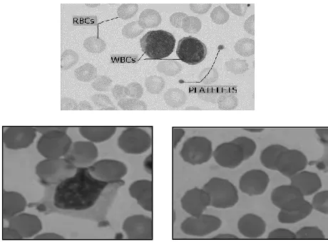
Cell images possess a complex nature and it is hard to segment them from background and count automatically, the aim of the blood cell segmentation is to make each component of the cell such as cytoplasm and nucleus of WBC, platelet, background and RBC be perfectly segmented. khou boon et al. examined nine different techniques for RBC image segmentation which consist of few operators and color transformation techniques, concluded that none of the methods are good enough for a perfect RBC Segmentation [1].R.adollah et al. mentioned that no single technique can be considered good to segment the blood cells [2].
There are several researches proposed some algorithms for blood segmentation, to get better result and solve the problem of blood cell segmentation. Chastine and Diana et al. reported recent algorithms to segment white blood cell (WBC) with overlapping condition [3]. Di Rubeto A.Dempster et al. proposed Segmentation of blood images using morphological operators. Which related of a malarial image processing system for detecting and classifying malaria parasites in images of Giemsa stained blood slides [4]. Also the algorithm of Cell segmentation with median filter and mathematical morphology operation has been proposed by D.Anoraganingrum .That describes a part of their
research work on an Automated Cell Tracking (ACT) project [5]. Mouroutis, A.Bharth et al. proposed robust cell nuclei segmentation using statistical modelling. In it they have proposed the multistage segmentation method for isolation of cell nuclei which is applicable to light microscope images of stained tissue sections6. Fabio scotti et.al proposed an automatic morphological analysis for acute leukemia identification in peripheral blood microscope images [7]. Mohamed and Amr Guaily et.al proposed an efficient technique for white blood cells nuclei automatic segmentation. The technique is based on gray scale contrast enhancement and filtering. Minimum segment size as implemented to remove false objects. The technique was tested on 365 blood images. The segmentation performance is quantitatively evaluated on the test set to be 79.7%. Blood cells namely, lymphocytes, monocytes and neutrophil in digital microscopic images [8].
J.M Sharif, Muhammad Mahadi bin Abdul Jamil et al. proposed to apply Masking and Watershed Algorithm for red blood cell segmentation and conclude that the technique is only capable of handling touched or small overlap in the image but unsuccessful for a big overlap[9].
3.0 IMAGE SEGMENTATION
Blood cell image possess a complicated environment with various type of components that differs in term of texture, color, size and morphology. In segmentation of RBC, these are the issues that needs to be considered. F.Sadeghian et.al stated, segmenting of blood cell is dealing with varying forms of shape, edge, position and size [10].S.Savker and S.P Narote stated that the use of Giemsa stain for blood slides preparation is good in appearance of RBCs colors. In contrast, it shows clearly also the parasites, (WBC), platelets, and various artefacts which makes the segmentation process trivial [11]. G.Diaz and A.Manzanera et al. Classified blood cell segmentation into region based and boundary based segmentation as a solution of complex appearance of blood cell image [12].
Figure 1 and 2 shows the image of the blood cell and it’s characteristic.
A) A good selection of segmentation method is important for next process. E.Monsteny et.al mentioned that segmentation process will affect the feature extraction and classification process, Also stated that previous works are mostly direct decision methods which lead to difficulties for correction process after a wrong decision has been made. As example in clustering process, the region between RBC, WBC and background keep mixing together as their color component are very close [13]. S.Chinwaraphat et.al Mentioned that a wrong clustering and scattering will cause a similar color pixel between cell and plasma as a background and also causing unclear boundary between them [14].Steven S.S.poon et.al Has identified that the segmentation of blood cell is more exposed to B) Color similarity: In other reviews, R.adollah et.al examined that Feature Space Clustering is a direct approach to make a classification. However, to determine the cluster number will be a difficult issue. For color image, the selection of color space is quite critical to cluster [16] F.Sadeghian et.al identified that close scales of color between the particle of cells will lead to a major error during thresholding method [10]. This problem applies to region growing, morphological method and thresholding each of the technique have weakness against the environments of the blood cell such as non-homogeneity of cells’ color, and inapplicable for multiple cases. G.diaz and A.Mazanera analyzed region based segmentation including Thresholding, Region Growing and Watershed. Thresholding and clustering also function as Automatic Thresholding have problems in identifying border between plasma, cytoplasm and earlier stage of parasites. For Region Growing, it have problems to make a good criterion to synchronize a neighbor pixel and the seed and also find the suitable seed [12]. Next, Classical Watershed Transform which mostly presenting regional minima which lead to over segmentation.
C) Weak edge boundary: Other problem which lies in image segmentation is defocused image as stated by Steven S.S Poon et.al where to define the boundary between cells might be difficult especially for nucleus and cytoplasm region if the transition of intensity levels is unclear. Moreover, there will be errors in cytoplasm segmentation if that region covered by the background [15]. R.adollah et.al mentioned that for segmentation, edge detection is unsuccessful to get the information and determine the location of the cells due to the weak boundaries of certain component in the blood cells 16. Said that the problems exist in the loss of protuberances pale tips and weak edge between the cell and background. He listed some techniques to overcome the problems like new edge operator and shape analysis but each of them has problem in dealing with blood cell image which has weak boundary and non-circle cells’ shape structure. This is also supported by G. Diaz and A. Manzanera et.al [12]. In analyzing the use of boundary based segmentation it is basically dealing with great problems as boundaries between cells are not clear and edge detection work bad for blood cell image. The other technique like Active Contours is good in cells cluster segmentation. However, it possess a high computational cost and resulting contours fail to respond over cells borders. D) Overlapping and clump cell: Overlapping and clumped cell is also another issue in blood cell segmentation as identified by E. Montseny et.al S. Chinwaraphat et.al Said that the overlap also may occur between the cell and plasma as the background[13],[14]. R. Adollah et.al mentioned that, normal human blood microscopic image has a high accumulation of red blood cells which could be observed and resulting the existence of touch and overlap between these cells16. Steven S.S. Poon et.al specified the overlapping cells as a major problematic task in blood cell segmentatio4. False negative and false positive may be resulted if a wrong indication is being made. False negative is the situation where an undetected important part of cell is ignored while false positive is where a detected ignored cell like Debris is being interpret as abnormal condition. Most major segmentation errors were due to cells with irregularly shaped nucleus and cells with atypical cytoplasm color which are characteristics of some types of normal cells in the bone marrow. Most debris have approximately equal intensity in all three color images and hence were easily eliminated by the subtraction of the blue image from the red. However, this process does not remove many regions in the image which belong to parts of red blood cells, platelets, and other debris. These were generally very small and were thus eliminated by the erosion process and the size criterion imposed on each isolated object. These include overlapping nucleated cells or cells which were so close together that even a human observer would have difficulty in segmenting.
[image:3.612.66.296.59.229.2]contour shape. In other work to get small overlap separated, Watershed algorithm is used on distance image but it is fails if the covered area is important. Next, Concavity analysis is involves in finding cut points on the boundary and joined them to make split lines. It is effective for separated pairs of cells but fail for multiple cluster of cells. Moreover, it is dependent on highly accurate segmentation. Work also done in producing multiple lines and the best split lines is being chosen. However it depends on implicit conditions of the cell shape or size. For Morphological operation which using centers of cell for separation, it is dependent on overlapping area between cells. Other methods like Template matching is computationally expensive and only covers separating small shape and size variations. In post processing, filling holes, artifacts and border touching cells were handled using morphological or connected operators [12].
e) Illumination: Another issue is the uncertainty images come from staining and illumination inconsistencies which make the task more challenging. For segmentation, the application of each method shows that a robust segmentation method can be evaluated by validating the accurateness and effectiveness of computational time for segmenting several types of blood cells from different human blood cell background and classifying it into different classes. It is in order to obtain the best result for human blood cell segmentation and cell counting. E. Montseny et.al said that microscopic images of blood cell contains uncertainties from varies level of cell maturity to be identified, inconsistence illumination, shadows and staining [13]. R. Adollah et.al also said that image uncertainty comes from inconsistencies staining and illumination, it makes blood cell image segmentation a difficult and challenging task2. Su M.-j. Et al Mentioned that “Cell images’ own complex nature (unevenly illuminated and low contrast), it is very difficult to separate cells from the background and count them automatically.” In certain images, they contain small non-cell objects in the background which cannot be subtracted by a normal de-noise technique and it effected cell counting [14]. The statement is supported by G. Diaz and A. Manzanera et.al which said that environment illumination conditions, dye duration, film thickness, or defects result in visual artifacts, non-uniform background, or different image luminance and color distribution in digitized images. There are two major solutions to the problems which is noise reduction by using filtering and feature enhancement by using tonal and spectral domain [12]. Steven S.S. Poon et.al stated that to make centrality and illumination in equilibrium point, it needs a good process of image acquisition. Although random noise can be minimized, fixed pattern noise is still exist and must be fixed. Basically there are three main sources of fixed pattern noise which is detector elements unequal sensitivity, systematic errors in camera control circuitry and shading and aberration effects resulted from optic.
To overcome this, optical densities decalibration and background subtraction are being used. One of the approach is involving bright image subtracted from the test image and an offset which have same value with bright image average value is added to produce a bright background image that have equal grey level value for all pixels. However, maximum 10 percent errors may exist from calculation of similar features and optical density which affected by absolute intensities multiplication or division [15]. Saravana Kumar Kumarasamy et.al stated that low contrast, poorly illuminated, unfocussed images and overlapped RBCs make a fully automated system for blood cell analysis development remains complicated. Detecting RBCs and parasites using Histogram based thresholding method is highly depend on quality of image and require distinct valleys to success. To separate RBCs from background using Zack’s histogram thresholding algorithm is not robust if histogram profiles is in wide range to characterize the digitized images. More robust segmentation is resulted from the level set approach which detecting objects in low contrast and noisy biomedical images. Unfortunately, it is computationally intensive and have poor convergence problem. In cell clumping condition, Morphological methods fail to get closed contour in overcome constituent cells contours result from poor and vary image contrast. The lack of robustness continue to be an issue in detecting parasites within the RBCs. It is still a problem to differentiate the blue and green channels for Giemsa stained nucleated due to variations in image contrast [12].
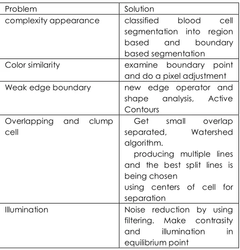
Table summarized the problems of blood cell image and proposed solutions as details in section 3.
Table 1 Problems related with red blood cell image
segmentation.
Problem Solution
complexity appearance classified blood cell
segmentation into region
based and boundary
based segmentation
Color similarity examine boundary point
and do a pixel adjustment
Weak edge boundary new edge operator and
shape analysis, Active
Contours Overlapping and clump
cell
Get small overlap
separated, Watershed
algorithm.
producing multiple lines and the best split lines is being chosen
using centers of cell for separation
Illumination Noise reduction by using
filtering. Make contrasity
and illumination in
[image:4.612.319.559.485.735.2]4.0 DISCUSSION
There are various methods for RBC segmentation, each of them have their own advantages and disadvantages. Some approaches are based on color differences others based on the shape, boundaries or region. Each one is poor to extract information from cell. In standalone. Some developed methods combine two or more combination approaches such as intensity and shape information of cell. In other issue of watershed clustering and scale-space filtering to avoid problems due to spatial variety and complexity in image space. Also morphological operations are used for the same reason. There are some previous researches which solve the problem of clump or overlapping by using recent algorithms. The degree of clump is the number of pixels in boundary. The extraction of blood cell's image from the background is the ultimate goal of blood cell segmentation. This paper summarizes majority of the accepted methodologies applicable for evaluation of image analysis. The narrowest in the segmentation method that has been proposed to separate each cell into structural or morphological components like nucleus, holes, cytoplasm and some others. The main task of this review is to propose an automated system on blood cell classification. There are many methods that can be used for blood cell segmentation and get good results. We should be researches endeavor to find absolute and robust solution to validation. Meanwhile new algorithms and systems are required with enhanced accuracy.
5.0 CONCLUSION
As conclusion, RBC segmentation in clump and overlapping condition must be handled smoothly by improving the past technique. The result from segmentation is also hoped to improve a fundamental step and create a novel RBC analysis like counting application, disease identification and health indicator. For this reason, more studies must be done towards the issue to build a strong analysis approach in the medical diagnosis area especially in hematology. In addition, it is hoped that a hybrid method will help to improve the current methods to be more capable, robust and effective. The automated segmentation can be applied to have a better support for future research. This will be an initial move towards the development of the automated analytic for the blood cell.
References
[1] Khoo Boon How, Alex See Kok Bin, Alex See Kok Bin, Ng
TeckSiong, Khoo Kong Soo :“Red Blood Cell Segmentation Utilizing Various Image Segmentation Techniques”, Proceeding of International Conference on Man-Machine Systems 2006, September 15-16, Langkawi, Malaysia.
[2] R. Adollah1, M.Y. Mashor2, N.F. Mohd Nasir, H. Rosline3, H.
Mahsin3, H. Adilah4 Blood Cell Image Segmentation: A Review 1 Biomedical Electronic Engineering Program.
[3] The Leukemia & Lymphoma Society, 1311 Mamaroneck
Avenue, White Plains, NY 10605 Information Resource Center (IRC) 800: 955.4572.
[4] Leukaemia Foundation, Fact Sheet: Related Blood
Disorders, October 2008.
[5] S.Shin, M.S.Park, J.H.Yang, Y.H.Ku, and J.S.Suh. 2004.
Measurement Of Red Blood Cell Aggregation By Analysis Of Light Transmission In A Pressure-Driven Slit Flow System, Dept. of Laboratory Medicine, Kyungpook National University, 1370 Sangyeok-dong, Buk-gu, Daegu 702-701 Korea, Korea-Australia Rheology Journal. 16(3): 129-134.
[6] Kim, K. 2001. Automatic Cell Classification in Human’s
Peripheral Blood Images Based on Morphological Image
Processing. Advances in Artificial Intelligence. 2001:
225-236.
[7] Sharif, J. M., Miswan, M., Ngadi, M., Salam, M. S. H, and
Mahadi, M. 2012. Red Blood Cell Segmentation Using Masking And Watershed Algorithm: A preliminary study .International Conference on Biomedical Engineering (ICoBE), 2012. 258-262.
[8] Wang, R., MacCane, B. and Fang, B. 2010. RBC Image
Segmentation based on Shape Reconstruction and
Multi-scale Surface Fitting. 3rd International Symposium. Of
Information Science and Engineering, 2010. 586-589.
[9] Sylvie Veriac, Valérie Perrone, Madeleine Avon,
Identification Of Red And White Blood Cells From Whole Blood Samples Using The Agilent 2100 Bio Analyzer.
[10] Steven S.S. Poon, Rabab K. Ward, and Branko Palcic,
1992. Automated Image Detection in Blood Smears and
Cytometry. 13: 766-774 .
[11] Su, M.-j., et al. A New Method For Blood Cell Image
Segmentation And Counting Based On PCNN And Auto Wave in Communications, Control and Signal Processing, 2008. (ISCCSP) 2008. 3rd International Symposium on 2008.
[12] Wang, R. and MacCane, B. 2008. Red Blood Cell
Classification through Depth Map and Surface Feature. International Symposium on Computer Science and Computational Technology. 339-342.
[13] ImageSegmentation using PCNN and template matching
for blood cell counting. IEEE International Conference on Computational Intelligence and Computing Research (ICCIC). 2013. 1-5.
[14] Otsu, N. 1979. A Threshold Selection Method from
Grat-Level Histograms. IEEE Transaction on System, Man and
Cybernetics. 9(1): 62-66.
[15] Zamani, F. and R. Safabakhsh 2006. An Unsupervised GVF
Snake Approach For White Blood Cell Segmentation
Based On Nucleus. In Signal Processing, 2006 8th