Single blind randomized Phase III trial to
investigate the benefit of a focal lesion ablative
microboost in prostate cancer (FLAME-trial): study
protocol for a randomized controlled trial
Lips
et al
.
S T U D Y P R O T O C O L
Open Access
Single blind randomized Phase III trial to
investigate the benefit of a focal lesion ablative
microboost in prostate cancer (FLAME-trial):
study protocol for a randomized controlled trial
Irene M Lips
1*, Uulke A van der Heide
2, Karin Haustermans
3, Emile NJT van Lin
4, Floris Pos
2, Stefan PG Franken
1,
Alexis NTJ Kotte
1, Carla H van Gils
5and Marco van Vulpen
1Abstract
Background:The treatment results of external beam radiotherapy for intermediate and high risk prostate cancer patients are insufficient with five-year biochemical relapse rates of approximately 35%. Several randomized trials have shown that dose escalation to the entire prostate improves biochemical disease free survival. However, further dose escalation to the whole gland is limited due to an unacceptable high risk of acute and late toxicity. Moreover, local recurrences often originate at the location of the macroscopic tumor, so boosting the radiation dose at the macroscopic tumor within the prostate might increase local control. A reduction of distant metastases and improved survival can be expected by reducing local failure. The aim of this study is to investigate the benefit of an ablative microboost to the macroscopic tumor within the prostate in patients treated with external beam radiotherapy for prostate cancer.
Methods/Design:The FLAME-trial (FocalLesionAblativeMicroboost in prostatEcancer) is a single blind
randomized controlled phase III trial. We aim to include 566 patients (283 per treatment arm) with intermediate or high risk adenocarcinoma of the prostate who are scheduled for external beam radiotherapy using fiducial markers for position verification. With this number of patients, the expected increase in five-year freedom from biochemical failure rate of 10% can be detected with a power of 80%. Patients allocated to the standard arm receive a dose of 77 Gy in 35 fractions to the entire prostate and patients in the experimental arm receive 77 Gy to the entire prostate and an additional integrated microboost to the macroscopic tumor of 95 Gy in 35 fractions. The secondary outcome measures include treatment-related toxicity, quality of life and disease-specific survival. Furthermore, by localizing the recurrent tumors within the prostate during follow-up and correlating this with the delivered dose, we can obtain accurate dose-effect information for both the macroscopic tumor and subclinical disease in prostate cancer. The rationale, study design and the first 50 patients included are described.
Trial registration:This study is registered at ClinicalTrials.gov: NCT01168479
Keywords:microboost, prostate cancer, external beam radiotherapy, FLAME-trial
* Correspondence: [email protected] 1
Department of Radiation Oncology, University Medical Center Utrecht, Utrecht, The Netherlands
Full list of author information is available at the end of the article
Lipset al.Trials2011,12:255
http://www.trialsjournal.com/content/12/1/255
TRIALS
Background
Localised prostate cancer can be treated by radical pros-tatectomy, brachytherapy or external beam radiotherapy. For low risk tumors the results of external beam radio-therapy are comparable to radical prostatectomy and brachytherapy with freedom from biochemical failure rates approximating 95% after 5- to 10-year follow-up [1]. For high risk patients, external beam radiotherapy is preferred. The outcome for intermediate and high risk patients is insufficient with freedom from biochemical failure ranging between 60% and 75% after 5- to 10-years follow-up [2-4].
Several randomized trials have proven that dose esca-lation in external beam radiotherapy improves the bio-chemical disease free survival [2,4-6]. Pollack et al. [2] compared the efficacy of 70 Gy versus 78 Gy on 305 patients with stage T1-3 prostate cancer. For patients with a pretreatment PSA > 10 ng/mL the freedom from biochemical failure rate at five year was 43% versus 62% respectively, in favor of the higher dose group. The ran-domized trial from Peeters et al. [4] compared 68 Gy versus 78 Gy on 669 patients with stage T1-4 prostate cancer. Five-year freedom from failure rate was signifi-cantly improved from 54% to 64%. Further increase in dose is considered to improve the treatment results even further [2,4,7]. Dose response models suggest that tumor areas with severe hypoxia will need very high (ablative) doses [8]. Moreover, local recurrences often occur at the site of the primary macroscopic tumor [9,10], so boosting the radiation dose to the macroscopic tumor might increase the local control. An improve-ment in distant metastases and survival can be expected by reducing local failure, due to the fact that local fail-ure is associated with distant metastases and mortality [11-13].
Dose escalation to the entire prostate is not consid-ered feasible by reason of unacceptable toxicity risks. This problem can be overcome by partial boosting stra-tegies. In this way, the macroscopic tumor can be irra-diated to a very high dose, while the dose constraints to the rectum and bladder can be maintained [14,15]. This approach is being used to deliver a microboost to the dominant tumor region in a number of pilot studies [16-19]. The dose to the macroscopic tumor is increas-ingly escalated. The highest dose was delivered in a fea-sibility study by Singh et al. [20] who treated 3 patients with an ablative dose of 95 Gy to the macroscopic tumor within the prostate with no severe toxicity (≥ grade 3).
To investigate the benefit of an ablative microboost to the macroscopic tumor within the prostate, we started a randomized controlled trial (the FLAME-trial: Focal
Lesion AblativeMicroboost in prostatEcancer, clinical
trials: study protocol number NCT01168479). The pur-pose of this trial is to assess whether a dose escalation to the macroscopic tumor increases the five-year free-dom from biochemical failure rate. Furthermore, we will assess the influence of this dose escalation on treat-ment-related toxicity, quality of life (QoL) and disease-specific survival.
Methods/Design
Study design
The FLAME-trial is a multicenter randomized con-trolled trial. Patients are recruited during the intake consultation at the Department of Radiation Oncology in one of the participating centers. After giving informed consent, patients are randomized either to the standard arm or to the experimental arm. Patients in the standard arm receive radiotherapy according to the current stan-dard [4], namely 77 Gy in 35 fractions of 2.2 Gy to the whole prostate. Patients in the experimental arm receive an additional integrated microboost to the macroscopic tumor to a total dose of 95 Gy in 2.7 Gy fractions.
To ensure unbiased assessment of QoL measurements, the patients are blinded to the actual treatment given (receiving an ablative microboost or not). The treating physician needs to be informed about the actual treat-ment, to be able to judge the treatment plans. This is unlikely to influence the assessment of the objective pri-mary endpoint of the trial.
Patients
Men with histological proven intermediate or high risk adenocarcinoma of the prostate, who will receive exter-nal beam radiotherapy using optimal position verifica-tion with implanted fiducial gold markers, are eligible for the study. Intermediate or high risk is defined according to the currently internationally accepted cri-teria from Ashet al. [21]. Intermediate risk is defined as patients having one factor of stage T2b-c, or Gleason score = 7, or initial prostate-specific antigen (iPSA) = 10-20 ng/mL. Patients having more than one of these factors or having stage T3, or Gleason score >7, or iPSA > 20 ng/mL are defined as high risk. Patients with low risk tumors are not included in this study, because the treatment outcome for this group is already excellent with a 10-year prostate cancer-specific survival approxi-mating 95% [1].
general contraindications for MRI (i.e. cardiac pace-maker, metal implants or history of severe allergic reac-tion after administrareac-tion of contrast agent) or the use of anti-coagulants that cannot be discontinued for the gold markers implantation.
The study protocol is approved by the Medical Ethical Committees of the participating hospitals. Written informed consent will be obtained from all patients.
Randomization
Randomization is performed by an independent trial center. If a patient meets the inclusion criteria and has provided informed consent, the physician contacts the trial center. To prevent randomly occurring differences in important prognostic factors across the two rando-mized groups, the randomization is stratified by TURP, hormonal treatment and by centre. A TURP prior to radiotherapy is associated with significantly more late genitourinary toxicity [23,24]. The likely mechanism of increased late toxicity is related to the relative devascu-larisation of the urethra after TURP and the decreased capability of the mucosa to repair sublethal damage after radiotherapy [24]. Hormonal treatment is a prog-nostic unfavorable factor for late genitourinary side effects [23,25] and erectile impotence [26-28]. Further-more, a protective effect for hormonal treatment is reported for acute gastrointestinal side effects [23,29,30]. To prevent small numbers of patients in a particular hospital from all receiving, by chance, the same treat-ment, the hospital is chosen as one of the factors for stratified randomization as well.
Time schedule
Between October 2009 and October 2010, the first 50 patients were included at the Department of Radiation Oncology of the University Medical Center Utrecht. Based on this accrual and the fact that other participat-ing centers recently started to include patients as well, we expect that the accrual will be completed within 5 years from start.
Radiotherapy
To minimize the positioning errors during treatment all patients are treated with an online position verification protocol using implanted fiducial gold markers [14,31,32]. Radiotherapy will be delivered with advanced radiotherapy techniques to be able to create adequate dose distributions.
A mean dose of 77 Gy in 35 fraction of 2.2 Gy is pre-scribed to the entire prostate gland [33,34]. The dose in the part of the PTV overlapping the rectum and bladder is limited to keep the risk of severe gastrointestinal and genitourinary toxicity acceptable.
To provide an accurate delineation of the prostate gland with respect to the surrounding tissues [35,36], the prostate is delineated on a computed tomography (CT) scan combined with a registered magnetic reso-nance imaging (MRI) scan. The rectum is contoured from the anus or ischial tuberosities to the rectosigmoid flexure or sacroiliac joints. The bladder is completely outlined from the bladder neck to the dome.
The precise execution of the radiotherapy treatment can differ in each participation center, but the treatment will always meet the above mentioned criteria.
Intervention
Patients randomized to the experimental arm are treated with the current standard of 77 Gy to the whole pros-tate and in addition receive an integrated microboost to the macroscopic tumor to reach a total dose of 95 Gy in 35 fractions of 2.7 Gy. To delineate the macroscopic tumor within the prostate, different MR imaging techni-ques are used. In addition to an anatomic T2 weighted sequence, a combination of the following functional imaging modalities can be used [37,38]. Dynamic con-trast-enhanced (DCE)-MRI gives a characterization of the tissue vasculature [39]. With this technique it is pos-sible to detect areas with macroscopic tumor, because tumors tend to contain higher density of leaky blood vessels. With diffusion-weighted imaging (DWI)-MRI the mobility of water molecules is measured [40]. Tumor tissue can be identified on DWI-MRI, because in tumor the extracellular volume is reduced, leading to reduced water diffusion in tumor tissue. MR spectro-scopic imaging (MRS) provides metabolic information with cancer regions showing higher choline and lower citrate levels [41].
Primary endpoint
To evaluate whether the addition of an ablative micro-boost to the macroscopic tumor within the prostate increases the five-year freedom from biochemical failure rate compared to the current standard of care. Biochem-ical failure is defined according to the Phoenix definition as a PSA rise of 2 ng/mL above the nadir PSA level [42]. The PSA level will be measured every six months until 10 years after treatment.
Secondary endpoints
Secondary endpoints are treatment-related toxicity, QoL and disease-specific survival. Treatment-related toxicity is measured by the Common Toxicity Criteria for adverse events version 3.0 (CTCAE) [43]. The following adverse events are scored: urinary frequency/urgency, urinary retention, bladder spasms, urinary incontinence, genitourinary hemorrhage, dysuria, rectal or perirectal pain, proctitis, diarrhea, flatulence hemorrhoids, anal
Lipset al.Trials2011,12:255
http://www.trialsjournal.com/content/12/1/255
incontinence, erectile dysfunction. The physician in attendance scores the complaints before treatment, acute toxicity (weekly during treatment and 4 weeks after treatment) and late toxicity (every six months until 10 years after treatment). All symptoms are registered even if they occur only on one single occasion. Grade > 2 is considered severe toxicity.General health-related QoL is measured using the RAND-36 generic health survey [44], cancer-specific QoL using the European Organization for Research and Treatment of Cancer (EORTC) core questionnaire (QLQ-C30) [45], and the prostate tumor-specific QoL using the EORTC prostate cancer module (QLQ-PR25) [46]. The disease- and treatment-related side effects are only a component of health-related quality of life [47]. To address the compo-nents of overall well-being, a general instrument is used in addition to the disease-specific questionnaires [48-50]. The RAND-36 assesses physical and social functioning, physical and emotional role restriction, mental health, vitality, pain, general health and change in health. The EORTC QLQ-C30 contains five func-tional scales, three symptoms scales, a global QoL scale and six single-items. The EORTC QLQ-PR25 assesses urinary, bowel and sexual symptoms and functioning, and the side effects of hormonal treatment. The first questionnaire is handed over to the patient one week before treatment at the Department of Radiation Oncol-ogy and the next questionnaires are sent to the patient every six months until 10 years after the completion of the treatment. It is important to measure QoL every 6 months after treatment, to be able to determine the point in time at which the QoL changes, for example due to side-effects after treatment.
For disease-specific survival, death with metastases is considered a death caused by the disease.
Safety
An independent data safety monitoring board (DSMB) will evaluate the toxicity and clinical outcome. The DSMB receives an update of the toxicity in total and per treatment arm every 3 months. Serious toxicity, defined as any acute or late toxicity requiring surgical intervention, any grade 4 toxicity, and any not-transient (duration >6 months) late toxicities grade 3, will imme-diately be reported to the DSMB. An overview of the percentage of biochemical recurrences per treatment arm together with the evaluated number of patients per arm will be sent to the DSMB yearly as well as a statisti-cal comparison of the incidence of serious and less ser-ious toxicities in the two arms. The DSMB decides on stopping or continuing the trial. Exact rules cannot be specified in advance, but an increase in the incidence of toxicity (grade 3-4) with 5% or a smaller, but statistically significant increase, are among the reasons to consider
stopping the trial. The DSMB can decide to prematurely stop the trial in case the improvement in the experi-mental arm is higher than anticipated in the trial design.
Sample size considerations
The statistical power of the study was calculated for the primary endpoint (five-year freedom from biochemical failure). Based on data of two randomized clinical trials [2,51] reporting the treatment results of patients treated with a radiation dose equal to the dose of the standard arm of our trial, we expect that the five-year freedom from biochemical failure of the standard arm will be approximately 64%. We expect that an additional abla-tive microboost to the macroscopic tumor will increase this number with at least 10%. A one-sided log rank sur-vival power analysis shows that the length of follow-up after accrual of the last patient should be 3.5 years to detect a difference of 10% (64% and 74% free from bio-chemical failure after 5 years, in the control arm and experimental arm, respectively) with a power of 80% at a one-sided 5% significance level. This is under the assumption that the patients enter the study during an accrual period of 5 years, 50% of the enrollment is com-plete when 70% of the accrual time has past and that 20% of the patients in both arms are lost to follow-up during the follow-up period of 5 years. The reason to choose a one sided p-value is that, although extremely improbable, an increase in biochemical failure in the experimental arm would lead to the same action as no difference at all between the two treatment arms. This is because the experimental treatment will only be implemented if it is significantly better than the usual treatment, due to the increased toxicity risk in the experimental arm.
Data analysis
significance [54]. For all other analyses, which do not include the QoL measures, a p-value of < 0.05 is consid-ered statistically significant.
Inclusion of the first 50 patients
The first 50 patients included in the FLAME- trial, were treated between October 2009 and October 2010 at the Department of Radiation Oncology of the UMC Utrecht. All patients received seven-beam intensity-modulated radiotherapy (IMRT). A mean dose of 77 Gy in 35 frac-tion of 2.2 Gy was prescribed to the planning target volume (PTV) and at least 70 Gy was prescribed to 99% of the PTV [33,34]. The radiation margin around the prostate depends on several uncertainties in the daily clinical practice [55-57]. For our institute based on minimal positioning errors [32] and a simulation of their impact on dose coverage [14], a PTV margin of 4 mm was chosen. We aimed at limiting the dose to the rectum and bladder so that ≤5% of the rectum and ≤ 10% of the bladder receives a dose of≥72 Gy. Further-more, a volume of 1 cc of the bladder and rectum receives a maximum dose of 80 Gy and 77 Gy, respec-tively, and ≤50% of the rectum receives a dose of ≥50 Gy [34]. All treatment plans were checked by two inves-tigators (UAH and MV) before start of the treatment. The beam directions were 0º, 50º, 100º, 155º, 205º, 260º and 310º. The location of the fiducial markers was determined by visualizing the markers using portal images of the first segment of the 0º beam and the 260º beam. A difference of more than 1 mm compared to the planning-CT was corrected online. After 5 fractions the average rotation of the prostate was calculated. A rota-tion of 3º around the anterior-posterior or the left-right axis and a rotation of 6º around the cranio-caudal axis was corrected by changing the gantry or table rotation or the collimator angle. The portal images of the first segment of the remaining beams were used to deter-mine the average intrafraction prostate motion [58]. For each patient the individual intrafraction and remaining rotational errors were used to calculate the actual deliv-ered doses to the target and the organs at risk.
The patients were treated in supine position. One hour before the pretreatment planning scans and the radiotherapy sessions, patients were instructed to drink 500 ml to create a full bladder. A full bladder during radiotherapy results in a decreased amount of bladder volume in the high dose region and a lower dose to bowel loops compared to treatment with an empty blad-der [59]. No antiflatulent diet or laxative was prescribed [60]. When the rectum filling on CT and MRI differed considerably, a new CT or MRI scan was performed, to minimize the registration uncertainty between these two imaging modalities.
For delineation of the macroscopic tumor within the prostate, defined as the gross tumor volume (GTV),
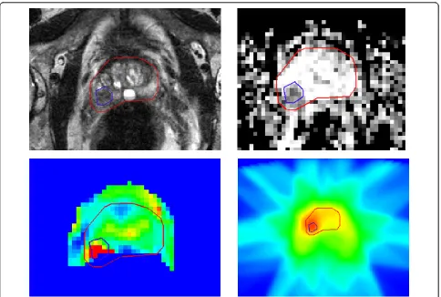
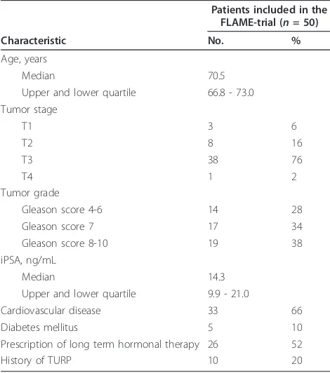
anatomical and functional imaging was performed on a 3 Tesla MRI scanner (Achieva Philips Medical Systems, Best, the Netherlands). The exam included 3 anatomical scans: a multislice T2 weighted turbo spin echo (TSE) sequence (TR/TE 8400/120 ms), a T1 weighted sequence and a balanced turbo field echo (TFE) sequence (TR/TE 2.8/1.4 ms, FOV = 25 cm, slice thick-ness = 1 mm). The DCE-MRI protocol consists of a 3D spoiled gradient echo sequence (TR/TE 4.0/1.0 ms, flip angle 6º). Scans were repeated 120 times at 2.4 s inter-val. A single acquisition consisted of 20 axial slices of 2.5 mm. The field of view was 40 × 40 cm2, the recon-struction matrix 160 × 160. For contrast enhancement, 0.1 mg/kg body weight gadobutrol (1.0 M (Gadovist, Schering) was injected intraveneously. Trace-kinetics modeling was done using the Tofts model [61] resulting in 3D maps of the transfer constant Ktrans. Diffusion-weighted imaging scans were performed using a multi-slice single shot SE-EPI sequence (FOV = 38 cm, multi-slice thickness = 3 mm, intersection gap = 1 mm, TR/TE = 5000/54 ms, acquisition matrix = 152 × 107, b values = 0, 300, 5000, 100 s/mm2). The delineation of the GTV was done by the treating physician and checked by two investigators (UAH and MV) before start of the treat-ment. To account for possible extracapsular extension, the delineation of the macroscopic tumor was expanded with an extra margin of 4 mm [62]. Figure 1 shows an example of the delineation of the GTV and the dose dis-tribution for a patient in the experimental arm. Table 1 shows the patient characteristics of the first 50 patients. Twenty-three patients were randomized into the experi-mental arm and 27 into the standard arm.
Discussion
The FLAME-trial is designed to investigate the effect of an ablative microboost to the macroscopic tumor for patients treated with external beam radiotherapy for prostate cancer.
Previous studies demonstrated that the rate of toxicity after high dose external beam radiotherapy with the use of accurate position verification is low and consequently high QoL is reported [34,63-66]. Planning studies showed that an ablative microboost to the macroscopic tumor was theoretically feasible within the currently used dose constraints for rectum and bladder [15,67,68]. Further-more, a feasibility study of Singh et al. [20] reported excellent early toxicity after simultaneous integrated IMRT boost of 95 Gy to the intraprostatic lesions. As a result, with the use of optimal position verification com-bined with the currently used dose constraints, the toxi-city in the experimental treatment arm with the ablative microboost of 95 Gy is expected to be acceptable.
Previous trials demonstrated a biochemical benefit of dose escalation. However, up to now none of the dose
Lipset al.Trials2011,12:255
http://www.trialsjournal.com/content/12/1/255
escalation trials were able to detect an improvement in disease specific or overall survival. However, all trials were designed for biochemical survival instead of overall or disease-specific survival due to the natural behavior of prostate cancer. For this reason the FLAME-trial is also powered for biochemical disease free survival. An improvement in local control without a proven benefit in overall survival is only acceptable when severe toxi-city remains limited. Furthermore, to establish whether a benefit in biochemical failure free survival also coun-terbalances the negative aspects of dose escalation, such as a small increase in toxicity, it is important that QoL is taken into account. Therefore, repeated QoL measure-ments are performed in patients included in this trial.
The precise delineation of the macroscopic tumor within the prostate is a topic of ongoing research [37,38,69]. In the FLAME-trial the delineation of the macroscopic tumor is based on anatomical and func-tional imaging according to the current opinion. The different imaging techniques might show conflicting results about the boundaries of the macroscopic tumor area, leading to difficult delineation decisions. Therefore,
it is of major importance to investigate the precise loca-tion of a recurrence and to establish what dose was pre-scribed to that location. When a patient shows a rising PSA without distant metastases, DCE-MRI and MRS can be used to detect the location of recurrent prostate cancer [70-74]. By correlating the dose distribution of the initial radiotherapy with the location of a local recurrence, accurate dose-effect information can be obtained. The dose-effect data generated from this ana-lysis will help us to evaluate the required dose for each cancer subunit and to provide a better understanding of the different imaging techniques [38]. The dose distribu-tions of the patients treated in the experimental arm, are inhomogeneous with very low and very high deliv-ered doses, and for that reason provide important dose-effect information to create a reliable dose-dose-effect curve.
[image:7.595.57.541.87.413.2]A randomized study design is indicated to resolve the problem of confounding effects. To our knowledge, no other randomized controlled trials are being performed to investigate the benefit of an ablative microboost to the macroscopic tumor in prostate cancer patients. Beside Singhet al. [20], three other groups performed a
pilot study in which a microboost to the dominant tumor region was delivered. Miralbell,et al. [16] treated 50 patients, after 64-64.4 Gy in 1.8-2 Gy fractions to the whole prostate, with a hypofractionated boost of 2 frac-tions of 5 to 8 Gy to the dominant tumor region, deli-neated by anatomical imaging. After a median follow up time of 63 months, a 5-year biochemical disease-free and disease-specific survival of 98% and 100%, respec-tively, were reported with acceptable long-term toxicity. Gaudetet al. [17] selectively delivered a brachytherapy hyperdosage of ≥216 Gy (150% of the prescribed dose) to the macroscopic tumor, defined according to positive areas on sextant biopsy, in 70 patients with localized prostate cancer treated with permanent seed prostate implant. No difference in acute or late toxicities com-pared to 120 patients with a standard plan were seen. Fonteyneet al. and De Meerleeret al. [18,19] performed the largest trial in which 230 patients were treated with a mean dose of 81-82 Gy to a dominant lesion, defined by T2 weighted MRI or MRI plus spectroscopy. With the use of IMRT and daily ultra-sound based prostate positioning, the acute toxicity remained low with no grade 3 or 4 acute gastrointestinal toxicity and 7% grade 3 genitourinary toxicity.
Analyses of the actual delivered dose in the first 50 patients included in the FLAME-trial, revealed that it is possible to deliver a high dose to the macroscopic tumor area without compromising the dose constraints
for the nearby organs at risk. The influence of the remaining intrafraction and rotational errors using an online position verification protocol is minimal.
In conclusion, the aim of the FLAME-trial is to assess, in patients treated with external beam radiotherapy for prostate cancer, the potential benefit of an additional ablative microboost to the macroscopic tumor on bio-chemical control. In addition, the subgroup of patients that will develop a local recurrence within the prostate after treatment can be used to obtain accurate dose-effect information for both the dominant lesion and subclinical disease in prostate cancer.
The study protocol was approved by the Medical Ethi-cal Committee. The trial is registered at http://CliniEthi-cal- http://Clinical-Trials.gov (Registration identification number: NCT01168479; URL: http://clinicaltrials.gov/ct2/show/ NCT01168479)
List of abbreviations used
CT: Computed Tomography; CTCAE: Common Toxicity Criteria for adverse events; DCE: dynamic contrast-enhanced; DSMB: data safety monitoring board; DWI: Diffusion-weighted imaging; EORTC QLQ-C30: European Organization for Research and Treatment of Cancer core questionnaire; EORTC QLQ-PR25: EORTC prostate cancer module; GTV: gross tumor volume; IMRT: intensity-modulated radiotherapy; IPSS: International Prostate Symptom Score; MRI: Magnetic Resonance Imaging; iPSA: initial prostate-specific antigen; PTV: planning target volume; QoL: quality of life; TURP: transurethral resection of the prostate; WHO: world health organization.
Acknowledgements and Funding
This study is supported by the Dutch Cancer Society (Grant No. UU2007-3893)
Author details
1Department of Radiation Oncology, University Medical Center Utrecht, Utrecht, The Netherlands.2Department of Radiotherapy, The Netherlands Cancer Institute - Antoni van Leeuwenhoek Hospital, Amsterdam, The Netherlands.3Department of Radiation Oncology, Leuven Cancer Institute, University Hospital Gasthuisberg, Leuven, Belgium.4Department of Radiation Oncology, Radboud University Nijmegen Medical Centre, Nijmegen, The Netherlands.5Julius Center for Health Sciences and Primary Care, University Medical Center Utrecht, Utrecht, The Netherlands.
Authors’contributions
IML participated in the design and data collection and drafted the manuscript. UAH participated in the study concept and design and revised the manuscript critically. ANTJK carried out the dose reconstruction. SPGF, KH, FP and ENL critically revised the manuscript CHG participated in the study design and performed the statistical calculations. MV participated in the study concept and its design and coordination and helped to draft the manuscript. All authors read and approved the final manuscript.
Competing interests
The authors declare that they have no competing interests.
Received: 5 September 2011 Accepted: 5 December 2011 Published: 5 December 2011
References
1. Lu-Yao GL, Yao SL:Population-based study of long-term survival in patients with clinically localised prostate cancer.Lancet1997,
349(9056):906-910.
[image:8.595.56.291.100.366.2]2. Pollack A, Zagars GK, Starkschall G, Antolak JA, Lee JJ, Huang E, von Eschenbach AC, Kuban DA, Rosen I:Prostate cancer radiation dose
Table 1 Patient characteristics
Patients included in the FLAME-trial (n= 50)
Characteristic No. %
Age, years
Median 70.5
Upper and lower quartile 66.8 - 73.0 Tumor stage
T1 3 6
T2 8 16
T3 38 76
T4 1 2
Tumor grade
Gleason score 4-6 14 28
Gleason score 7 17 34
Gleason score 8-10 19 38
iPSA, ng/mL
Median 14.3
Upper and lower quartile 9.9 - 21.0
Cardiovascular disease 33 66
Diabetes mellitus 5 10
Prescription of long term hormonal therapy 26 52
History of TURP 10 20
Abbreviations: TURP = transurethral resection of the prostate; iPSA = initial prostate-specific antigen.
Lipset al.Trials2011,12:255
http://www.trialsjournal.com/content/12/1/255
response: results of the M. D. Anderson phase III randomized trial.Int J Radiat Oncol Biol Phys2002,53(5):1097-1105.
3. Widmark A, Klepp O, Solberg A, Damber JE, Angelsen A, Fransson P, Lund JA, Tasdemir I, Hoyer M, Wiklund F, Fossa SD, Scandinavian Prostate Cancer Group Study 7, Swedish Association for Urological Oncology 3:
Endocrine treatment, with or without radiotherapy, in locally advanced prostate cancer (SPCG-7/SFUO-3): an open randomised phase III trial.
Lancet2009,373(9660):301-308.
4. Peeters ST, Heemsbergen WD, Koper PC, van Putten WL, Slot A, Dielwart MF, Bonfrer JM, Incrocci L, Lebesque JV:Dose-response in radiotherapy for localized prostate cancer: results of the Dutch multicenter randomized phase III trial comparing 68 Gy of radiotherapy with 78 Gy.J Clin Oncol2006,24(13):1990-1996.
5. Zietman AL, DeSilvio ML, Slater JD, Rossi CJ Jr, Miller DW, Adams JA, Shipley WU:Comparison of conventional-dose vs high-dose conformal radiation therapy in clinically localized adenocarcinoma of the prostate: a randomized controlled trial.JAMA2005,294(10):1233-1239.
6. Dearnaley DP, Sydes MR, Graham JD, Aird EG, Bottomley D, Cowan RA, Huddart RA, Jose CC, Matthews JH, Millar J, Moore AR, Morgan RC, Russell JM, Scrase CD, Stephens RJ, Syndikus I, Parmar MK, RT01 collaborators:Escalated-dose versus standard-dose conformal radiotherapy in prostate cancer: first results from the MRC RT01 randomised controlled trial.Lancet Oncol2007,8(6):475-487. 7. Zelefsky MJ, Fuks Z, Hunt M, Yamada Y, Marion C, Ling CC, Amols H,
Venkatraman ES, Leibel SA:High-dose intensity modulated radiation therapy for prostate cancer: early toxicity and biochemical outcome in 772 patients.Int J Radiat Oncol Biol Phys2002,53(5):1111-1116. 8. Nahum AE, Movsas B, Horwitz EM, Stobbe CC, Chapman JD:Incorporating
clinical measurements of hypoxia into tumor local control modeling of prostate cancer: implications for the alpha/beta ratio.Int J Radiat Oncol Biol Phys2003,57(2):391-401.
9. Pucar D, Hricak H, Shukla-Dave A, Kuroiwa K, Drobnjak M, Eastham J, Scardino PT, Zelefsky MJ:Clinically significant prostate cancer local recurrence after radiation therapy occurs at the site of primary tumor: magnetic resonance imaging and step-section pathology evidence.Int J Radiat Oncol Biol Phys2007,69(1):62-69.
10. Cellini N, Morganti AG, Mattiucci GC, Valentini V, Leone M, Luzi S, Manfredi R, Dinapoli N, Digesu’C, Smaniotto D:Analysis of intraprostatic failures in patients treated with hormonal therapy and radiotherapy: implications for conformal therapy planning.Int J Radiat Oncol Biol Phys 2002,53(3):595-599.
11. Coen JJ, Zietman AL, Thakral H, Shipley WU:Radical radiation for localized prostate cancer: local persistence of disease results in a late wave of metastases.J Clin Oncol2002,20(15):3199-3205.
12. Kupelian PA, Ciezki J, Reddy CA, Klein EA, Mahadevan A:Effect of increasing radiation doses on local and distant failures in patients with localized prostate cancer.Int J Radiat Oncol Biol Phys2008,71(1):16-22. 13. Jacob R, Hanlon AL, Horwitz EM, Movsas B, Uzzo RG, Pollack A:The
relationship of increasing radiotherapy dose to reduced distant metastases and mortality in men with prostate cancer.Cancer2004,
100(3):538-543.
14. Lips IM, van der Heide UA, Kotte AN, van Vulpen M, Bel A:Effect of translational and rotational errors on complex dose distributions with off-line and on-line position verification.Int J Radiat Oncol Biol Phys2009,
74(5):1600-1608.
15. van Lin EN, Futterer JJ, Heijmink SW, van der Vight LP, Hoffmann AL, van Kollenburg P, Huisman HJ, Scheenen TW, Witjes JA, Leer JW, Barentsz JO, Visser AG:IMRT boost dose planning on dominant intraprostatic lesions: gold marker-based three-dimensional fusion of CT with dynamic contrast-enhanced and 1H-spectroscopic MRI.Int J Radiat Oncol Biol Phys 2006,65(1):291-303.
16. Miralbell R, Molla M, Rouzaud M, Hidalgo A, Toscas JI, Lozano J, Sanz S, Ares C, Jorcano S, Linero D, Escude L:Hypofractionated boost to the dominant tumor region with intensity modulated stereotactic radiotherapy for prostate cancer: a sequential dose escalation pilot study.Int J Radiat Oncol Biol Phys2010,78(1):50-57.
17. Gaudet M, Vigneault E, Aubin S, Varfalvy N, Harel F, Beaulieu L, Martin AG:
Dose escalation to the dominant intraprostatic lesion defined by sextant biopsy in a permanent prostate I-125 implant: a prospective
comparative toxicity analysis.Int J Radiat Oncol Biol Phys2010,
77(1):153-159.
18. Fonteyne V, Villeirs G, Speleers B, De Neve W, De Wagter C, Lumen N, De Meerleer G:Intensity-modulated radiotherapy as primary therapy for prostate cancer: report on acute toxicity after dose escalation with simultaneous integrated boost to intraprostatic lesion.Int J Radiat Oncol Biol Phys2008,72(3):799-807.
19. De Meerleer G, Villeirs G, Bral S, Paelinck L, De Gersem W, Dekuyper P, De Neve W:The magnetic resonance detected intraprostatic lesion in prostate cancer: planning and delivery of intensity-modulated radiotherapy.Radiother Oncol2005,75(3):325-333.
20. Singh AK, Guion P, Sears-Crouse N, Ullman K, Smith S, Albert PS, Fichtinger G, Choyke PL, Xu S, Kruecker J, Wood BJ, Krieger A, Ning H:
Simultaneous integrated boost of biopsy proven, MRI defined dominant intra-prostatic lesions to 95 Gray with IMRT: early results of a phase I NCI study.Radiat Oncol2007,2:36.
21. Ash D, Flynn A, Battermann J, de Reijke T, Lavagnini P, Blank L, ESTRA/EAU Urological Brachytherapy Group, EORTC Radiotherapy Group:ESTRO/EAU/ EORTC recommendations on permanent seed implantation for localized prostate cancer.Radiother Oncol2000,57(3):315-321.
22. Oken MM, Creech RH, Tormey DC, Horton J, Davis TE, McFadden ET, Carbone PP:Toxicity and response criteria of the eastern cooperative oncology group.Am J Clin Oncol1982,5(6):649-655.
23. Peeters ST, Heemsbergen WD, van Putten WL, Slot A, Tabak H, Mens JW, Lebesque JV, Koper PC:Acute and late complications after radiotherapy for prostate cancer: results of a multicenter randomized trial comparing 68 Gy to 78 Gy.Int J Radiat Oncol Biol Phys2005,61(4):1019-1034. 24. Sandhu AS, Zelefsky MJ, Lee HJ, Lombardi D, Fuks Z, Leibel SA:Long-term
urinary toxicity after 3-dimensional conformal radiotherapy for prostate cancer in patients with prior history of transurethral resection.Int J Radiat Oncol Biol Phys2000,48(3):643-647.
25. Schultheiss TE, Lee WR, Hunt MA, Hanlon AL, Peter RS, Hanks GE:Late GI and GU complications in the treatment of prostate cancer.Int J Radiat Oncol Biol Phys1997,37(1):3-11.
26. van der Wielen GJ, van Putten WL, Incrocci L:Sexual function after three-dimensional conformal radiotherapy for prostate cancer: results from a dose-escalation trial.Int J Radiat Oncol Biol Phys2007,68(2):479-484. 27. Zelefsky MJ, Cowen D, Fuks Z, Shike M, Burman C, Jackson A,
Venkatramen ES, Leibel SA:Long term tolerance of high dose three-dimensional conformal radiotherapy in patients with localized prostate carcinoma.Cancer1999,85(11):2460-2468.
28. Turner SL, Adams K, Bull CA, Berry MP:Sexual dysfunction after radical radiation therapy for prostate cancer: a prospective evaluation.Urology 1999,54(1):124-129.
29. Christie D, Denham J, Steigler A, Lamb D, Turner S, Mameghan H, Joseph D, Matthews J, Franklin I, Atkinson C, North J, Poulsen M, Spry NA, Tai KH, Wynne C, Duchesne G, Kovacev O, Francis L, Kramar A, D’Este C, Bill D:Delayed rectal and urinary symptomatology in patients treated for prostate cancer by radiotherapy with or without short term neo-adjuvant androgen deprivation.Radiother Oncol2005,77(2):117-125. 30. Vavassori V, Fiorino C, Rancati T, Magli A, Fellin G, Baccolini M, Bianchi C,
Cagna E, Mauro FA, Monti AF, Munoz F, Stasi M, Franzone P, Valdagni R:
Predictors for rectal and intestinal acute toxicities during prostate cancer high-dose 3D-CRT: results of a prospective multicenter study.Int J Radiat Oncol Biol Phys2007,67(5):1401-1410.
31. Moman MR, van der Heide UA, Kotte AN, van Moorselaar RJ, Bol GH, Franken SP, van Vulpen M:Long-term experience with transrectal and transperineal implantations of fiducial gold markers in the prostate for position verification in external beam radiotherapy; feasibility, toxicity and quality of life.Radiother Oncol2010,96(1):38-42.
32. van der Heide UA, Kotte AN, Dehnad H, Hofman P, Lagenijk JJ, van Vulpen M:Analysis of fiducial marker-based position verification in the external beam radiotherapy of patients with prostate cancer.Radiother Oncol2007,82(1):38-45.
33. Nederveen AJ, van der Heide UA, Hofman P, Welleweerd H, Lagendijk JJ:
Partial boosting of prostate tumours.Radiother Oncol2001,61(2):117-126. 34. Lips IM, Dehnad H, van Gils CH, Boeken Kruger AE, van der Heide UA, van
Vulpen M:High-dose intensity-modulated radiotherapy for prostate cancer using daily fiducial marker-based position verification: acute and late toxicity in 331 patients.Radiat Oncol2008,3:15.
36. Usmani N, Sloboda R, Kamal W, Ghosh S, Pervez N, Pedersen J, Yee D, Danielson B, Murtha A, Amanie J, Monajemi T:Can Images Obtained with High Field Strength Magnetic Resonance Imaging Reduce Contouring Variability of the Prostate?Int J Radiat Oncol Biol Phys2010,80(3):728-734. 37. Groenendaal G, van den Berg CA, Korporaal JG, Philippens ME, Luijten PR,
van Vulpen M, van der Heide UA:Simultaneous MRI diffusion and perfusion imaging for tumor delineation in prostate cancer patients.
Radiother Oncol2010,95(2):185-190.
38. van der Heide UA, Korporaal JG, Groenendaal G, Franken SP, van Vulpen M:
Functional MRI for tumor delineation in prostate radiation therapy.
Imaging Med2011,3(2):219-231.
39. Padhani AR:Dynamic contrast-enhanced MRI in clinical oncology: current status and future directions.J Magn Reson Imaging2002,16(4):407-422. 40. Hosseinzadeh K, Schwarz SD:Endorectal diffusion-weighted imaging in prostate cancer to differentiate malignant and benign peripheral zone tissue.J Magn Reson Imaging2004,20(4):654-661.
41. Scheidler J, Hricak H, Vigneron DB, Yu KK, Sokolov DL, Huang LR, Zaloudek CJ, Nelson SJ, Carroll PR, Kurhanewicz J:Prostate cancer: localization with three-dimensional proton MR spectroscopic imaging– clinicopathologic study.Radiology1999,213(2):473-480.
42. Roach M, Hanks G, Thames H Jr, Schellhammer P, Shipley WU, Sokol GH, Sandler H:Defining biochemical failure following radiotherapy with or without hormonal therapy in men with clinically localized prostate cancer: recommendations of the RTOG-ASTRO Phoenix Consensus Conference.Int J Radiat Oncol Biol Phys2006,65(4):965-974. 43. Trotti A, Colevas AD, Setser A, Rusch V, Jaques D, Budach V, Langer C,
Murphy B, Cumberlin R, Coleman CN, Rubin P:CTCAE v3.0: development of a comprehensive grading system for the adverse effects of cancer treatment.Semin Radiat Oncol2003,13(3):176-181.
44. Hornbrook MC, Goodman MJ:Assessing relative health plan risk with the RAND-36 health survey.Inquiry1995,32(1):56-74.
45. Aaronson NK, Ahmedzai S, Bergman B, Bullinger M, Cull A, Duez NJ, Filiberti A, Flechtner H, Fleishman SB, de Haes JC:The European Organization for Research and Treatment of Cancer QLQ-C30: a quality-of-life instrument for use in international clinical trials in oncology.J Natl Cancer Inst1993,85(5):365-376.
46. Borghede G, Sullivan M:Measurement of quality of life in localized prostatic cancer patients treated with radiotherapy. Development of a prostate cancer-specific module supplementing the EORTC QLQ-C30.
Qual Life Res1996,5(2):212-222.
47. Litwin MS, Fitzpatrick JM, Fossa SD, Newling DW:Defining an international research agenda for quality of life in men with prostate cancer.Prostate 1999,41(1):58-67.
48. Nout RA, van de Poll-Franse LV, Lybeert ML, Warlam-Rodenhuis CC, Jobsen JJ, Mens JW, Lutgens LC, Pras B, van Putten WL, Creutzberg CL:
Long-term outcome and quality of life of patients with endometrial carcinoma treated with or without pelvic radiotherapy in the post operative radiation therapy in endometrial carcinoma 1 (PORTEC-1) trial.
J Clin Oncol2011,29(13):1692-1700.
49. Kornblith AB, Lan L, Archer L, Partridge A, Kimmick G, Hudis C, Winer E, Casey R, Bennett S, Cohen HJ, Muss HB:Quality of life of older patients with early-stage breast cancer receiving adjuvant chemotherapy: A companion study to cancer and leukemia group B 49907.J Clin Oncol 2011,29(8):1022-1028.
50. Inoue S, Shiina H, Hiraoka T, Wake K, Sumura M, Honda S, Urakami S, Igawa M, Usui T:Five-year longitudinal effect of radical perineal prostatectomy on health-related quality of life in japanese men, using general and disease-specific measures.BJU Int2009,104(8):1077-1084. 51. Peeters ST, Heemsbergen WD, Koper PC, van Putten WL, Slot A,
Dielwart MF, Bonfrer JM, Incrocci L, Lebesque JV:Dose-response in radiotherapy for localized prostate cancer: results of the Dutch multicenter randomized phase III trial comparing 68 Gy of radiotherapy with 78 Gy.J Clin Oncol2006,24(13):1990-1996.
52. Vickers AJ, Altman DG:Statistics notes: Analysing controlled trials with baseline and follow up measurements.BMJ2001,323(7321):1123-1124. 53. Osoba D, Bezjak A, Brundage M, Zee B, Tu D, Pater J, Quality of Life
Committee of the NCIC CTG:Analysis and interpretation of health-related quality-of-life data from clinical trials: basic approach of The National Cancer Institute of Canada Clinical Trials Group.Eur J Cancer2005,
41(2):280-287.
54. Movsas B, Moughan J, Sarna L, Langer C, Werner-Wasik M, Nicolaou N, Komaki R, Machtay M, Wasserman T, Bruner DW:Quality of life supersedes the classic prognosticators for long-term survival in locally advanced non-small-cell lung cancer: an analysis of RTOG 9801.J Clin Oncol2009,
27(34):5816-5822.
55. Mutanga TF, de Boer HC, van der Wielen GJ, Hoogeman MS, Incrocci L, Heijmen BJ:Margin Evaluation in the Presence of Deformation, Rotation, and Translation in Prostate and Entire Seminal Vesicle Irradiation with Daily Marker-Based Setup Corrections.Int J Radiat Oncol Biol Phys. 56. Meijer GJ, de Klerk J, Bzdusek K, van den Berg HA, Janssen R, Kaus MR,
Rodrigus P, van der Toorn PP:What CTV-to-PTV margins should be applied for prostate irradiation? Four-dimensional quantitative assessment using model-based deformable image registration techniques.Int J Radiat Oncol Biol Phys2008,72(5):1416-1425. 57. Rijkhorst EJ, Lakeman A, Nijkamp J, de Bois J, van Herk M, Lebesque JV,
Sonke JJ:Strategies for online organ motion correction for intensity-modulated radiotherapy of prostate cancer: prostate, rectum, and bladder dose effects.Int J Radiat Oncol Biol Phys2009,75(4):1254-1260. 58. Kotte AN, Hofman P, Lagendijk JJ, van Vulpen M, van der Heide UA:
Intrafraction motion of the prostate during external-beam radiation therapy: analysis of 427 patients with implanted fiducial markers.Int J Radiat Oncol Biol Phys2007,69(2):419-425.
59. Pinkawa M, Asadpour B, Gagel B, Piroth MD, Holy R, Eble MJ:Prostate position variability and dose-volume histograms in radiotherapy for prostate cancer with full and empty bladder.Int J Radiat Oncol Biol Phys 2006,64(3):856-861.
60. Lips IM, Kotte AN, van Gils CH, van Leerdam ME, van der Heide UA, van Vulpen M:Influence of Antiflatulent Dietary Advice on Intrafraction Motion for Prostate Cancer Radiotherapy.Int J Radiat Oncol Biol Phys. 61. Tofts PS, Brix G, Buckley DL, Evelhoch JL, Henderson E, Knopp MV,
Larsson HB, Lee TY, Mayr NA, Parker GJ, Port RE, Taylor J, Weisskoff RM:
Estimating kinetic parameters from dynamic contrast-enhanced T(1)-weighted MRI of a diffusable tracer: standardized quantities and symbols.J Magn Reson Imaging1999,10(3):223-232.
62. Chao KK, Goldstein NS, Yan D, Vargas CE, Ghilezan MI, Korman HJ, Kernen KM, Hollander JB, Gonzalez JA, Martinez AA, Vicini FA, Kestin LL:
Clinicopathologic analysis of extracapsular extension in prostate cancer: should the clinical target volume be expanded posterolaterally to account for microscopic extension?Int J Radiat Oncol Biol Phys2006,
65(4):999-1007.
63. Lips I, Dehnad H, Kruger AB, van Moorselaar J, van der Heide U,
Battermann J, van Vulpen M:Health-related quality of life in patients with locally advanced prostate cancer after 76 Gy intensity-modulated radiotherapy vs. 70 Gy conformal radiotherapy in a prospective and longitudinal study.Int J Radiat Oncol Biol Phys2007,69(3):656-661. 64. Lips IM, van Gils CH, van der Heide UA, Kruger AE, van Vulpen M:
Health-related quality of life 3 years after high-dose intensity-modulated radiotherapy with gold fiducial marker-based position verification.BJU Int2009,103(6):762-767.
65. Marchand V, Bourdin S, Charbonnel C, Rio E, Munos C, Campion L, Bonnaud-Antignac A, Lisbona A, Mahe MA, Supiot S:No impairment of quality of life 18 months after high-dose intensity-modulated radiotherapy for localized prostate cancer: a prospective study.Int J Radiat Oncol Biol Phys2010,77(4):1053-1059.
66. Zelefsky MJ, Chan H, Hunt M, Yamada Y, Shippy AM, Amols H:Long-term outcome of high dose intensity modulated radiation therapy for patients with clinically localized prostate cancer.J Urol2006,176(4 Pt 1):1415-1419.
67. Pickett B, Vigneault E, Kurhanewicz J, Verhey L, Roach M:Static field intensity modulation to treat a dominant intra-prostatic lesion to 90 Gy compared to seven field 3-dimensional radiotherapy.Int J Radiat Oncol Biol Phys1999,44(4):921-929.
68. Xia P, Pickett B, Vigneault E, Verhey LJ, Roach M:Forward or inversely planned segmental multileaf collimator IMRT and sequential tomotherapy to treat multiple dominant intraprostatic lesions of prostate cancer to 90 Gy.Int J Radiat Oncol Biol Phys2001,51(1):244-254. 69. Langer DL, van der Kwast TH, Evans AJ, Trachtenberg J, Wilson BC,
Haider MA:Prostate cancer detection with multi-parametric MRI: logistic regression analysis of quantitative T2, diffusion-weighted imaging, and dynamic contrast-enhanced MRI.J Magn Reson Imaging2009,30(2):327-334.
Lipset al.Trials2011,12:255
http://www.trialsjournal.com/content/12/1/255
70. van Vulpen M, van den Berg CA, Moman MR, van der Heide UA:Difficulties and potential of correlating local recurrences in prostate cancer with the delivered local dose.Radiother Oncol2009,93(2):180-184. 71. De Visschere PJ, De Meerleer GO, Futterer JJ, Villeirs GM:Role of MRI in
follow-up after focal therapy for prostate carcinoma.AJR Am J Roentgenol 2010,194(6):1427-1433.
72. Kim CK, Park BK, Park W, Kim SS:Prostate MR imaging at 3T using a phased-arrayed coil in predicting locally recurrent prostate cancer after radiation therapy: preliminary experience.Abdom Imaging2010,
35(2):246-252.
73. Rouviere O:MR assessment of recurrent prostate cancer after radiation therapy.Radiology2007,242(2):635-6, author reply 636-7.
74. Haider MA, Chung P, Sweet J, Toi A, Jhaveri K, Menard C, Warde P, Trachtenberg J, Lockwood G, Milosevic M:Dynamic contrast-enhanced magnetic resonance imaging for localization of recurrent prostate cancer after external beam radiotherapy.Int J Radiat Oncol Biol Phys2008,
70(2):425-430.
doi:10.1186/1745-6215-12-255
Cite this article as:Lipset al.:Single blind randomized Phase III trial to investigate the benefit of a focal lesion ablative microboost in prostate cancer (FLAME-trial): study protocol for a randomized controlled trial.
Trials201112:255.
Submit your next manuscript to BioMed Central and take full advantage of:
• Convenient online submission
• Thorough peer review
• No space constraints or color figure charges
• Immediate publication on acceptance
• Inclusion in PubMed, CAS, Scopus and Google Scholar
• Research which is freely available for redistribution