MEDICAL IMAGE COMPRESSION BASED ON SET
PARTITIONING IN HIERARCHICAL TREES USING
QUANTIZED COEFFICIENTS OF SELF ORGANIZING
FEATURE MAP FOR MR IMAGES
*S. SRIDEVI1, V.R.VIJAYAKUMAR2,V.SUTHA JEBAKUMARI3
1. Dept of Computer Science and Engg, Sethu Insitute of Technology, India 2. Dept. of Electronics and Communication Engg, Anna University, India
3. Dept of Computer Science and Engg, Kamaraj College of Engineering and Technology, India.
Email: [email protected]
ABSTRACT
Medical imaging plays a vital role in medical diagnosis. These medical images available in hospitals and medical organizations occupy a lot of space. The massive use of digitized images has led to the compression allowing economical storage and fast data transfer. Over the years, JPEG compression schemes based on Discrete Cosine Transform have been proposed and standardized. The input image has to be blocked which results in blocking artifacts. In recent years wavelet transform has gained widespread acceptance in image compression. Many compression algorithms using wavelets like EZW, SPIHT and SPECK have been proposed and they can be used for lossy or lossless compression. SPIHT is an efficient compression algorithm which has better performance over the others. In this paper, SPIHT based medical image compression algorithm using the quantized coefficients of Self Organizing Feature Map (SOFM) was brought in and proved to have better performance over existing methods.
Keywords: SPIHT, SOFM, DWT, Image Compression, PSNR, CR
1. INTRODUCTION
Medical image compression plays an important role in the health care services like teleconsultation, telemedicine, medical science, e-health etc. They will be effective if the compression technique preserves all the relevant and important image information needed. The main goal of image compression is to represent a compact representation [3] of an digital images while maintaining all necessary information. The large volumes of data needed to describe the images are not possible for transmission and also for storage[11]. The main aim is to preserve the data for storage and transmission and to remove the redundant data present in the image.
Image compression can be classified into two types: Lossless compression and Lossy compression. Lossless compression enables complete recovery of the original image from the compressed image but is limited in terms of compression ratio. It is mainly used in medical application where any loss can affect the accuracy of diagnosis. Lossy compression gives better compression ratio but produces some distortion[8] in the reconstructed image Nowadays many methods are available for image compression like predictive coding, vector quantization, sub band coding[6], transform coding etc. Over the past decades, Wavelet Transform for image compression technique has been widely accepted and gained popularity. The traditional algorithms like JPEG[16] and MPEG
are widely accepted for still and image motion pictures and they have made use of Discrete Cosine Transform (DCT). JPEG is used mainly for lossy compression and it comprises of encoding and decoding of images. JPEG uses DCT to get the coefficients which is followed by quantization. The
Performance of the techniques are measured using Compression Ratio(CR) and Peak Signal Noise Ratio (PSNR).
Trees (SPIHT) coding and Kohonen’s Self Organizing Feature Map (SOFM) for quantization. In this scheme, the input is a dyadic square MRI image. The Input image is fed to Biorthogonal filter to get co efficients. The coefficients are quantized using SOFM quantization. The LD and HD co efficients are presented to discrete wavelet transform and LR and HR coefficients are applied to Inverse Wavelet transform. Finally, SPIHT compression is applied to get the compressed image. Using SPIHT decompression, the reconstructed image is obtained. The performance of the proposed scheme is measured in terms of Peak Signal to Noise Ratio (PSNR) and the Compression Ratio (CR) attained.
This paper is organized as follows: In section 2, related works are discussed and Section 3 covers the over view of wavelet based compression techniques. Section 4 deals with a proposed method which uses quantized coefficients. Performance metrics are discussed in section 5. The comparison of results obtained using the proposed method and the SPIHT is given in Section 6. Finally, conclusion of the research is given in Section 7.
2. RELATED WORK
In wavelet based image compression technique, the image is decorrelated by applying a wavelet transform[5]. Then the wavelet coefficients obtained from the transformation are quantized using any one of the quantization techniques such as Vector Quantization [7], Scalar Quantization etc. Finally, the quantized coefficients are coded. There are many coders such as Embedded Zero-Tree Wavelets (EZW)[1], Morphological Representation of wavelet Date (MRWD)[13], Set Partitioning in Hierarchical Trees (SPIHT)[2], Modulated Wavelet Subband Image Coding[14], etc. From the above, Set Partitioning in Hierarchical Tress (SPIHT) achieves better performance compared to other methods like vector quantization using JPEG and Quantization using Wavelets. In the recent years, developing hybrid schemes for improving compression ratio and image quality in image compression has achieved a tremendous popularity from researchers.
Many researchers have investigated the use of neural network concepts for compression and they have also succeeded. One of the most interesting neural network concepts is Kohonen's Self Organizing Feature Map[9] . The main objective of the SOFM algorithm is to store a large
set of input vectors by finding a set of weights in order to serve a good approximation to the original output space. Some recent papers show that the integration of neural network based approach[4] and transform based approach lead to better compression ratio and image quality.
In paper [15], Lossy compression scheme for the low depth-of-field (DOF) images, where the quality factor is altered based on compressing object-of-interest (OOI) or the background. The method involves segmentation of OOI and then the application of lossy scheme. In paper [10], the authors presented a compression scheme for SPIHT-Based Coding of the Shape and Texture of Arbitrarily Shaped Visual Objects. The coding is based on SPIHT which uses shape-adaptive discrete wavelet transform (SA-DWT).
In paper [12], they have presented a coding scheme for Fingerprint Compression Using Contourlet Transform and Self Organizing Feature Map which had better compression ratio.
3. OVERVIEW OF WAVELET BASED
IMAGE COMPRESSION TECHNIQUES
.
3.1 Wavelet Transform
Wavelet based techniques are the recent developments in medical image compression. A wavelet is a small wave which allows time and frequency analysis. The discrete wavelet transform used in various medical image applications decomposes an image into a set of successively smaller orthonormal images. The low pass and high pass filters are applied to the image in both the horizontal and vertical direction generating HH, HL, LH high pass sub bands and LL a low pass sub band. To achieve improved compression, integer wavelet transform[6] is used where the reconstructed image quality is better than wavelet transform which achieves good performance by exploiting the spatial dependencies of pixels in different sub bands of a scalar wavelet transform
3.2 The SPIHT Algorithm
Set Partitioning in Hierarchical Trees in an embedded coding algorithm was developed by Said and Pearlman in 1996 .It is useful for a transmission over noisy channel. This algorithm is a refinement of EZW proposed by Shapiro.
2. List of Insignificant Pixels (LIP) 3. List of Insignificant Sets(LIS)
The original signal is first decomposed into several sub bands by DWT. The spatial orientation tree is defined according to the similarity which exists among coefficients across bands.
3.3 SOFM Algorithm:
A organizing map (SOM)[17] or self-organizing feature map (SOFM) is a type of artificial neural network which is trained using unsupervised learning to produce discretized representation of the input space of the training samples, called a map.
SOMs operate in two modes: training and mapping. Training builds the map using input examples. It is a competitive process, also called vector quantization. Mapping automatically classifies a new input vector.
A self-organizing map consists of components called nodes or neurons. Associated with each node is a weight vector of the same dimension as the input data vectors and a position in the map space. The usual arrangement of nodes is a regular spacing in a hexagonal or rectangular grid. The self-organizing map describes a mapping from a higher dimensional input space to a lower dimensional map space. The procedure for placing a vector from data space onto the map is to first find the node with the closest weight vector to the vector taken from data space. Once the closest node is located it is assigned the values from the vector taken from the data space.
Algorithm:
1. Randomize the map's nodes' weight vectors 2. Grab an input vector
3. Traverse each node in the map
• Use Euclidean distance formula to find similarity between the input vector and the map's node's weight vector
• Track the node that produces the smallest distance (this node is the best matching unit, BMU)
4. Update the nodes in the neighborhood of BMU by pulling them closer to the input vector
W(t + 1) = W(t) +
Θ
(t)α(t)(I(t) - W(t))5. Increase t and repeat from 2 while t<λ
• t denotes current iteration
• λ is the limit on time iteration
• W is the current weight vector
• I is the target input
•
Θ
(t) is restraint due to distance from BMU, usually called the neighborhood function, and• α (t) is learning restraint due to time.
4. PROPOSED METHOD
[image:3.612.328.503.344.520.2]Most of the coding techniques are based on wavelet compression techniques. Our work is based on Set Partitioning in Hierarchal Trees. The present work uses bi-orthogonal wavelet filter to obtain coefficients and the coefficients are quantised. The reason for quantisation is to improve the compression ratio. The block diagram of the proposed scheme is given in Figure 1.
Figure 1. Flow diagram of proposed scheme
Algorithm:
1. Load the test image.
2. Apply bi-orthogonal wavelet filter to obtain the filter coefficients.
3. SOFM Quantization was then applied to the filter coefficients.
4. Discrete Wavelet Transformation (DWT) is applied to LD and HD coefficients.
5. The resultant from DWT and LR and HR co efficient are given to Inverse Wavelet Transform (IWT).
7. SPIHT Decompression produces the reconstructed original image.
4.1 Bi-Orthogonal Wavelet Filters
Bi-orthogonal filter is used to get the coefficients for decomposition and reconstruction. The LD and HD coefficients are used for decomposition and LR, HR are used for reconstruction.
5. PERFORMANCE METRICS
The quality of the image can be obtained after compression using metrics. Two error metrics are listed below.
1. Peak Signal to Noise Ratio (PSNR)
PSNR=20* log10 (255 /
MSE
(1)2. Compression Ratio
CR=
image
ed
uncompress
the
of
Size
image
compressed
the
of
Size
(2)
6. RESULTSThis section presents the results obtained from the experiments that illustrate the effectiveness of the proposed scheme in medical image compression. Our analysis was implemented using MATLAB (Matlab 7.9) and has been simulated on various grayscale images size of 512x512. The MRI images of ten patients are considered for experimentation. The quality of the reconstructed images is evaluated in terms of PSNR value and the efficiency of the algorithm is measured using compression ratio. [image:4.612.302.564.122.357.2]
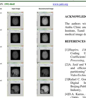
The original image slices and the reconstructed image slices are obtained using the proposed scheme is given in Figure.5 and the comparison of performance measures are listed in Table 1.
[image:4.612.314.569.391.517.2]Table 1. Comparison Of CR, PSNR For SPIHT And The Proposed Method
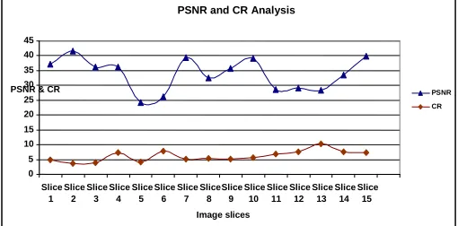
Figure 2: Chart Of PSNR Vs CR For Patient 1 Using Proposed Method
7. DISCUSSIONS
From the above table, it is clearly shown that the proposed scheme gives better performance than SPIHT. Our method’s advantageous over SPIHT is more evident as the compression ratio increases. For example, For patient 1, slice 4 and 13, the compression ratio is very high and the PSNR ratio is more than 10 db. In patient 2, for the slice 6 and 13, outperforms in terms of PSNR and CR .
Patient 1 Patient 2
Slices SPIHT method Proposed SOM with SPIHT SPIHT method Proposed SOM with SPIHT
CR PSNR CR PSNR CR PSNR CR PSNR
1 2.31 21.2 4.90 37.19 3.21 19.42 5.04 39.01
2 3.67 16.21 3.64 41.60 4.01 21.31 4.16 24.23
3 2.97 18.43 3.85 36.15 2.89 19.52 3.01 41.03
4 3.01 22.76 7.26 36.15 3.41 21.48 5.83 39.78
5 3.11 19.32 4.16 24.23 2.39 18.54 3.56 41.54
6 3.56 24.11 7.71 26.20 5.20 23.74 6.88 28.63
7 3.10 20.08 5.21 39.27 4.11 19.95 4.93 31.25
8 4.2 25.51 5.34 32.58 3.94 23.11 4.17 31.01
9 3.96 19.22 5.24 35.68 2.10 19.75 5.33 32.57
10 4.18 21.37 5.71 39.22 2.69 18.43 3.24 41.20
11 4.25 16,23 6.95 28.72 3.58 20.17 4.64 31.45
12 5.34 26.12 7.53 29.13 4.90 19.99 5.74 39.26
13 4.26 19.3 10.34 28.47 5.87 23.52 6.37 28.21
14 5.2 21.3 7.64 33.43 3.17 19.74 4.82 37.11
15 5.55 23.1 7.25 39.86 2.87 18.57 5.33 32.56
PSNR and CR Analysis
0 5 10 15 20 25 30 35 40 45 Slice 1 Slice 2 Slice 3 Slice 4 Slice 5 Slice 6 Slice 7 Slice 8 Slice 9 Slice 10 Slice 11 Slice 12 Slice 13 Slice 14 Slice 15 Image slices
PSNR & CR PSNR
Figure 5. Original Image Slices And The Reconstructed Image Slices Using Proposed Method.
The graphs of PSNR versus Compression Ratio for the test images are shown in Figure.2, which shows our proposed method is far better in visual quality as compared to the SPIHT compression image
8.CONCLUSIONS
Here, we have presented a novel compression algorithm based on SPIHT with quantized coefficients of SOFM to improve the image quality as well as Compression ratio, which can be applied to numerous medical applications, particularly MRI medical Images. The results presented in this paper, provide enough evidence to conclude that SPIHT based SOFM is an efficient algorithm in compressing MRI images.. The present work will be helpful to compress MRI medical images with improved performance in the medical sector. Our method is very suitable to obtain high compression ratio compared to SPHIT and can be used for lossy and lossless coding. We have tested our results with the radiologists and the reconstructed image maintain the good visual quality for diagnosis. Moreover it can reduce the storage and transmission cost of large volumes of medical data.
ACKNOWLEDGEMENTS
The authors wish to thank Dr. Amba Bhavani, Amba Clinic and Dr. Devaki from Devaki Cancer Institute, Tamil nadu, India, for the provision of medical image data sets used in this study.
REFERENCES
[1]Shapiro, J.M.; (1993), “Embedded Image
Coding Using Zerotrees of Wavelet Coefficients”, IEEE Transactions on Signal Processing, 41(12), Pages: 3445 – 3462 [2]A. Said and W. Pearlman, (1996), “A new, fast
and efficient image codec based on set partitioning”, IEEE Trans. Circuits Syst. VideoTechnol., 6, pp. 243-250,.
[3]Rafael C. Gonzalez, Richard E. Woods, (2007), Digital Image Processing, 2nd ed., Beijing:Publishing House of Electronics Industry,
[4]D.A. Karras, S.A. Karkanis and D.E. Maroulis, (2000), “Efficient Image Compression of Medical Images Using the Wavelet Transform and Fuzzy c-means Clustering on Regions of Interest”, in Proc. of the 26th EUROMICRO Conf., 2, pp. 2469-2473.
[5]A. S. Lewis and G. Knowles, (1992), “Image Compression using the 2-D Wavelet Transform”, IEEE Trans. Image Processing, 1, pp. 244–250.
[6]S. Hsiang and J. W.Woods, (2000), “Embedded Image Coding using Zeroblocks of subband/wavelet Coefficients and Context Modeling”, in Proc. 2000 IEEE Int. Symp. Circuits and Systems, 3, pp. 662–665,
[7]A. Laha, N.R. Pal, and B. Chanda, (2004), “Design of Vector Quantizer for image compression using Self Organizing Feature Map and Surface Fitting”, IEEE Transactions on Image Processing, 13(10), pp. 1291-1303, [8] Cazuguel, A.Cziho, B.Solaiman and C.Roux,
(1998), “Medical Image Compression and Feature Extraction using Vector Quantization,
[9]T. Kohonen, (1989), “Self-Organization and associative Memory”, 3d ed, Springer-Verlag. [10]Martin, K. Lukac, R. Plataniotis, K.N. (2006),
“SPIHT-Based Coding of the Shape and Texture of Arbitrarily Shaped Visual Objects”, IEEE Transactions on Circuits and Systems for Video Technology, 16(10), pp. 1196-1208, [11]Yen-Yu Chen, (2007), “Medical images
compression for remote diagnosis using modified SPIHT data organization and fidelity enhancement filter”, International Journal of Imaging Systems and Technology, 17(2), pp. 49 – 61.
[12]T. Veerakumar, S. Esakkirajan, R. Sudhakar, and V. Senthil Murugan, (2007), “Fingerprint Compression Using Contourlet Transform and Self Organizing Feature Map”, IRANIAN Journal of Electrical and Computer Engineering, 6(2), pp. 133-140, Summer-Fall. [13]Servetto, S.D., Ramchandran, K. and Orchard,
M.T., (1999), “Image Coding based on a Morphological Representation of Wavelet Data”, IEEE Transactions on Image Processing, 8(9), pp. 1161-1174.18.
[14]Hsin, H.-C., (2005), “Adaptive Modulated Subband Image Coding”, Pattern Recognition Letters, 26(6), pp.809-818,
[15]S. Kavitha, S. Mohammed Mansoor Roomi, N. Ramaraj, (2009), “Lossy Compression through segmentation on low depth-of-field images”, Digital Signal Processing, 19 pp.59–65,. [16]Wallace, G. K., (1991), “The JPEG Still Picture
Compression Standard”, in Commun. ACM, 34, pp. 30 – 44,