PAX6 CONTROLS ASTROCYTE POSITIONAL IDENTITY IN THE SPINAL CORD
Thesis by Christian Hochstim
In Partial Fullfillment of the Requirements for the Degree of
Doctor of Philosophy
California Institute of Technology Pasadena, California
2007
Acknowledgements
I would like to thank my advisor David Anderson for his unstoppable passion for new data and results and his contagious drive to learn and discover. Equally
important was his insistence on excellence and doing things the right way. I soon learned how experiments without proper controls would be received! Importantly, he always led by example, demanding more of himself than he demanded of those of us in the lab. For all of these reasons he will always have my utmost respect and my gratitude for the experience of training in his lab.
I would also like to thank the other members of my thesis committee: Marianne Bronner-Fraser, Paul Sternberg, and Kai Zinn. They each offered a different perspective in our meetings and I thank them for their constructive pragmatic approach and valuable insights and ideas.
Several other members of the Caltech community have been very helpful. Rochelle Diamond of the Sorting Facility was an enormous help and a pleasure to work with. Jennifer Alex, Shirley Pease, Bruce Kennedy and all of the technical straff of the Caltech Animal Facility were invaluable with maintaining mouse lines and with timed matings. Peter Lwigale was very helpful in my ability to
an important mentor and collaborator from whom I learned a great deal. His contribution to my education and progression cannot be understated. Zhou and I worked together on the Olig Microarray experiments and early analysis of candidate genes. Gloria Choi was a great friend whose intelligence and wise instincts helped guide me along. I also thank Yosuke Mukoyama for many important discussions and for always taking the time and interest to advise me about my project and helping instruct me in the art of FACS sorting and many other techniques. I thank LiChing Lo who is an authority on many techniques in the lab and who helped me on many occasions with valuable insights based on her vast experience. Donghun Shin, Laurent Van Trigt, Kenji Orimoto, Xinzhong Dong, Limor Gabay, Sally Lowell, Emma Dormand and Sebastian Gerety were also very helpful past lab members whom I count as friends.
Hochstim), Peng Wu, and Monica Martinez have all been a very big help with genotyping mouse tails. Celso Perez, Jung Sook Chang, and Ritchie Ho have been very helpful with cell culture support and maxipreps. I also thank my two great rotation students, Melanie Lee and Janet Chow who were very productive and helpful to me and a pleasure to work with. And thanks to all of the Anderson Lab Members past and present with whom I have overlapped, since all of you have helped me in one way or another.
I can’t thank Gaby enough for the many, many occasions on which she has so readily helped me with anything I needed. I have never met someone who works harder and with more energy to get things done than Gaby. Problems with lab equipment, orders or anything were always rapidly and decisively addressed which allowed me to focus on experiments and made my time so much easier. Likewise Gina Mancuso was a big help with administrative issues, scheduling, etc, and I thank her so much for always being so ready to help and so good at taking care of things. I want to thank my parents for always making education the only priority besides family and for leading me to believe in myself and embrace challenges. I thank my sister and the rest of my extended family for their unconditional love and support, which I have never doubted. Finally, I especially want to thank another former member of the lab, my wife Joana, for her continual patience, love and
Abstract
While astrocytes play many diverse and important roles in the vertebrate
CNS, little is known about the molecular diversity of these cells or the factors
specifying such diversity. We found that the secreted signaling molecules Reelin
and Slit1 mark 3 positionally defined subsets of astrocytes in the ventral white
matter of the spinal cord: Reelin+/Slit1- astrocytes in the lateral white matter (L
type), Reelin+/Slit1+ astrocytes in the ventral-lateral white matter (VL type),
and Slit1+/Reelin- astrocytes in the ventral-medial white matter (VM type). The
homeodomain transcription factor Pax6 is specifically expressed in Reelin+
astrocytes (L and VL type). We found that Pax6 plays a necessary and
instructive role in specifying these populations via its actions of promoting
Reelin and repressing Slit1 expression. We additionally show that the
homeodomain transcription factor Nkx6.1 specifically marks Slit1+ astrocytes
(VM and VL type), and provide evidence that VL type astrocytes
(Pax6+/Nkx6.1+) are derived from the p2 domain of the ventricular zone
(Pax6+/Nkx6.1+). These data are consistent with a model whereby these
astrocyte populations are prespecified in the ventricular zone. Importantly, we
provide the first evidence that molecularly distinct subtypes of astrocytes are
produced during development, each with a defined positional identity in the
white matter. While positional identity is an important property of many
neuronal subtypes, it has not been previously described in glial cells and may be
Table of contents
Acknowledgements
iii
Abstract
v
Table
of
Contents
vi
Chapter 1
Introduction
1
References
16
Chapter 2
Microarray Screen for Olig Target genes in Gliogenesis
23
References
32
Chapter 3
Pax6 controls Astrocyte positional Identity in the Spinal Cord
38
References
59
Appendix
I. The role of Pax6 in pMN progenitors
76
II. Tet-Inducible Avian retroviruses
77
III. The role of Shox2 in V2/MN and OPC specification
78
List of Tables and Illustrations
Chapter 1
Figure 1 -
Neural Pattterning in the ventral spinal cord9
Figure 2 –
Cell Fate Transformation in the Olig1,2 -/- mutant12
Chapter 2
Figure 1 –
Isolation of Olig +/- and -/- cells for Gene ExpressionProfiling
33
Figure 2 –
Analysis of Differential Gene Expression34
Table 1 –
Differentially expressed transcription factors in E13/E14.5Combined analysis
35
Figure 3 –
HFH4/Foxj1 in situ on Olig +/+ and -/- spinal cord36
Figure 4 –
Shox2 in situ on Olig +/+ and -/- spinal cord37
Chapter 3
Figure 1 –
Reelin and Slit1 mark subsets of astrocytes70
Figure 2 –
Pax6 is coexpressed with Reelin in astrocytes71
Figure 3 –
Loss of Reelin+ astrocytes in Pax6 -/- mice72
Figure 4 –
Astrocyte subtype conversion in Pax6 -/- mice73
Figure 6 –
Model of ventral astrocyte specification75
Appendix
Figure I – 1
Loss of pMN progenitors with Pax6 siRNA83
Figure I – 2
Pax6 siRNA alters spatial distribution of MN lineagemarkers
84
Figure II – 1
Inducible Gain of Function (Tet On System)85
Figure II – 2
In ovo induciion of Tet-Inducible GFP by Dox86
Figure III – 1
Shox2 is expressed in pMN domain during earlygliogenesis
87
Figure III – 2
Shox2 is sufficient to repress motor neurogenesis88
Figure III – 3
Shox2 and Olig2 co-misexpression promotesectopic and precocious oligodendrocyte precursors
89
Figure IV – 1
Increased overlap of HFH4 with Olig2-GFP inOlig -/- spinal cord
90
Chapter1
All multicellular organisms begin from a single cell which proliferates and gives rise to several distinct cell types organized in a complex three-dimensional architecture. This cellular diversity and organization enables multicellular organisms to achieve a wide degree of functional capabilities. A striking example is illustrated by the vertebrate central nervous system where a vast array of neuronal and glial subtypes, organized and interconnected together, provide the necessary substrate for fascinating higher order functions such as perception, behavior, cognition and emotion.
It is estimated that there are thousands of different kinds of neurons in the vertebrate central nervous system (Hall 1992). These neurons can be distinguished by different morphologies as well as molecular criteria such as
neurotransmitter/neuropeptide secretion and expression of different ion channels, receptors or molecular markers (Hall 1992). Different neurons are also spatially organized so as to have a positional identity. Positional identity is particularly important property of these neuronal subtypes because it establishes a spatial
organization which allows for neurons to find and connect with their proper partners to form circuits. This is evident in the cerebral cortex where different types of neurons are arranged in both layers and columns, as well in the spinal cord where subclasses of motor neurons are arranged in columns and pools in the ventral horn (Jessell, 2000).
Oligodendrocytes have been primarily associated with one critical supportive role, that of wrapping axons with myelin sheaths to enable rapid saltatory conduction. By contrast, astrocytes have been associated with several diverse functions including both passive supportive functions such as balacing pH and ion concentrations,
recycling neurotransmitters, storing energy, controlling blood vessels and forming the blood-brain barrier, as well as actively modulating CNS function by communicating with neurons at synapses, providing trophic signals for adult neurogenesis and serving as a source of stem cells (Dani et al., 1992; Doetsch, 2003; Gee and Keller, 2005; Parpura and Haydon, 2000; Song et al., 2002). Given this functional diversity an important question is whether there are molecularly distinct subsets of astrocytes and whether these astrocyte subtypes have different functional responsibilities.
Furthermore, since spatial segregation and organization are important for the proper function of neuronal subtypes, an important question is whether different types of astrocytes are also spatially organized so as to have positional identity and whether postional identity is important for their functions.
Astrocyte Diversity
Diversity of Astrocyte functions
Astrocytes are the most abundant cell type in the central nervous system (CNS) and perform a wide variety of diverse roles. Many of these functions are involved with passively providing a supportive environment for neurons. These include balacing pH and ion concentrations, recycling neurotransmitters, storing energy, controlling blood vessels and forming the blood-brain barrier (Gee and Keller, 2005). However, there is also increasing evidence that, like neurons, astrocytes may play an active role in information processing in the CNS (Fields and Stevens-Graham, 2002). Hippocampal astrocytes respond to glutamatergic firing with actively propagating Ca2+
waves in organotypic slice cultures (Dani et al., 1992). A higher resolution version of this experiment, where photo-release of glutamate onto single astrocytes resulted in calcium elevations in some of the neighboring astrocytes, suggests that astrocytes might have specific connectivity with one another (Sul, Orosz et al. 2004). The full functional significance of such astrocytic networks or circuits as well as their specificity and prevalance within various regions of the CNS remains to be
elucidated.
plasticity by releasing D-serine which acts on the glycine binding site of NMDA receptor, and contribute to heterosynaptic depression by their release of ATP which is extracellularly converted to the inhibitory molecule adenosine (Haydon and
Carmignoto, 2006).
In addition to affecting synaptic function, astrocytes impact ongoing
Molecular, Morphological and Positional Diversity
Several morphologically distinct subclasses of astrocytes have been described both in vitro (Miller and Szigeti, 1991) and in vivo (Mary S. Bailey, 1993).
Additionally, the morphologically distinct type 1 (protoplasmic) and type 2 (fibrous) astrocytes observed in rat optic nerve and cortical cultures have been shown to have different antigenic profiles. Type 1 astrocytes express lower levels of GFAP and are negative for A2B5 and tetanus toxin, while type 2 astrocytes express high levels of GFAP and bind both A2B5 and tetanus toxin (Miller and Raff, 1984). There is also evidence that type 1 and type 2 astrocytes may have different distribution between the white and grey matter: cultures from developing gray matter generate only type 1 astrocytes, while white matter derived cultures generate both type 1 and type 2 astrocytes (Miller and Raff, 1984). However because these types are characterized after in vitro culture, it is not clear whether they are truly differentially distributed in vivo. Hippocampal astrocytes in the CA1 and CA3 layers have different
electrophysiological properties, suggesting they may represent functionally distinct subtypes. (D'Ambrosio et al., 1998). Astrocytes within the CA1 layer display heterogeneous expression of glutamate transporter and AMPA receptor (Zhou and Kimelberg, 2001). More recently, gene expression profiling experiments were conducted on various in vitro astrocyte cultures and astrocyte-rich CNS tissues in an effort to uncover information about the molecular diversity of astrocytes (Bachoo et al., 2004).
with distinct molecular and positional identities are specified during development. An alternative explanation for the observed heterogeneity is that a single type of astrocyte is produced initially from a uniform population of progenitors but that astrocytes can subsequently alter their phenotypes (including gene expression, morphology, electrophysiological properties, etc.) in response to local signals in the region in which they settle. Genetic studies and lineage tracing experiments can distinguish between these possibilities. If a gene can instructively promote a particular astrocyte identity this argues against a model of passive acquisition of heterogeneity due to regional cues and for a model of cell intrinsic identity
specification. Furthermore if cells derived from a particular domain of progenitors, as marked with a lineage marker, give rise to astrocytes with a distinct molecular and positional identity this also argues that these astrocytes are pre-specified.
Generation of Cellular diversity in the Spinal cord
Spatial patterning and Neuronal subtype specification
In the ventral spinal cord, molecularly distinct subtypes of neurons are generated from spatially segregated domains of progenitor cells along the
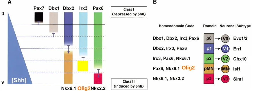
and Irx3 (Briscoe et al., 2000; Jessell, 2000; Novitch et al., 2001). Each of these transcription factors is differentially sensitive to Shh concentration, which leads to each gene having a unique and restricted expression pattern along the dorsoventral axis. For example, a higher concentration of Shh is required to induce Nkx2.2 compared to that required to induce Nkx6.1, thus Nkx6.1 expression extends more dorsally where Shh concentrations are lower. This logic also holds true for Class I genes, for example higher concentrations of Shh are needed to repress Pax6 compared with those required to repress Dbx2, thus Pax6 expression extends more ventrally into regions with higher Shh concentration (Jessell, 2000). In addition to the Shh gradient, dorsally derived Bone Morphogenic Proteins (BMPs) exert an opponent influence on these patterning factors (Jessell, 2000).
Pairs of Class I and Class II genes exert cross repressive interactions on one another which allows for the refinement of these extracellular signals into sharp boundaries. For example Pax6 and Nkx2.2 cross repress each other, and Nkx6.1 and Dbx2 cross repress each other (Ericson et al., 1997; McMahon, 2000) These
code of patterning molecules expressed in each progenitor domain are illustrated in Figure 1.
Figure 1 – Neural Patterning in the Ventral Spinal Cord
Olig2
Olig2
- modified from: (McMahon, 2000)
result in an even greater diversity of neurons generated just within the ventral spinal cord.
Neuron-glia transition
In all vertebrates in all regions of the CNS, neurons are generated prior to glial cells. In the spinal cord, this involves a temporal transition in ventricular zone precursors from producing neurons to producing glial cells. This “Gliogenic” switch involves both the inhibition of neurogenesis and the induction of gliogenesis.
Recently, the NFI family of transcription factors was found to be both necessary and sufficient for this transition (Deneen et al., 2006). GLAST is a marker of radial-glia / astrocyte precursors which is not expressed in ventricular zone progenitors during neurogenesis but which turns on at the onset of the transition to gliogenesis (Shibata et al., 1997). Like GLAST, NFI genes are also upregulated in ventricular zone at the onset of gliogenesis. Moreover, NFIA/B gain of function drives precocious
expression of GLAST, and conversely the normal temporal induction of GLAST fails to occur in the presence of NFIA siRNA (Deneen et al., 2006). Thus the pro-Glial aspect of this transition appears to be controlled by NFI genes.
NFIA siRNA experiments, and it was discovered that Notch signaling is dependant on NFI gene function during gliogenesis (Deneen et al., 2006).
Another important aspect of the transition from neurogenesis to gliogenesis is that it appears to be an irreversible transition, at least within ventricular zone
progenitors from the pMN domain. While isochronic transplantation of neurogenic stage (mouse E9.5) FACS isolated Olig2-GFP + progenitors into the neurogenic environment of the E2 chick embryonic spinal cord, these cells made neurons. By contrast, heterochronic transplantation of gliogenic stage (mouse E13.5) Olig2-GFP+ progenitors into the neurogenic E2 chick spinal cord did not yield any detectable neurons (Mukouyama et al., 2006). These in vivo experiments provide a more accurate assessment of the intrinsic potential of these cells than can be obtained by in vitro experiments. In fact these results contradict the in vitro observation that E13.5 glial stage Olig2-GFP+ cells can produce neurospheres which subsequently yield neurons (Mukouyama et al., 2006). The in vitro neurogenic capacity of these cells is likely due to reprogramming in neurosphere culture which has been previously observed with regard to patterning information (Gabay et al., 2003).
Spatial patterning and Gliogenesis
The strategy of spatial patterning which is essential in the generation of
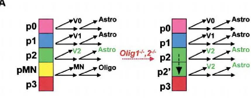
development (Lu et al., 2000; Zhou et al., 2000). This domain generates motor neurons during neurogenesis and Olig2 is necessary and sufficient for both motor neuron and oligodendrocyte fate (Lu et al., 2002; Novitch et al., 2001; Zhou and Anderson, 2002; Zhou et al., 2001). In Olig1,2 -/- embryos, the progenitors of the erstwhile pMN domain acquire the identity of the immediately dorsal p2 domain, as Irx3 expands ventrally during the phase of ventral patterning (Zhou and Anderson, 2002). This transformed ectopic p2 domain generates V2 interneurons and then subsequently astrocytes. This fate transformation is illustrated in Figure 2. Olig2 controls the specification of a neuronal subtype and glial subtype from the same progenitor domain during different phases of development.
Figure 2: Cell Fate Transformation in the Olig1,2 -/- mutant
Whether other factors involved in neuronal patterning are also important in glial subtype specification is less clear. While Nkx2.2 represses Olig2 during neurogenesis, it is co-expressed with Olig2 in the oligodendrocyte lineage and collaborates with Olig2 in promoting oligodendrocyte fate (Zhou et al., 2001).
Olig2 antagonizes the ability of NFI genes to induce astrocyte differentiation and GFAP expression (Deneen et. al., 2006). Thus the pro-glial program of NFI genes is coupled to their later pro-astrocytic functions, and while Olig2 is dependant on the early pro-glial function it is also sufficient to override the later pro-astrocytic functions.
The above results suggest that astrocyte specification can proceed under the control of NFI genes so long as Olig2 is repressed in astrocyte precursors. Further evidence of this comes from the discovery that the bHLH transcription factor SCL is a p2 domain specific factor which promotes astrocyte fate and represses
oligodendrocyte generation within this domain by repressing Olig2 expression (Muroyama et al., 2005). These studies also show that Olig2 can repress SCL. Such cross-repressive interactions can ensure the silencing of Olig2 expression in astrocyte precursors and allow for astrocye specification and differentiation.
has led to speculation over whether there are distinct subtypes of oligodendrocytes (Richardson et al., 2006).
Outline of the Thesis
This thesis aims to address the how astrocyte diversity is specified in the developing spinal cord. Chapter 2 describes Microarray experiments comparing Olig -/- and Olig +/- populations during early gliogenesis which identified genes
differentially expressed in early astrocyte vs. oligodendrocyte progenitor populations. I will discuss how these microarray data were analyzed and how among the candidate genes with increased in the Olig -/- astrocytic population we identified Reelin, Slit1 and Pax6, which we confirmed to be expressed in subsets of white matter astrocytes but not in oligodendrocytes. In Chapter 3, I will discuss our finding that the partially overlapping pattern of Reelin and Slit1 expression defines 3 molecularly and
positionally distinct subsets of astrocytes in the ventral white matter and that this positional identity is controlled by Pax6. Additionally, I will discuss our discovery that while Pax6 specifically marks Reelin expressing astrocytes, Nkx6.1 specifically labels Slit1 expressing astrocytes. I will present our evidence for the prespecification of astrocyte identity using these markers along with the Olig2-GFP in the Olig -/- mutant as marker of p2 derived astrocytes. Importantly, the regulation of astrocyte positional identity in the spinal cord by Pax6 provides the first evidence that different types of astrocytes are produced during development.
examining the role of Pax6 in pMN progenitors during motor neurogenesis, cloning and testing Tet inducible RCAN retroviral vectors for inducible misexpression of Pax6 in ovo, investigating the role of Shox2 in motor-neuron vs. V2 interneuron fate and oligodendrocyte precursor induction, and functional studies of HFH4 during gliogenesis.
References
Bachoo, R.M., Kim, R.S., Ligon, K.L., Maher, E.A., Brennan, C., Billings, N., Chan, S., Li, C., Rowitch, D.H., Wong, W.H., and DePinho, R.A. (2004).
Molecular diversity of astrocytes with implications for neurological disorders. PNAS 101, 8384-8389.
Briscoe, J., Pierani, A., Jessell, T.M., and Ericson, J. (2000). A Homeodomain Protein Code Specifies Progenitor Cell Identity and Neuronal Fate in the Ventral Neural Tube. Cell 101, 435-445.
Cafferty, W.B.J., Yang, S.-H., Duffy, P.J., Li, S., and Strittmatter, S.M. (2007). Functional Axonal Regeneration through Astrocytic Scar Genetically Modified to Digest Chondroitin Sulfate Proteoglycans. J. Neurosci. 27, 2176-2185.
Cai, J., Qi, Y., Hu, X., Tan, M., Liu, Z., Zhang, J., Li, Q., Sander, M., and Qiu, M. (2005). Generation of Oligodendrocyte Precursor Cells from Mouse Dorsal Spinal Cord Independent of Nkx6 Regulation and Shh Signaling. Neuron 45, 41-53.
Dani, J.W., Chernjavsky, A., and Smith, S.J. (1992). Neuronal activity triggers calcium waves in hippocampal astrocyte networks. Neuron 8, 429-440.
Deneen, B., Ho, R., Lukaszewicz, A., Hochstim, C.J., Gronostajski, R.M., and Anderson, D.J. (2006). The Transcription Factor NFIA Controls the Onset of Gliogenesis in the Developing Spinal Cord. Neuron 52, 953-968.
Doetsch, F. (2003). The glial identity of neural stem cells. Nat Neurosci 6, 1127-1134.
Doetsch, F., Caille, I., Lim, D.A., Garcia-Verdugo, J.M., and Alvarez-Buylla, A. (1999). Subventricular Zone Astrocytes Are Neural Stem Cells in the Adult
Mammalian Brain. Cell 97, 703-716.
Elisabet Berglöf, S.A.B.I.S. (2007). Glial influence on nerve fiber formation from rat ventral mesencephalic organotypic tissue cultures. The Journal of Comparative Neurology 501, 431-442.
Ericson, J., Rashbass, P., Schedl, A., Brenner-Morton, S., Kawakami, A., van Heyningen, V., Jessell, T.M., and Briscoe, J. (1997). Pax6 Controls Progenitor Cell Identity and Neuronal Fate in Response to Graded Shh Signaling. Cell 90, 169-180.
Fields, R.D., and Stevens-Graham, B. (2002). NEUROSCIENCE: New Insights into Neuron-Glia Communication. Science 298, 556-562.
Gee, J.R., and Keller, J.N. (2005). Astrocytes: regulation of brain homeostasis via apolipoprotein E. The International Journal of Biochemistry & Cell Biology 37, 1145-1150.
Hall, Z ed. (1992). Introduction to Molecular Neurobiology. Sinauer Assoc.
Haydon, P.G., and Carmignoto, G. (2006). Astrocyte Control of Synaptic
Transmission and Neurovascular Coupling. Physiological Reviews 86, 1009-1031.
Jessell, T.M. (2000). NEURONAL SPECIFICATION IN THE SPINAL CORD: INDUCTIVE SIGNALS AND TRANSCRIPTIONAL CODES. Nature Reviews Genetics 1, 20-29.
Karunaratne, A., Hargrave, M., Poh, A., and Yamada, T. (2002). GATA Proteins Identify a Novel Ventral Interneuron Subclass in the Developing Chick Spinal Cord. Developmental Biology 249, 30-43.
Kettenmann, H.R., BR. (1995). Neuroglia. New York. Oxford University Press.
Laywell, E.D., Rakic, P., Kukekov, V.G., Holland, E.C., and Steindler, D.A. (2000). Identification of a multipotent astrocytic stem cell in the immature and adult mouse brain. PNAS 97, 13883-13888.
Louvi, A., and Artavanis-Tsakonas, S. (2006). Notch signalling in vertebrate neural development. Nat Rev Neurosci 7, 93-102.
Lu, Q.R., Sun, T., Zhu, Z., Ma, N., Garcia, M., Stiles, C.D., and Rowitch, D.H. (2002). Common Developmental Requirement for Olig Function Indicates a Motor Neuron/Oligodendrocyte Connection. Cell 109, 75-86.
Mary S. Bailey, M.T.S. (1993). Astrocyte subtypes in the rat olfactory bulb: Morphological heterogeneity and differential laminar distribution. The Journal of Comparative Neurology 328, 501-526.
Masahira, N., Takebayashi, H., Ono, K., Watanabe, K., Ding, L., Furusho, M., Ogawa, Y., Nabeshima, Y.-i., Alvarez-Buylla, A., Shimizu, K., and Ikenaka, K. (2006). Olig2-positive progenitors in the embryonic spinal cord give rise not only to motoneurons and oligodendrocytes, but also to a subset of astrocytes and ependymal cells. Developmental Biology 293, 358-369.
McMahon, A.P. (2000). Neural patterning: The role of Nkx genes in the ventral spinal cord. Genes Dev. 14, 2261-2264.
Miller, R.H., and Raff, M.C. (1984). Fibrous and protoplasmic astrocytes are biochemically and developmentally distinct. J. Neurosci. 4, 585-592.
Miller, R.R.H., and Szigeti, V.V. (1991). Clonal analysis of astrocyte diversity in neonatal rat spinal cord cultures. Development 113, 353-362.
Mukouyama, Y.-s., Deneen, B., Lukaszewicz, A., Novitch, B.G., Wichterle, H., Jessell, T.M., and Anderson, D.J. (2006). Olig2+ neuroepithelial motoneuron progenitors are not multipotent stem cells in vivo. PNAS 103, 1551-1556.
Muroyama, Y., Fujiwara, Y., Orkin, S.H., and Rowitch, D.H. (2005).
Novitch, B.G., Chen, A.I., and Jessell, T.M. (2001). Coordinate Regulation of Motor Neuron Subtype Identity and Pan-Neuronal Properties by the bHLH Repressor Olig2. Neuron 31, 773-789.
Ogawa, Y., Takebayashi, H., Takahashi, M., Osumi, N., Iwasaki, Y., and Ikenaka, K. (2005). Gliogenic Radial Glial Cells Show Heterogeneity in the Developing Mouse Spinal Cord. Developmental Neuroscience 27, 364-377.
Parpura, V., Basarsky, T.A., Liu, F., Jeftinija, K., Jeftinija, S., and Haydon, P.G. (1994). Glutamate-mediated astrocyte-neuron signalling. Nature 369, 744-747.
Parpura, V., and Haydon, P.G. (2000). From the Cover: Physiological astrocytic calcium levels stimulate glutamate release to modulate adjacent neurons. PNAS 97, 8629-8634.
Paton, J.A., and Nottebohm, F. (1984). Neurons Generated in the Adult Brain Are Recruited into Functional Circuits. Science 225, 1046-1048.
Pringle, N.P., Yu, W.-P., Howell, M., Colvin, J.S., Ornitz, D.M., and Richardson, W.D. (2003). Fgfr3 expression by astrocytes and their precursors: evidence that astrocytes and oligodendrocytes originate in distinct neuroepithelial domains. Development 130, 93-102.
Qi, Y., Cai, J., Wu, Y., Wu, R., Lee, J., Fu, H., Rao, M., Sussel, L., Rubenstein, J., and Qiu, M. (2001). Control of oligodendrocyte differentiation by the Nkx2.2 homeodomain transcription factor. Development 128, 2723-2733.
Seri, B., Garcia-Verdugo, J.M., McEwen, B.S., and Alvarez-Buylla, A. (2001). Astrocytes give rise to new neurons in the adult mammalian hippocampus. Journal of Neuroscience 21, 7153-7160.
Shibata, T., Yamada, K., Watanabe, M., Ikenaka, K., Wada, K., Tanaka, K., and Inoue, Y. (1997). Glutamate Transporter GLAST Is Expressed in the Radial Glia-Astrocyte Lineage of Developing Mouse Spinal Cord. J. Neurosci. 17, 9212-9219.
Song, H., Stevens, C.F., and Gage, F.H. (2002). Astroglia induce neurogenesis from adult neural stem cells. Nature 417, 39-44.
Sun, T., Pringle, N.P., Hardy, A.P., Richardson, W.D., and Smith, H.K. (1998). Pax6 Influences the Time and Site of Origin of Glial Precursors in the Ventral Neural Tube. Molecular and Cellular Neuroscience 12, 228-239.
Vallstedt, A., Klos, J.M., and Ericson, J. (2005). Multiple Dorsoventral Origins of Oligodendrocyte Generation in the Spinal Cord and Hindbrain. Neuron 45, 55-67.
van Praag, H., Schinder, A.F., Christie, B.R., Toni, N., Palmer, T.D., and Gage, F.H. (2002). Functional neurogenesis in the adult hippocampus. Nature 415, 1030-1034.
Zhou, M., and Kimelberg, H.K. (2001). Freshly isolated hippocampal CA1 astrocytes comprise two populations differing in glutamate transporter and AMPA receptor expression. Journal of Neuroscience 21, 7901-7908.
Zhou, Q., Choi, G., and Anderson, D.J. (2001). The bHLH Transcription Factor Olig2 Promotes Oligodendrocyte Differentiation in Collaboration with Nkx2.2. Neuron 31, 791-807.
Chapter 2
Microarray Screen for Olig target genes in gliogenesis
Introduction
The bHLH transcription factor Olig2 is the first gene identified which regulates glial subtype specification. Olig2 is necessary and sufficient for
oligodendrocyte formation and in the absence of Olig2, cells marked by a knockin Olig2-GFP generate astrocytes (Zhou and Anderson, 2002; Zhou et al., 2001). This fate conversion suggests that in addition to promoting oligodendrocyte fate, Olig2 plays an important role in suppressing astrocyte fate (Lu et al., 2002; Zhou and Anderson, 2002). Given evidence that Olig2 acts as a transcriptional repressor (Zhou et al., 2001), we postulated that important astrocyte regulatory genes might be targets of Olig2 repression and designed a screen to identify these target genes.
Our strategy was to use Olig2-GFP to FACS isolate glial progenitors from Olig +/- and Olig -/- embryos and to compare their gene expression profiles on Affymetrix cDNA microarrays. We hoped to identify new pro-astrocytic or
Results
Isolation and Microarray comparison of E14.5 Olig +/- and Olig -/- glial
progenitors
We performed our first microarray comparison with cells isolated from E14.5 embryos. The major reason for selecting this stage was to avoid contamination of our samples by neurons. Olig2 is normally expressed in motor neuron precursors but is rapidly downregulated as precursors migrate out of the ventricular zone and
differentiate into motor neurons. Expression of Olig2 protein is not detectable in postmitotic motor neurons. By contrast, due to the stability of the GFP protein, Olig2-GFP expression perdures into postmitotic motor neurons (+/-) or V2
interneurons (-/-). Some of this GFP protein perdurance in neurons is still detectable in the early stages of gliogenesis (E12-E13). By E14.5, there is no detectable GFP expression in neurons and we were confident that all GFP+ cells isolated for comparison would be glial precursors.
significant differences in gene expression between these two populations which are unrelated to the question of oligodendrocyte vs. astrocyte fate.
A Second Microarray comparison of Olig +/- and Olig -/- progenitors: E13.0
Screen
To complement our analysis and address our concerns, we performed a second microarray comparison with cells isolated at E13.0. At this stage we were able to obtain very comparable populations of ventricular zone cells for the comparison, by using PDGFRa staining as a negative selection marker to exclude the few migrating oligodendrocyte precursors in the Olig1,2 +/- heterozygote (see FACS plots, Figure 1). The possibility of neuronal contamination due to GFP perdurance, as discussed above, was the potential caveat at this earlier stage. We performed independent replicates at this timepoint, as was done at E14.5.
MAS4 and Rosetta Resolver Analysis: selection of candidate genes
The results of the E14.5 and E13.0 screens revealed several interesting candidates common to both screens. For both screens we initially analyzed the data using Affymetrix Microarray Suite software (MAS4). We set our threshold for differential expression at a minimum 3 fold change in expression between samples and a
minimum average difference change between samples of 50 (with the target intensity normalized to 200). In the E14.5 screen of the A, B, and C chips over 1300
cell cycle factors were differentially expressed in the E14.5 screen, and this
background (presumably due to fundamental differences in ventricular vs. migrating cells) was eliminated in the E13.0 screen. Likewise, factors involved in neurogenesis such as Irx3, Chx10 and Islet1 were identified as differentially expressed in the E13.0 screen but not the E14.5 screen. Importantly, both screens found oligodendrocyte precursor markers (PDGFRa, sox10 and NG2) to be strongly downregulated in the Olig1,2 -/- mutant, while the astrocyte marker glutathione transferase Yb is strongly upregulated in the Olig 1,2 -/- mutant. The consistency of these genechip results with our previous observations in the Olig1,2 -/- mutant is an important positive control and indicates that our screens can identify expected differences in gene expression between the cell types being compared.
In order to eliminate likely false positives and focus on genes whose
differential expression is really due to the presence or absence of Olig expression, we decided to focus on genes differentially expressed in both screens. For this we
performed analysis with Rosetta Resolver, which enabled us to perform a 4 way cross comparison, using both replicates of both the E14.5 and E13.0 screens. This analysis yielded around 350 genes across the A, B and C chips with at least a 3 fold change and a p value less than .01. The log intensity plot for the A chip is shown in Figure 2. Genes with higher expression in the Olig -/- population are shown in blue, while
total of 41 transcription factors were differentially expressed across the A, B and C chips in this analysis and these are shown in Table 1.
Candidate gene validation
We next began using in situ hybridization to examine the expression of candidate genes through different stages of gliogenesis. Priority for follow up analysis by in situ was given to transcription factors and genes with known functions in the nervous system with strong fold changes and average difference changes. 35 candidate genes were tested by in situ hybridization of Olig1,2 +/- heterozygous and Olig1,2 -/- homozygous embryos during early gliogenesis (E12.5), mid gliogenesis (E14.5) and late gliogenesis (E18.5). Sample in situ panels are shown for 2 candidate
transcription factors: HFH4 (Figure 3) and Shox2 (Figure 4). From these analyses we selected 5 genes whose expression patterns suggest they may be regulated by Olig and are expressed in glial cells or their precursors: HFH4, Shox2, Pax6, Reelin and Slit1. We performed further analysis and experiments with these genes. Experiments with Pax6, Reelin and Slit1 are discussed in Chapter 3, while experiments involving HFH4 and Shox2 are discussed in the Appendix. All of these genes were upregulated in the Olig1,2 -/- mutant, which is interesting given Olig2 appears to act as a
transcriptional repressor.
Discussion
factor Pax6, all of which were upregulated in the Olig -/- “astrocyte progenitor” population. Our expression data confirmed that these genes are expressed in specific subsets of astrocytes in the ventral white matter and are not expressed in
oligodendrocytes. Studies with these genes led to our discovery of how astrocyte positional identity is regulated in the spinal cord (Chapter 3).
Functional experiments were also performed 2 other candidate genes
upregulated in the Olig -/- population, both of which were expressed in a restricted subset of ventricular zone progenitors during early gliogenesis. First, the
homeodomain transcription factor Shox2, which upon closer examination was
expressed in the pMN domain at the onset of oligodendrocyte formation, as well as in V2 interneurons and whose gain of function phenotypes include repression of MN fate, promotion of V2 interneuron fate, and promoting precocious oligodendrocytes together with Olig2. Second, was the forkhead transcription factor HFH4, whose gain of function and knockout analysis had no phenotypes with respect to general oligodendrocyte and astrocyte specification markers. These experiments are discussed in further detail in the Appendix.
was recently shown to play a role in both V2b interneuron and astrocyte generation from the p2 domain via cross-repressive interactions with Olig2 (Muroyama et al., 2005). Unfortunately, SCL was identified as upregulated in the Olig -/- mutant population in our screen (see Table I), but we chose not to pursue it due to published data at the time that it was only expressed in V2 interneurons and not in any other cells at stages of early gliogenesis (Emma Smith, 2002). Despite our inability to find a pro-astrocytic regulator, we were able to take advantage of the identification of Pax6, Reelin and Slit1 expression in subsets of astrocytes to uncover the first evidence of astrocyte subtype specification during development.
Experimental Procedures
Spinal cord dissociation and FACS
We devised a method to rapidly genotype embryos using Xgal staining of the head. This was done to speed up the process of dissection and sorting with minimal wait time, thus minimizing cell death. Olig1,2 -/- homozygotes show very bright Xgal staining (due to the Olig1-lacZ knockin allele), Olig1,2 +/- heterozygotes show weak staining, and wild type embryos have no staining. PCR was then done
RNA isolation, amplification and preparation
Immediately following FACS, RNA was isolated from the cells using the Stratagene microRNA isolation kit. The RNA was then subjected to two rounds of amplification using the MessageAmp aRNA kit (Ambion). The aRNA was biotin labeled during the second round and fragmented to an average size of 80-100 bp, as recommended by Affymetrix. Using these procedures we could generate around 100 ug of aRNA probe from a starting material of 3000-5000 FACS isolated cells.
In situ hybridization
Non-radioactive in situ hybridization using DIG-labelled probes was performed on frozen sections of mouse spinal cord, as previously described (Zhou et al., 2000).
Acknowledgements
References
Deneen, B., Ho, R., Lukaszewicz, A., Hochstim, C.J., Gronostajski, R.M., and Anderson, D.J. (2006). The Transcription Factor NFIA Controls the Onset of Gliogenesis in the Developing Spinal Cord. Neuron 52, 953-968.
Emma Smith, M.H.T.Y.C.G.B.M.H.L. (2002). Coexpression of SCL and GATA3 in the V2 interneurons of the developing mouse spinal cord. Developmental Dynamics 224, 231-237.
Lu, Q.R., Sun, T., Zhu, Z., Ma, N., Garcia, M., Stiles, C.D., and Rowitch, D.H. (2002). Common Developmental Requirement for Olig Function Indicates a Motor Neuron/Oligodendrocyte Connection. Cell 109, 75-86.
Muroyama, Y., Fujiwara, Y., Orkin, S.H., and Rowitch, D.H. (2005). Specification of astrocytes by bHLH protein SCL in a restricted region of the neural tube. Nature 438, 360-363.
Zhou, Q., and Anderson, D.J. (2002). The bHLH Transcription Factors OLIG2 and OLIG1 Couple Neuronal and Glial Subtype Specification. Cell 109, 61-73.
Zhou, Q., Choi, G., and Anderson, D.J. (2001). The bHLH Transcription Factor Olig2 Promotes Oligodendrocyte Differentiation in Collaboration with Nkx2.2. Neuron 31, 791-807.
Chapter 3
Pax6 controls astrocyte positional identity in the spinal cord
Abstract
While astrocytes play many diverse and important roles in the vertebrate
CNS, little is known about the molecular diversity of these cells or the factors
specifying such diversity. We found that the secreted signaling molecules Reelin
and Slit1 mark 3 positionally defined subsets of astrocytes in the ventral white
matter of the spinal cord: Reelin+/Slit1- astrocytes in the lateral white matter (L
type), Reelin+/Slit1+ astrocytes in the ventral-lateral white matter (VL type),
and Slit1+/Reelin- astrocytes in the ventral-medial white matter (VM type). The
homeodomain transcription factor Pax6 is specifically expressed in Reelin+
astrocytes (L and VL type). We found that Pax6 plays a necessary and
instructive role in specifying these populations via its actions of promoting
Reelin and repressing Slit1 expression. We additionally show that the
homeodomain transcription factor Nkx6.1 specifically marks Slit1+ astrocytes
(VM and VL type), and provide evidence that VL type astrocytes
(Pax6+/Nkx6.1+) are derived from the p2 domain of the ventricular zone
(Pax6+/Nkx6.1+). These data are consistent with a model whereby these
astrocyte populations are prespecified in the ventricular zone. Importantly, we
provide the first evidence that molecularly distinct subtypes of astrocytes are
produced during development, each with a defined positional identity in the
white matter. While positional identity is an important property of many
neuronal subtypes, it has not been previously described in glial cells and may be
Introduction
Astrocytes are the most abundant cell type in the central nervous system (CNS) and perform a wide variety of diverse roles. Many of these functions are involved with passively providing a supportive environment for neurons, including balacing pH and ion concentrations, recycling neurotransmitters, storing energy, controlling blood vessels and forming the blood-brain barrier (Gee and Keller, 2005). However, there is also evidence suggesting that, like neurons, astrocytes may play an active role in information processing in the CNS (Fields and Stevens-Graham, 2002).
Hippocampal astrocytes respond to glutamatergic firing with actively propagating Ca2+ waves in organotypic slice cultures (Dani et al., 1992). Perisynaptic astrocytes
and grey matter: cultures from developing gray matter generate only type 1 astrocytes, while white matter derived cultures generate both type 1 and type 2 astrocytes (Miller and Raff, 1984). However because these types are characterized after in vitro culture, it is not clear whether they are truly differentially distributed in vivo. Hippocampal astrocytes in the CA1 and CA3 layers have different
electrophysiological properties, suggesting they may represent functionally distinct subtypes. (D'Ambrosio et al., 1998). Astrocytes within the CA1 layer display heterogeneous expression of glutamate transporter and AMPA receptor (Zhou and Kimelberg, 2001). More recently, gene expression profiling experiments were conducted on various in vitro astrocyte cultures and astrocyte-rich CNS tissues in an effort to uncover information about the molecular diversity of astrocytes (Bachoo et al., 2004). While there is evidence supporting the notion of morphological,
molecular and regional heterogeneity among astrocytes, it is not clear whether this is indicative of distinct pre-specified astrocyte subtypes or merely passively acquired phenotypic differences due to regional cues acting on a single uniformly specified astrocyte population.
2000). The neuronal subtypes generated from this spatial patterning are distinguished both by their expression of specific molecular markers and by their positional identity along the dorsoventral axis. There is a hierarchical determination of neuronal subtype identity in the motor neuron lineage, where LIM homeodomain proteins act to further sub-specify motor neurons into distinct columns, divisions and pools, each of which is distinguished by a distinct cell body positioning within the motor column and by projections to distinct targets (Jessell, 2000).
Oligodendrocytes and astrocytes are generated from ventricular zone progenitors following a cell intrinsic temporal switch from neurogenesis to gliogenesis. In the spinal cord, this switch involves the upregulation of the pro-glial NFI family of transcription factors as well as the inhibition of neurogenesis by Notch signalling (Deneen et al., 2006; Louvi and Artavanis-Tsakonas, 2006). Oligodendrocytes are generated from the pMN domain of the ventral ventricular zone, which is marked by the expression of the bHLH transcription factor Olig2 (Lu et al., 2000; Zhou et al., 2000). This domain generates motor neurons during neurogenesis and Olig2 is necessary and sufficient for both motor neuron and oligodendrocyte fate (Lu et al., 2002; Novitch et al., 2001; Zhou and Anderson, 2002; Zhou et al., 2001). In the absence of Olig2, the progenitors acquire the identity of the immediately dorsal p2 domain and they generate V2 interneurons and then astrocytes. Thus, Olig2 controls the specification of a neuronal and glial subtype from the same progenitor domain during different phases of development.
with this it has been shown that the pro-glial NF1 genes are sufficient to promote astrocyte differentiation in the absence of Olig2 antagonism (Deneen et al., 2006). Also, astrocytes appear to be broadly generated from VZ progenitors outside of pMN, as assayed by the migration of NFIA+, GLAST+, and FGFR3+ cells (Deneen et al., 2006; Pringle et al., 2003; Shibata et al., 1997). Further evidence that repression of Olig2 is essential in astrocyte specification comes from the discovery that the bHLH transcription factor SCL is a p2 domain specific factor which promotes astrocyte fate and represses oligodendrocyte generation within this domain by repressing Olig2 expression (Muroyama et al., 2005).
We sought to investigate the issue of astrocyte diversity and its developmental specification in the spinal cord. As a first step in addressing this question it was necessary to identify molecular markers which label specific populations of
astrocytes. Here we describe 3 molecularly and positionally distinct subpopulations of astrocytes in the spinal cord using the secreted signaling molecules Reelin and Slit1 as molecular markers: Reelin+/Slit1- in the lateral white matter, Reelin+/Slit1+ in the ventral-lateral white matter, and Slit1+/Reelin- in the ventral-medial white matter.
The homeodomain transcription factor Pax6 is specifically expressed in Reelin+ astrocytes. Pax6 has been previously shown to play an essential role as fate determinant in the development of many organs including cortex, spinal cord, eye and pancreas (Ashery-Padan et al., 2000; Gotz et al., 1998; Marquardt et al., 2001; Muzio et al., 2002; St-Onge et al., 1997) In the spinal cord, Pax6 is part of the
V2 interneuron generation (Ericson et al., 1997). We found that Pax6 is necessary and sufficient to promote Reelin and repress Slit1 in astrocytes of the ventral white matter. These results suggest that, like Olig2, Pax6 plays a dual role and in regulating both neuronal and glial subtype specification and provides additional evidence that the same factors can impact the specification of cell types from a particular pool of VZ progenitors in both the neurogenic and gliogenic phases.
Results
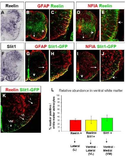
Reelin and Slit1 mark subpopulations of astrocytes in ventral white matter During gliogenesis, the bHLH transcription factor Olig2 controls the
oligodendrocyte vs. astrocyte fate decision, with progenitors in the Olig2 mutant mouse generating astrocytes instead of oligodendrocytes (Zhou and Anderson, 2002). We used Affymetrix cDNA microarrays to compare the gene expression profiles of FACS isolated Olig2-GFP expressing glial progenitors from the Olig1,2 -/- and Olig1,2 +/- spinal cord. Reelin and Slit1 were among the candidate genes with higher expression in the Olig1,2 -/- ‘converted astrocyte progenitor’ population. As our initial analysis, we performed in situ hybridization on spinal cord sections and found Reelin and Slit1 were expressed in the white matter at E18.5, indicating these genes were likely expressed in glial cells (Figure 1 A, F). We then examined the
NFIA+ astrocytes (Figure 1) and were not expressed in Olig2+ oligodendrocytes (data not shown). Both Reelin and Slit1 were also expressed in many neurons in the grey matter, but in the white matter their expression was astrocyte specific.
Morphologically, both Reelin+ and Slit1+ astrocytes are characterized by their cell bodies localized at the subpial surface (as identified by NF1A+ nuclei) with radially oriented GFAP+ processes projecting inward. This morphology has been previously characterized as the most common for astrocytes in the white matter (Liuzzi and Miller, 1987).
Interestingly, we found that Reelin and Slit1 did not label all astrocytes, but each was spatially restricted to a subset of astrocytes in the ventral white matter. Reelin was expressed in astrocytes of the lateral and ventral-lateral white matter, but not in astrocytes close to the ventral midline (Figure 1). Slit1 on the other hand is expressed in astrocytes in the ventral-medial and ventral-lateral white matter but not in
astrocytes of the lateral white matter. Double labelling of Reelin and Slit1 shows that their expresion overlaps in the ventral-lateral white matter (Figure 1 K).
Quantification of this overlap shows that around 50% of Reelin+ astrocytes are also Slit1+, and around 50% of Slit1+ astrocytes are also Reelin+. Thus astrocytes in the ventral white matter can be divided into 3 positionally and molecularly defined subpopulations which are approximately equal in abundance: Slit1+ only astrocytes in the ventral-medial, Slit1+/Reelin + co-expressing astrocytes in the ventral-lateral, and Reelin + only astrocytes in the lateral white matter (see Figure 1 L). For
as L type (lateral white matter – Reelin+), VL type (ventral-lateral white matter – Reelin+, Slit1+), and VM type (ventral-medial white matter – Slit1+).
Reelin expressing astrocytes co-express Pax6
Pax6 was also upregulated in the Olig1,2 -/- population in our microarray screen. We found that Pax6 marked a subpopulation of astrocytes in the ventral-lateral white matter very similar in distribution to that of Reelin. The Pax6+ fraction represents approximately 40% of GFAP+ and NF1A+ white matter astrocytes (Figure 2 A-D, I). Double labeling confirmed that Pax6 and Reelin are colocalized and mark L and VL type astrocytes (Figure 2 E,F,I). We confirmed that, like Reelin, Pax6 was co-expressed with Slit1 in the VL type astrocyte population (Figure 2 G,H, I). Since Pax6 has been shown to be a cell fate determinant in many systems, including during neurogenesis in the ventral spinal cord, we next sought to examine whether Pax6 played any role in regulating the identity of these astrocyte subpopulations.
Pax6 is required for Reelin expression in astrocytes
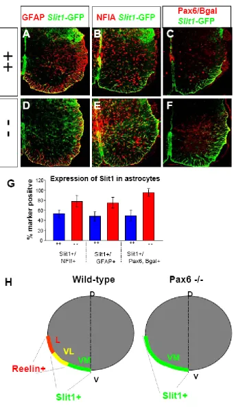
Pax6 knockout mice have been previously generated with LacZ knocked into the Pax6 locus (St-Onge et al., 1997). We examined E18.5 spinal cord sections from Pax6 -/- and +/+ mice and found that there were a normal number of NF1A and GFAP+ astrocytes in the Pax6 -/- spinal cord (Figure 3). Thus Pax6 does not appear to be required for generic migration and differentiation of astrocytes.
in the Pax6 mutant. This loss of astrocytic Reelin was evident both by in situ
hybridization (Figure 3 A,B,E,F) and immunostaining (Figure 3 C,D,G,H). We found that the total number of Reelin+/GFAP+ and Reelin+/NFIA+ cells was significantly reduced, as were the percentages of GFAP+ and NFIA+ cells which are Reelin+ (Figure 3 I, J). One possible explanation for the loss of Reelin+ astrocytes is that the loss of Pax6 leads to the selective cell death of this subpopulation or to a failure to migrate or differentiate normally, coupled with a non cell-autonomous compensation for the number of GFAP+ and NFIA+ astrocytes. This is not the case because there were a normal number of Bgal+ astrocytes in the white matter of the Pax6 -/- mutant. The percentage of Bgal+ cells expressing Reelin in the Pax6 -/- mutant was severely reduced compared to the percentage of Pax6+ cells which normally express Reelin in the wild-type (Figure 3J). Thus Pax6 is required for the expression of Reelin in L and VL type astrocytes, but not for their formation and localization.
Astrocyte subtype conversion in the absence of Pax6
Since L type astrocytes express Pax6 and Reelin but not Slit1, we sought to examine whether Pax6 might also play a role in preventing Slit1 expression in these cells. To this end, we crossed the Pax6-lacZ mice with Slit1-GFP mice and analyzed the expression of Slit1-GFP in Pax6 mutants. We found that in addition to the loss of Reelin, GFAP+ and NFIA+ astrocytes in the lateral white matter of the Pax6 mutant upregulate Slit1 expression. While around 50% of Pax6+ cells normally co-express Slit1 (VL type), nearly all of the Bgal+ cells in the Pax6 -/- co-express Slit1,
required for the normal repression of Slit1 in L type astrocytes. The expansion of Slit1 expression together with the loss of Reelin expression in astrocytes of the Pax6 -/- mutant results in the molecular conversion of the L and VL type astrocytes (marked by Bgal expression) to the VM identity (Figure 4 H). Taken together these data demonstrate that Pax6 is required for the generation of L and VL type astrocytes in the white matter of the ventral spinal cord via its regulation of Reelin and Slit1 expression.
least some of this increase can be attributed to positionally ectopic Reelin expression in ventral astrocytes (Figure 5 C). The total number of NFIA+ astrocytes was not affected by Pax6 electroporation (data not shown). Thus Pax6 is sufficient to promote Reelin expression in astrocytes while not affecting general astrocyte fate. Interestingly, we only detected a mild increase in astrocytic Reelin when comparing the electroporated (56% Reelin+/NFIA+) vs. control sides (42%
Reelin+/NFIA+) of Pax6 electroporated embryos despite a visibly much higher level of Pax6 expression on the electroporated side. One possible explanation for this finding is that low levels of Pax6 misexpression on the control side, due to secondary infection by the replication competent virus, are sufficient to promote an increase in Reelin expression.
We also examined the effects of Pax6 misexpression on Slit1. We found a significant reduction in the percentage of Slit1+/NFIA+ astrocytes with Pax6 misexpression compared with the GFP control (Figure 5). Thus Pax6 is also sufficient to repress Slit1 expression in astrocytes. Interestingly, the reduction of Slit1 expression was only seen on the electroporated side of Pax6 electroporated embryos. This suggests that the low levels of Pax6 misexpression on the control side, due to viral spread, are not sufficient to repress Slit1. Taken together these results show that Pax6 is sufficient to promote Reelin expression and repress Slit1 expression in astrocytes. Through these functions Pax6 plays an instructive role in regulating astrocyte identity.
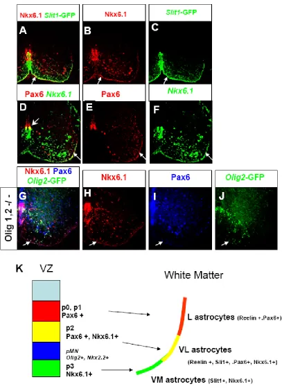
Given our results with Pax6, we hypothesized that another regulatory gene may specifically mark Slit1+ astrocytes. Such a gene would also partially overlap with Pax6 and Reelin in type VL astrocytes, and represent the functional antagonist of Pax6 in regulating the identity of these populations. Nkx6.1 is a homeodomain transcription factor expressed in the ventral ventricular zone which normally partially overlaps with Pax6 in the pMN and p2 domains. The ventral p3 domain only
expresses Nkx6.1 but not Pax6, while the more dorsal p0 and p1 domains express Pax6 but not Nkx6.1 (Jessell, 2000). This partially overlapping pattern of Pax6 and Nkx6.1 expression in the ventricular zone bears an intriguing resemblance to the partially overlapping pattern of Reelin and Slit1 expressing astrocytes in the white matter. We examined Nkx6.1 expression in E18.5 spinal cord and found that Nkx6.1 specifically marks Slit1 expressing astrocytes (Figure 6 A-C). Double labeling with Pax6 and Nkx6.1 revealed that these genes partially overlap in astrocytes of the ventral-lateral white matter, recapitulating the pattern of Reelin and Slit1 expression (Figure 6 D-F). Thus Pax6 partially overlaps with Nkx6.1 both in the ventricular zone and in ventral white matter astrocytes, such that these factors mark 3 populations of glial progenitors in the ventricular zone and 3 positionally defined types of
astrocytes along the subpial surface of the ventral white matter.
Pax6+/Nkx6.1+ astrocytes are derived from the p2 domain
The correlation between the pattern of Nkx6.1 and Pax6 expression in
ventral-medial white matter (VM type) could be derived from Nkx6.1+ progenitors from the p3 domain, Nkx6.1+, Pax6+ astrocytes of the ventral-lateral white matter (VL type) could be derived from the Nkx6.1+, Pax6+ progenitors of the p2 domain, and Pax6+ astrocytes of the lateral white matter (L type) could be derived from the Pax6+ progenitors of the p0 and p1 domains. Although Pax6 and Nkx6.1 also overlap in the pMN domain in the ventricular zone, the pMN domain is unlikely to contribute significantly to these astrocyte subtypes since pMN progenitors give rise almost exclusively to oligodendrocytes, under the direction of Olig2 (Zhou and Anderson, 2002).
In order to test our hypothesis that these astrocyte subpopulations may be
prespecified from particular ventricular zone domains, we took advantage of the fact that Olig2-GFP expressing cells in the Olig1,2 -/- mutant are respecified to p2 domain identity and generate p2 derived astrocytes (Zhou and Anderson, 2002). We sought to test our prediction that the Nkx6.1+, Pax6+ VL type astrocyte population was derived from the p2 domain, using Olig2-GFP in the Olig1,2 -/- mutant as a p2 lineage marker. We performed triple-labeling with Nkx6.1, Pax6 and Olig2-GFP on E18.5 Olig1,2 -/- mutant spinal cord. In this assay we found that the p2 derived Olig2-GFP+ astrocytes are Nkx6.1+ and Pax6+ (Figure 6 G-J). Not all of the Nkx6.1+, Pax6+ cells are Olig2-GFP+ since Olig2-GFP only marks the super-numerary copy of p2 replacing pMN.
positional identity of astrocytes in the ventral white matter. Further lineage tracing experiments are necessary to formally determine whether the Pax6+, Reelin+
astrocytes of the lateral white matter are derived from p0-p1 progenitors and whether the Nkx6.1+, Slit1+ astrocytes of the ventral medial white matter are derived from the p3 domain.
Discussion
While astroycte diversity has been described in various contexts of the adult CNS, the generation of this diversity has not been examined in vivo during
development. We identified 3 molecularly and positionally defined astrocyte
subpopulations in the ventral white matter of the E18.5 mouse spinal cord. We found that the homeodomain transcription factor Pax6 is an essential factor and plays an instructive role in specifying the positional identity of these populations. We
additionally provide evidence that at least one of these populations is prespecified in the ventricular zone, and propose a general model of how these subtypes are
generated based on the correlation between the partially overlapping patterns of expression of Pax6 and Nkx6.1 in both the ventricular zone and the white matter.
Molecular and Positional Identity of Astrocytes in the ventral white matter of the spinal cord
It has been shown that progenitors of the ventral ventricular zone give rise to at least 5 general classes of neurons, each arising from distinct domain of VZ
pMN domain generate oligodendrocytes (Zhou and Anderson, 2002; Zhou et al., 2000), while progenitors from other ventral domains make astrocytes (Deneen et al., 2006; Pringle et al., 2003; Shibata et al., 1997). Prior to this study, it was not clear whether a single type of astrocyte was generated from all of the domains outside of pMN or whether molecularly distinct subtypes of astrocytes are produced.
Microarray analysis of Olig mutant mice led us to identify Reelin and Slit1 as new markers which are specific for astrocytes and not oligodendrocytes. Importantly, Reelin and Slit1 were expressed in subsets of astrocytes. We found that astrocytes of the ventral white matter of the spinal cord are divided into 3 positionally and
molecularly defined subpopulations which are approximately equal in abundance: VM type (Slit1+, Nkx6.1+, ventral-medial white matter), VL type (Slit1+,Reelin +, Pax6+, Nkx6.1+, ventral-lateral white matter), and L type (Reelin +, Pax6+, lateral white matter).
1997). Likewise, markers of differentiated astrocytes such as GFAP and S100B are expressed broadly throughout the white matter. Using the molecular markers Reelin and Slit1, along with Pax6 and Nkx6.1, we were able to identify the L, VL and VM type astrocyte populations distributed along the arc of ventral white matter. The positional identity of these astrocytes may be important for region specific functions and represents a new level of both molecular diversity and organizational complexity of spinal cord glia.
Pax6 is necessary for the positional identity of spinal cord white matter astrocytes Analysis of Pax6 -/- mutant embryo spinal cord revealed that Pax6 plays 2 roles which allow for the specification of astrocyte positional identity. First, Pax6 is
required for Reelin expression in both VL and L type astrocytes. Second, Pax6 is required for the repression of Slit1 expression in L type astrocytes. These functions may or may not be mechanistically related. Since VL type astrocytes are
Reelin+/Slit1+, the functions of Pax6 in repression of Slit1 and the promotion of Reelin need not be coupled in a fate switching mechanism. On the contrary, the fact that Pax6 fails to repress Slit1 in VL type astrocytes, yet still serves to promote Reelin in this population suggests that these 2 functions can be uncoupled.
be able to repress Slit1 which is only present in L type but not VL type astrocytes. Finally, a dose dependant mechanism is possible where repression of Slit1 is highly sensitive to Pax6 concentration and where hypothetically, higher levels of Pax6 expression in L type astrocytes compared with VL type astrocytes could lead to Slit1 repression in only the L type population. While there is no evidence of such a difference in Pax6 levels of expression by immunostaining, such a model has precedent since it has been shown that Pax6 concentration can be critical in other developmental systems with a high degree of haploinsufficiency being observed in Pax6 +/- mice with respect to eye development (Davis-Silberman et al., 2005). However this possibility is unlikely as we did not detect any happloinsufficiency in Pax6 +/- spinal cord with regard to either loss of Reelin or upregulation of Slit1 (data not shown).
Pax6 is sufficient to promote Reelin and repress Slit1 expression in white matter astrocytes
experiment argues against a model where Pax6 requires a cofactor to repress Slit1, which is normally present in L type but not VL type astrocytes.
The most likely explanation for why Pax6 normally represses Slit1 in lateral but not ventral-lateral white matter astrocytes is the presence of a factor in VL but not L type astrocytes which interferes with the ability of Pax6 to repress Slit1. This interference could be overridden in a gain of function experiment due to the high levels of Pax6 misexpression. Nkx6.1 is a candidate factor for this role, as it is expressed in VL but not L type astrocytes. Additionally, while Nkx6.1 and Pax6 do not transcriptionally repress each other during patterning, there is precedent for their functional antagonism in the determination of α and β cell fates in the pancreas (Hill et al., 1999; Schisler et al., 2005). A competition experiment in which Nkx6.1 is misexpressed together with Pax6 would reveal if indeed Nkx6.1 can block the ability of Pax6 to repress Slit1 expression.
Prespecification of the ventral-lateral astrocyte subpopulation
the Pax6+/Nkx6.1+ astrocytes in this experiment were Olig2-GFP+, however, since the Olig2-GFP only marks the pMN Æ p2 converted domain and not the endogenous
p2 domain.
Spatial patterning during gliogenesis
a critical regulator of this process. The full extent and functional significance of this new aspect of glial diversity remains to be explored, yet given the genetic investment in specifying these distinct astrocyte subtypes it is likely that they have some region specific functions.
Experimental Procedures
Mouse mutants
Olig1,2 -/- mice (Zhou and Anderson, 2002), Pax6-LacZ mice (St-Onge, Sosa-Pineda et al., 1997) and Slit1-GFP mice (Plump, Erskine et al. 2002) were genotyped by PCR using lacZ and GFP primers. Pax6-LacZ mice were crossed into the Slit1-GFP background to generate Pax6 -/-, Slit1-Slit1-GFP +/- and Pax6 +/-, Slit1-Slit1-GFP +/- embryos for analysis.
In situ hybridization / Immunohistochemistry
In situ hybridization was performed on frozen sections as previously described (Zhou, Wang et al 2000). Antibodies were used against Pax6 (rabbit polyclonal, Covance and IgG1 monoclonal, DHSB), Reelin (G10 IgG1 monoclonal, Novus and 142 IgG1 monoclonal, Novus), GFP (chick polyclonal, Abcam), GFAP (rabbit polyclonal, DAKO and IgG1 monoclonal, Chemicon), NFIA (rabbit polyclonal, Active Motif), S100B (IgG1 monoclonal, Sigma), Olig2 (rabbit polyclonal, a kind gift of Tom Jessell), Nkx6.1 (IgG1 monoclonal, DHSB)