ORIGINAL RESEARCH
FUNCTIONAL
The Contribution of Common Surgically Implanted Hardware
to Functional MR Imaging Artifacts
A.A. Desai, M.K. Strother, C.C. Faraco, V.L. Morgan, T.R. Ladner, L.M. Dethrage, L.C. Jordan, and M.J. Donahue
ABSTRACT
BACKGROUND AND PURPOSE: Blood oxygenation level–dependent MR imaging is increasingly used clinically to noninvasively assess cerebrovascular reactivity and/or language and motor function. However, many patients have metallic implants, which will induce susceptibility artifacts, rendering the functional information uninformative. Here, we calculate and interpret blood oxygenation level– dependent MR imaging artifact impact arising from surgically implanted hardware.
MATERIALS AND METHODS: A retrospective analysis of all blood oxygenation level–dependent MRIs (n⫽343; B0⫽3T; TE⫽35 ms; gradient echo EPI) acquired clinically (year range⫽2006 –2014) at our hospital was performed. Blood oxygenation level–dependent MRIs were most commonly prescribed for patients with cerebrovascular disease (n⫽80) or patients undergoing language or motor localization (n⫽263). Artifact volume (cubic centimeters) and its impact on clinical interpretation were determined by a board-certified neuroradiologist.
RESULTS: Mean artifact volume associated with intracranial hardware was 4.3⫾3.2 cm3(range⫽1.1–9.4 cm3). The mean artifact volume from extracranial hardware in patients with cerebrovascular disease was 28.4⫾14.0 cm3(range⫽6.1– 61.7 cm3), and in patients with noncerebrovascular disease undergoing visual or motor functional mapping, it was 39.93⫾27.0 cm3(range⫽6.9 –77.1 cm3). The mean artifact volume for ventriculoperitoneal shunts was 95.7⫾39.3 cm3(range⫽64.0 –139.6 cm3). Artifacts had no-to-mild effects on clinical interpretability in all patients with intracranial implants. Extracranial hardware artifacts had no-to-moderate impact on clinical interpret-ability, with the exception of 1 patient with 12 KLS-Martin maxDrive screws with severe artifacts precluding clinical interpretation. All examined ventriculoperitoneal shunts resulted in moderate-to-severe artifacts, limiting clinical interpretation.
CONCLUSIONS: Blood oxygenation level–dependent MR imaging yields interpretable functional maps in most patients beyond a small (30 – 40 cm3) artifact surrounding the hardware. Exceptions were ventriculoperitoneal shunts, particularly those with programmable valves and siphon gauges, and large numbers of KLS-Martin maxDrive screws.
ABBREVIATIONS:BOLD⫽blood oxygenation level–dependent; CVR⫽cerebrovascular reactivity; VP⫽ventriculoperitoneal
B
lood oxygenation level–dependent (BOLD) fMRI is becoming increasing used for presurgical mapping of eloquent cortex be-fore resection of brain tumors1or epileptogenic foci2and to assesscerebrovascular reactivity (CVR) when combined with hypercapnic gas stimuli.3,4The main advantage of BOLD fMRI is the lack of
ion-izing radiation or exogenous contrast required, which makes it a
par-ticularly appealing approach for longitudinal monitoring of patients or for evaluating short-term responses to therapy in situations in which exogenous contrast agents may be dose-restricted.
A known limitation of BOLD fMRI over alternative ap-proaches for assessing hemodynamics is the sensitivity of BOLD to magnetic field inhomogeneities, including those produced by implants. At clinical field strengths of 1.5T to 3T, BOLD fMRI requires a relatively long TE (30 –55 ms) to sensitize the sequence to susceptibility differences between oxygenated (diamagnetic) and deoxygenated (paramagnetic) blood. However, this
require-Received October 22, 2014; accepted after revision March 26, 2015.
From the Departments of Radiology and Radiological Sciences (A.A.D., M.K.S., C.C.F., V.L.M., T.R.L., L.M.D., M.J.D.), Pediatrics (L.C.J.), Division of Pediatric Neurol-ogy, Psychiatry (M.J.D.), and Neurology (M.J.D.), Vanderbilt University School of Medicine, Nashville, Tennessee; and Department of Physics and Astronomy (M.J.D.), Vanderbilt University, Nashville, Tennessee.
This work was supported by the National Institutes of Health/National Institute of Neurological Disorders and Stroke (5R01NS078828) and the American Heart Asso-ciation (14GRNT20150004).
Paper previously presented at: American Society of Neuroradiology Annual Meet-ing and the Foundation of the ASNR Symposium, May 17–22, 2014; Montreal, Que-bec, Canada.
Please address correspondence to Aditi A. Desai, MD, Department of Radiology and Radiological Sciences, 1161 21st Ave South, Medical Center North, Suite CCC-1121, Nashville, TN 37232-2675; e-mail: [email protected]
Indicates open access to non-subscribers at www.ajnr.org
Indicates article with supplemental on-line tables.
ment also sensitizes the sequence to susceptibility variations be-tween implants and surrounding tissue. This feature adds vari-ability to the static magnetic field (B0) and may manifest as distortion or signal voids.
The influence of implants on the quality of the diagnostic in-formation has been historically defined as “MR imaging compat-ibility.” Medical devices labeled “MR imaging compatible” were MR imaging–safe and did not affect the quality of the diagnostic information. However, due to ongoing confusion with medical labeling, in 2005, the US Food and Drug Administration recog-nized a new set of terms by the American Society for Testing and Materials international, which included the influence of implants on image quality, and requested manufacturers to use the termi-nology for new products. However, there remains a fundamental need to define the extent of image artifacts, because manufactur-ers frequently provide only general statements regarding the pos-sible impact on image quality.5Furthermore, such MR imaging
compatibility is most commonly considered in the context of con-ventional structural MR imaging sequences that use short TEs,6-8
avoid single-shot echo-planar imaging readouts, and/or may not use parallel imaging, all of which are commonly used in BOLD fMRI. Thus, the impact of implants specifically on BOLD fMRI signal quality is not well-characterized.
For cerebral hemodynamic measurements derived from BOLD MR imaging to be reliable in routine clinical practice, sus-ceptibility-induced influences of endovascular and surgical hard-ware on BOLD MR imaging must be characterized, particularly in studies that require imaging before and after implantation of sur-gical hardware. The purpose of this study was to review BOLD fMRI examinations performed clinically at 3T and to calculate the volume and artifact extent in patients with implants, as well as the degree to which the artifact impacts the clinical utility of the study. This information is intended to serve as an exemplar for when BOLD fMRI scans may remain interpretable, even in the presence of surgically implanted hardware.
MATERIALS AND METHODS
Patient Demographics and Ethical Considerations
This study was approved by our local institutional review board and conformed to the requirements of the United States Health Insur-ance Portability and Accountability Act. We conducted a retrospec-tive analysis of all BOLD MRI acquired clinically (January 2006 to March 2014) at our institution as part of 2 separate institutional re-view board–approved studies. In one study, patients were prospec-tively recruited to undergo BOLD MR imaging by using a CVR pro-tocol (institutional review board No. 110468,n⫽80). Additionally, clinical BOLD MR imaging was performed by using a protocol with functional tasks for language or motor localization (functional local-izer protocol,n⫽263). An institutional review board exemption was obtained to retrospectively review data from the functional localizer protocol (institutional review board No. 150190).
Implant manufacturer and model were obtained when avail-able in the electronic medical record, and all patients were screened per departmental policy to determine the MR imaging compatibility and safety of implants before scanning, following the American College of Radiology recommendations for safe MR imaging practices.9
MR Imaging Parameters
All patients in the CVR protocol were scanned by using 3T MR im-aging (Achieva; Phillips Healthcare, Best, the Netherlands) with body coil transmission and sensitivity encoding 8-array coil reception with a multimodal protocol, which included hypercarbic BOLD: spatial resolution⫽3.4⫻3.4⫻5 mm3, single-shot gradient-echo EPI,
TR/TE⫽2000/35 ms, flip angle⫽80°, sensitivity encoding factor⫽ 2, 30 sections; and a block paradigm of 3/3 minutes baseline (room air)/5% carbogen (5% CO2, 95% O2) breathing repeated twice.
Pa-tients undergoing the functional localizer protocol were scanned by using the same scanner with a slightly modified protocol: spatial res-olution⫽3.75⫻3.75⫻5.00 mm3, single-shot gradient-echo EPI,
TR/TE⫽2000/35 ms, flip angle⫽78°, sensitivity encoding factor⫽ 1.8, 30 sections. The TE and readout type (single-shot gradient-echo EPI) were identical for both protocols; the total voxel volume (57.8 versus 52.7 mm3) and parallel imaging factor (2.0 versus 1.8) were
nearly identical between protocols and within the range of common fMRI acquisition parameters (see “Discussion”).
Analysis
All fMRI data were corrected for motion and baseline drift and were coregistered to T1 and standard space (Montreal Neurolog-ical Institute, 2 mm) by using linear registration routines from the fMRI of the Brain Software Library (FSL; http://www.fmrib.ox. ac.uk/fsl).10Dephasing artifacts associated with metallic objects
on BOLD MR imaging were overseen by a board-certified neuro-radiologist (M.K.S. with 13 years’ experience) and were quantified on all patients with implanted hardware by manually drawing ROIs demarcating areas of signal loss on each affected image sec-tion (Fig 1). These data were then summed to yield a total artifact volume (cubic centimeters). In patients with multiple ipsilateral plants, artifact volume per hemisphere was summated, because im-plant proximity precluded the ability to distinguish the extent of ar-tifacts associated with each individual implant in subjects with bilateral implants. In patients with bilateral implants, right and left hemisphere artifacts were calculated separately. Additional analysis was performed to calculate the time course signal-to-noise ratio in each voxel by measuring the ratio of the mean baseline signal to the SD of the baseline signal with time.
All BOLD MRIs were reviewed by a fellowship-trained, board-certified neuroradiologist (M.K.S) and a MR imaging physicist (V.L.M. with 15 years’ experience) to characterize the degree that artifact volume impacted clinical interpretability by using the fol-lowing scale: none⫽no compromise of functional interpretation
[image:2.594.301.531.47.140.2]from hardware artifact (eg, hardware artifacts do not project onto the parenchyma); mild⫽hardware artifacts present but do not compromise clinical interpretation; moderate⫽hardware arti-facts somewhat limit, but do not preclude, clinical interpretation; severe⫽hardware artifacts preclude clinical interpretation.
RESULTS
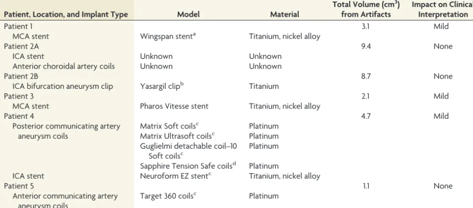
In patients in the CVR protocol, 26 of 80 (32.5%) had surgical hardware: intracranial implants (n⫽5), extracranial hardware (n⫽19), and ventriculoperitoneal (VP) shunts (n⫽2). Intracra-nial implant characteristics are summarized inTable 1. Implanted extracranial hardware (On-line Table 1) primarily included clo-sure hardware composed of titanium alloy and 1 patient with a polyethylene implant. Six of 19 patients had bilateral closure hardware with artifact sizes calculated separately by hemisphere, for a total of 25 hemispheres. Implanted VP shunts are described in On-line Table 2.Artifacts from surgically implanted hardware are shown inTable 2.
[image:3.594.54.535.59.271.2]In patients in the functional localizer protocol, 14 of 263 (5.3%) had surgical hardware: extracranial hardware (n⫽13) and VP shunt (n⫽1). Extracranial hardware comprised surgical closure hardware, and implant records were not available for these patients. The implanted VP shunt is included in On-line Table 2.
Mean artifact volume associated with intracranial hardware in patients undergoing the CVR protocol was 4.3⫾3.2 cm3; range⫽
1.1–9.4 cm3. As a point of reference, the average volume of the
adult human brain is approximately 1380 cm3.11The small
arti-fact volume associated with implanted intracranial hardware did not compromise the functional interpretation of cerebral hemo-dynamic data, with intracranial implants considered to have no (n⫽3) or mild (n⫽3) effect on the clinical interpretability of the examination in the affected hemisphere.Figure 2shows a repre-sentative image maximally affected by implanted intracranial hardware and hemodynamic data from a patient with a left MCA stent with in-stent restenosis, demonstrating the impact of the artifacts on the reactivity map.
Mean artifact volume of extracranial hardware in patients in the CVR protocol was 28.4⫾14.0 cm3; range⫽6.1– 61.7 cm3;
and in patients in the functional localizer protocol, it was 39.9⫾ 27.0 cm3(range⫽6.9 –77.1 cm3). Artifact volume associated with
titanium closure hardware is typically small and peripheral, with 19 of the evaluated hemispheres (76%) considered to have mild artifacts, which did not compromise clinical interpretation of ce-rebral hemodynamics (Fig 3). Closure hardware artifacts in 5 of the evaluated hemispheres (20%) had a moderate effect on the clinical interpretation. However, in the patient (patient 10) with the largest number of maxDrive screws (KLS-Martin, Jacksonville, Florida),12artifact size (61.7 cm3) was rated as severe and precluded
ipsilateral CVR interpretation in the region of the indirect revascu-larization that underlay the closure hardware (Fig 4). These artifacts were larger than those in other patients with fewer KLS-Martin LP maxDrive screws (patient 20: 5 screws, artifact volume⫽17.1 cm3;
Table 1: Intracranial implants
Patient, Location, and Implant Type Model Material
Total Volume (cm3) from Artifacts
Impact on Clinical Interpretation
Patient 1 3.1 Mild
MCA stent Wingspan stenta Titanium, nickel alloy
Patient 2A 9.4 None
ICA stent Unknown Unknown
Anterior choroidal artery coils Unknown Unknown
Patient 2B 8.7 None
ICA bifurcation aneurysm clip Yasargil clipb Titanium
Patient 3 2.1 Mild
MCA stent Pharos Vitesse stent Titanium, nickel alloy
Patient 4 4.7 Mild
Posterior communicating artery aneurysm coils
Matrix Soft coilsc Platinum Matrix Ultrasoft coilsc Platinum Guglielmi detachable coil–10
Soft coilsc
Platinum
Sapphire Tension Safe coilsd Platinum
ICA stent Neuroform EZ stentc Titanium, nickel alloy
Patient 5 1.1 None
Anterior communicating artery aneurysm coils
Target 360 coilsc Platinum
a
Manufactured by Stryker, Kalamazoo, Michigan for Boston Scientific, Natick, Massachusetts.
b
Aesculap, Center Valley, Pennsylvania.
c
Stryker, Kalamazoo, Michigan.
d
[image:3.594.55.533.328.393.2]Micro Therapeutics, Irvine, California.
Table 2: Artifacts from surgically implanted hardware
Hardware Type
Average Artifact
Volume (cm3) SD Range (cm3)
Intracranial hardware 4.3 3.2 1.1–9.4
Extracranial hardware (CVR protocol) 28.4 14.0 6.1–61.7
Extracranial hardware (functional localizer protocol) 39.9 27.0 6.9–77.1
patient 21: 8 screws, artifact volume⫽24.6 cm3). The number of
screws did not correlate directly with artifact volume across screw types, with up to 19 LP Cross-Drive screws (KLS-Martin) in 1 patient causing fewer artifacts (23.5 cm3) than the 12 KLS-Martin maxDrive
screws (61.7 cm3).
The average artifacts associated with VP shunts were 95.7⫾ 39.3 cm3, range⫽64.0 –139.6 cm3, and were rated as severe in 2 of
3 patients (Fig 5, CVR protocol) and moderate in the remaining patient (functional localizer protocol).
DISCUSSION
Specificity of Different Implants on BOLD fMRI Artifact Volume
Dephasing artifacts on MR imaging associated with implanted devices are caused by perturbations in the magnetic field due to differences in magnetic susceptibility or the degree of magnetiza-tion of an object in the presence of a magnetic field between the implanted device and surrounding tissue. Larger differences in
FIG 2. A representative patient (patient 3) with an intracranial im-plant. Signal drop-out from a left MCA Pharos Vitesse stent (Codman Neurovascular) is apparent on the magnitude BOLD fMRI image (A, white arrow), resulting in a total artifact volume of 2.1 cm3, which only mildly affected clinical interpretation of the examination. The patient was evaluated 2 years following implantation of the Pharos Vitesse stent in a stenosed left MCA. DSA (B) shows in-stent restenosis (black arrow), with corresponding decreased cerebrovascular reactivity (normalized CVR: voxel CVR normalized to cerebellar CVR) in the left MCA territory (C). In contrast, there is relative symmetry of the tem-poral signal-to-noise ratio (tSNR) map (D), suggesting that the asym-metric hemodynamic findings are not attributable to artifacts.
FIG 3. A patient with Moyamoya disease and asymmetric right-sided intracranial stenosis who underwent right encephaloduroanrterio-synangiosis. Comparison of pre- (Pre-Op) and postoperative (Post-Op) magnitude BOLD images (A) shows minimal artifacts (26.5 cm3) associated with the closure hardware (LP Plate 0.6⫻15 mm, Low Profile Micro Plate, and Cross-Drive 1.5⫻4 mm screws; KLS-Martin) at the corresponding surgical site, which was determined to only mildly impact clinical interpretation of the study. Both pre- and postopera-tive temporal signal-to-noise ratio (tSNR) maps (B) demonstrate rela-tively symmetric tSNR, while pre- and postoperative CVR maps (C) show decreased CVR in the right hemisphere along the right frontal cortex, which improves following revascularization.
FIG 4. Pre- (Pre-Op) and postoperative (Post-Op) magnitude and he-modynamic images in a patient with left-sided idiopathic Moyamoya disease who underwent a left encephaloduroarteriosynangiosis with implantation of closure hardware. Magnitude images (A) demonstrate the large artifacts (61.7 cm3) associated with surgical closure hardware, attributable to the large number of KLS-Martin maxDrive screws. Ar-tifacts were thought to severely impact clinical interpretation of the study, and hemodynamic evaluation with temporal signal-to-noise ratio maps (B) and reactivity maps (C) shows that interpretable hemo-dynamic data were not obtainable near the closure hardware. tSNR indicates temporal signal-to-noise ratio.
[image:4.594.54.285.46.265.2] [image:4.594.300.533.48.163.2] [image:4.594.300.535.282.547.2] [image:4.594.53.287.395.514.2]magnetic susceptibility between an implanted device and sur-rounding tissue lead to larger perturbations of the magnetic field and greater spectral dispersion of spins within a voxel, resulting in more extensive dephasing artifacts. In BOLD fMRI, long-TE, T2*-weighted imaging is used to sensitize the sequence to susceptibil-ity differences between oxygenated and deoxygenated blood, but these imaging parameters also render the sequence exquisitely sensitive to susceptibility variations from magnetic field inhomo-geneities, exacerbating artifact extent.6-8The effect of increased
dephasing artifacts associated with implants on the quality of data obtained specifically from BOLD MR imaging has not been pre-viously well-documented, to our knowledge. Our results demon-strate that BOLD MR imaging is interpretable in the presence of most implants evaluated, except for VP shunts, particularly those with programmable valves and siphon gauges and 1 patient with multiple KLS-Martin maxDrive screws.
The intracranial implants and extracranial hardware analyzed are composed of materials with low magnetic susceptibilities, such as titanium, nickel, platinum, or their alloys. All intracranial implanted devices produced small artifacts that had no-to-mild effects on clinical interpretability, and closure hardware typically resulted in small, peripheral artifacts that did not preclude inter-pretation. The single notable exception was a patient with a large number of KLS-Martin maxDrive screws, which were associated with more artifacts than the KLS-Martin Cross-Drive screws. While both the maxDrive and Cross-Drive screws are made of a titanium and nickel alloy, the maxDrive screw has a larger head profile. The number of screws did not correlate directly with ar-tifacts across screw types.
In contrast, VP shunts resulted in large artifacts that produced a moderate-to-severe impact on clinical interpretation, an effect attributable to the shunt valve composition. The valve portion of the VP shunt is composed of 316L stainless steel in addition to unalloyed titanium and tantalum in some models.12,13Stainless
steel contains iron, a ferromagnetic substance. While 316L stain-less steel contains a larger amount of nickel, which stabilizes iron in its nonmagnetic state and reduces its magnetic susceptibility, its magnetic susceptibility remains larger relative to titanium, re-sulting in larger perturbations of the magnetic field and more extensive artifacts.14,15 In addition, the Certas programmable
valve (Codman & Shurtleff, Raynham, Massachusetts) contains neodymium magnets, which caused extensive signal loss13,16,17
and likely contributed to the large artifact volume (139.6 cm3)
relative to other VP shunts evaluated (64.0 and 83.5 cm3).
Studies have described the effect of previously implanted sur-gical hardware on BOLD fMRI in the preoperative evaluation of brain tumor resection, demonstrating that language lateralization and primary motor cortex activation can be ascertained despite a reduction in total volume activation due to susceptibility arti-facts.18,19Our study confirms the feasibility of obtaining data
de-spite implants, which is particularly important in patients with cerebrovascular disease because longitudinal monitoring of pa-tients before and after revascularization may be desired.
Imaging Parameters and Artifact Volume
Artifact volume will vary with imaging readout and scan param-eters.20 We have used scan parameters that are common for
BOLD fMRI on all major vendor platforms, including single-shot gradient-echo EPI, voxel volume⫽50 – 60 mm3, and TE centered
on the approximate 3T tissue T2* (eg, 35 ms).21,22We also used
image-based parallel imaging (sensitivity encoding) with an ac-celeration factor of 1.8 –2.0, which reduces the EPI readout train by an approximate factor of 2. Geometric distortions from off-resonant spins and signal drop-out are largely affected by suscep-tibility gradients, and parallel imaging can reduce these issues by increasing the per-voxel bandwidth in the phase-encoding direc-tion,23thereby resulting in a shorter EPI readout (eg,
approxi-mately 36 ms without parallel imaging versus 18 ms with parallel imaging by using scan parameters in our sequences). While we used sensitivity encoding in our protocol, which is common on Philips scanners,k-space parallel imaging by using Generalized Autocalibrating Partial Parallel Acquisition or Autocalibrating Reconstruction for Cartesian sampling is more common on Sie-mens and GE Healthcare scanners, respectively, and performs comparably for reducing geometric distortions when similar ac-celeration factors are used. In the absence of parallel imaging, partialk-space acquisitions may also be used to reduce geometric distortions.
Limitations
First, information regarding implant manufacturer and type is not known for all implants considered, precluding a rigorous analysis in all patients. However, the composition of implants evaluated is similar to that in other commonly deployed implants; thus, results may generalize. Of the patients with intracranial im-plants analyzed, 1 (patient 2A) had incomplete information avail-able regarding embolization coils and an ipsilateral stent, though this patient had an artifact size (9.4 cm3) similar to that in another
patient (patient 4A) with known platinum embolization coils and an ipsilateral titanium and nickel alloy stent (4.7 cm3). Indeed,
most embolization coils are composed of platinum, with only the earliest models containing stainless steel,24,25and most
intracra-nial stents are composed of titanium, titanium alloys, or stainless steel.24,26,27Thus, our finding that the small artifacts produced by
intracranial implants do not impact clinical interpretability of BOLD fMRI is likely generalizable, with the exception of those models of intracranial implants that contain stainless steel, which were not evaluated in our study population and may produce larger artifacts due to higher magnetic susceptibility.
Information regarding specific implant manufacturers and composition was not available for a subset of patients with ex-tracranial implants (CVR protocol,n⫽2; functional localizer protocol,n⫽13). However, those patients with unknown closure hardware had artifact extent and clinical interpretability similar to those in patients with known extracranial hardware, which pri-marily included closure hardware composed of titanium alloy. Most implanted neurosurgical hardware is composed of titanium or titanium alloys similar to those evaluated in our study,24,25
suggesting that results are likely generalizable. We did not evalu-ate any issues relevalu-ated to the safety of metallic devices; however, this topic has been studied extensively, and all patients were screened by using standard MR imaging safety screening procedures.9
fMRI application, additional methods such as arterial spin-label-ing are bespin-label-ing increasspin-label-ingly used.28We did not specifically evaluate
the impact of artifacts on arterial spin-labeling image quality; however, arterial spin-labeling generally uses readouts similar to those in BOLD (eg, single-shot EPI with comparable spatial reso-lution), however with a shorter TE (eg, 10 –20 ms). Thus, the impact of image distortion on similar data should be reduced relative to BOLD, and findings from this study could provide a conservative reference for guiding similar studies.
Third, this study focused on patients undergoing BOLD fMRI for clinical purposes, primarily those with cerebrovascular dis-ease, epilepsy, and brain tumors. Thus, implants considered were specific to these populations, and a different population may have a higher fraction of implants not considered in this study. There-fore, while we hope that the findings of this study are useful for guiding imaging decisions in some of the most common types of patients undergoing BOLD fMRI, future studies incorporating a broader range of patients and implant types would be useful.
CONCLUSIONS
Three-Tesla single-shot gradient-echo EPI BOLD MR imaging performed on patients with a variety of implanted intracranial and extracranial hardware yields interpretable image quality in most patients beyond a small (30 – 40 cm3) volume surrounding
the hardware. Exceptions were VP shunts, particularly those with programmable valves and siphon gauges, and large numbers of KLS-Martin maxDrive screws.
Disclosures: Aditi A. Desai—RELATED:Grant: National Institutes of Health,* Com-ments: The study was funded by a National Institutes of Health grant. Megan K. Strother—RELATED:Grant: National Institutes of Health/National Institute of Neu-rological Disorders and Stroke R01 NS078828 – 01A1.* Carlos C. Faraco—RELATED:
Grant: National Institutes of Health,*Comments: T-32 Postdoctoral Fellow Training Grant. Lori C. Jordan—UNRELATED:Grants/Grants Pending: American Heart Asso-ciation Collaborative Science Award,*Comments: to study novel MRI techniques in sickle cell disease. Manus J. Donahue—RELATED:Grant: National Institutes of Health grants (National Institutes of Health/National Institute of Neurological Disorders and Stroke and National Institutes of Health/National Institute of Nursing Re-search).* *Money paid to the institution.
REFERENCES
1. Håberg A, Kvistad KA, Unsgård G, et al.Preoperative blood oxygen level-dependent functional magnetic resonance imaging in pa-tients with primary brain tumors: clinical application and out-come.Neurosurgery2004;54:902–14; discussion 914 –15
2. Binder JR.Functional MRI is a valid noninvasive alternative to Wada testing.Epilepsy Behav2011;20:214 –22
3. Mandell DM, Han JS, Poublanc J, et al.Mapping cerebrovascular reactivity using blood oxygen level-dependent MRI in patients with arterial steno-occlusive disease: comparison with arterial spin la-beling MRI.Stroke2008;39:2021–28
4. Donahue MJ, Dethrage LM, Faraco CC, et al.Routine clinical evalu-ation of cerebrovascular reserve capacity using carbogen in pa-tients with intracranial stenosis.Stroke2014;45:2335– 41
5. Shellock FG, Woods TO, Crues JV 3rd.MR labeling information for implants and devices: explanation of terminology.Radiology2009; 253:26 –30
6. Schenck JF.The role of magnetic susceptibility in magnetic reso-nance imaging: MRI magnetic compatibility of the first and second kinds.Med Phys1996;23:815–50
7. Port JD, Pomper MG.Quantification and minimization of
mag-netic susceptibility artifacts on GRE images.J Comput Assist Tomogr
2000;24:958 – 64
8. Koch KM, Hargreaves BA, Pauly KB, et al.Magnetic resonance im-aging near metal implants.J Magn Reson Imaging2010;32:773– 87 9. Kanal E, Barkovich AJ, Bell C, et al; Expert Panel on MR Safety.ACR
guidance document on MR safe practices: 2013.J Magn Reson Imag-ing2013;37:501–30
10. Jenkinson M, Beckmann CF, Behrens TE, et al.FSL.Neuroimage
2012;62:782–90
11. Filipek PA, Richelme C, Kennedy DN, et al.The young adult human brain: an MRI-based morphometric analysis.Cereb Cortex1994;4: 344 – 60
12. Shellock FG, Bedwinek A, Oliver-Allen M, et al.Assessment of MRI issues for a 3-T “immune” programmable CSF shunt valve.AJR Am J Roentgenol2011;197:202– 07
13. Shellock FG, Wilson SF, Mauge CP.Magnetically programmable shunt valve: MRI at 3-Tesla.Magn Reson Imaging2007;25:1116 –21 14. Vaccaro AR, Chesnut RM, Scuderi G, et al.Metallic spinal artifacts in
magnetic resonance imaging. Spine (Phila Pa 1976) 1994;19: 1237– 42
15. Khursheed F, Rohlffs F, Suzuki S, et al.Artifact quantification and tractography from 3T MRI after placement of aneurysm clips in subarachnoid hemorrhage patients.BMC Med Imaging2011;11:19 16. Lavinio A, Harding S, Van Der Boogaard F, et al.Magnetic field
interactions in adjustable hydrocephalus shunts.J Neurosurg Pedi-atr2008;2:222–28
17. Toma AK, Tarnaris A, Grieve JP, et al.Adjustable shunt valve-in-duced magnetic resonance imaging artifact: a comparative study.
J Neurosurg2010;113:74 –78
18. Kim MJ, Holodny AI, Hou BL, et al.The effect of prior surgery on blood oxygen level-dependent functional MR imaging in the preop-erative assessment of brain tumors. AJNR Am J Neuroradiol
2005;26:1980 – 85
19. Peck KK, Bradbury M, Petrovich N, et al.Presurgical evaluation of language using functional magnetic resonance imaging in brain tu-mor patients with previous surgery.Neurosurgery2009;64:644 –52; discussion 652–53
20. Hargreaves BA, Worters PW, Pauly KB, et al.Metal-induced artifacts in MRI.AJR Am J Roentgenol2011;197:547–55
21. Donahue MJ, Hoogduin H, van Zijl PC, et al.Blood oxygenation level-dependent (BOLD) total and extravascular signal changes and A¨ R2* in human visual cortex at 1.5, 3.0 and 7.0 T.NMR Biomed
2011;24:25–34
22. Rane S, Mason E, Hussey E, et al.The effect of echo time and post-processing procedure on blood oxygenation level-dependent (BOLD) functional connectivity analysis. Neuroimage 2014;95: 39 – 47
23. Schmiedeskamp H, Newbould RD, Pisani LJ, et al.Improvements in parallel imaging accelerated functional MRI using multiecho echo-planar imaging.Magn Reson Med2010;63:959 – 69
24. Shellock FG.Reference Manual for Magnetic Resonance Safety, Im-plants, and Devices: 2014 Edition.Los Angeles: Biomedical Research Publishing Group; 2014
25. Shellock FG.Metallic neurosurgical implants: evaluation of mag-netic field interactions, heating, and artifacts at 1.5-Tesla.J Magn Reson Imaging2001;14:295–99
26. Lo¨vblad KO, Yilmaz H, Chouiter A, et al.Intracranial aneurysm stenting: follow-up with MR angiography.J Magn Reson Imaging
2006;24:418 –22
27. Choi JW, Roh HG, Moon WJ, et al.Optimization of MR parameters of 3D TOF-MRA for various intracranial stents at 3.0T MRI. Neu-rointervention2011;6:71–77