Diffusion Abnormality of Deep Gray Matter in
External Capsular Hemorrhage
Won-Jin Moon, Dong Gyu Na, Sam Soo Kim, Jae Wook Ryoo, and Eun Chul Chung
BACKGROUND AND PURPOSE:To our knowledge, diffusion abnormality of the unaffected deep gray matter during striatocapsular hemorrhage has not been previously described in the literature. We report the presence of the diffusion abnormality separated from hematoma in patients with external capsular (lateral striatocapsular) hemorrhage and suggest the plausible mechanisms of diffusion signal intensity change.
METHODS:We retrospectively reviewed MR images in 28 consecutive patients with sponta-neous striatocapsular hemorrhage and evaluated signal intensity changes at sites separated from the hemorrhage and the lesions on diffusion-weighted (DW) images. Apparent diffusion coefficients (ADCs) of the lesions were measured, and volume changes in the deep gray matter were assessed at follow-up.
RESULTS: On DW images, hyperintensity of deep gray matter was found in nine patients (25%). In all patients with DW imaging abnormality, the hemorrhage was located in the external capsule, and the interval from hemorrhagic ictus to MR imaging study was 8 –54 days. Hyperintensity of the deep gray matter was seen in the caudate (n ⴝ 8), putamen (n ⴝ 7), thalamus (n ⴝ 5), and substantia nigra (n ⴝ 2). Mean relative ADC ratios of the diffusion abnormality were 0.76ⴞ 0.10 in the caudate, 0.79ⴞ 0.07 in the putamen, and 0.85ⴞ0.11 in the thalamus. DW imaging abnormality disappeared with mild atrophy in two patients who underwent follow-up imaging.
CONCLUSION: External capsular hemorrhage may be uncommonly accompanied by diffu-sion abnormality in the striatum or thalamus at follow-up, and the lediffu-sion should not be misdiagnosed as new-onset infarction. Secondary neuronal degeneration may play an important role in the development of diffusion abnormality.
Spontaneous intracerebral hemorrhage (ICH) ac-counts for 10% of all strokes and is associated with mortality and morbidity rates higher than those of cerebral infarction (1). Spontaneous ICH can be sified into two types: deep hemorrhage, which is clas-sically associated with hypertension, and lobar hem-orrhage, which may result from cerebral amyloid angiopathy (1). The hypertensive hemorrhage of the striatocapsule is most frequently encountered in daily practice (2).
Although nonenhanced CT has been the diagnostic technique of choice for ICH, the use of MR imaging has recently expanded. The different stages of ICH
can be easily determined by analyzing the appearance its various patterns on MR images. With the advent of diffusion-weighted (DW) imaging, valuable addi-tional information about ICH, especially features to differentiate hemorrhage from infarction in hyper-acute stroke, can be obtained (3– 6).
In addition to the study of the hematoma itself, the accompanying abnormality of ICH on DW imaging has been investigated (7–11). Wallerian degeneration of the corticospinal tract after ICH can be depicted earlier on DW imaging than on conventional MR imaging (9, 10). Several reports suggest that perihe-matoma ischemia or periheperihe-matoma edema can be identified on DW imaging (7, 8, 11).
We observed diffusion abnormalities in the caudate nucleus and putamen of patients with external capsu-lar (lateral striatocapsucapsu-lar) hemorrhage; these abnor-malities were separated from the bleeding site and the rim of perihematoma edema. To our knowledge, only one report has described this diffusion abnor-mality separated from the hematoma (12). We per-formed this retrospective study to determine the pres-ence of the diffusion abnormality separated from
Received December 31, 2003; accepted after revision June 3, 2004.
From the Department of Radiology, Kangbuk Samsung Hospital (W.-J.M., E.C.C.) and Samsung Medical Center (S.S.K., J.W.R.), Sungkyunkwan University School of Medicine, Seoul National Uni-versity Hospital (D.G.N.), Seoul, Korea.
Address reprint requests to Dong Gyu Na, MD, Department of Radiology, Seoul National University Hospital, 28 Yongon-Dong, Chongno-gu, Seoul 110-744, Korea, email: [email protected].
©American Society of Neuroradiology
hematoma in patients with striatocapsular hemor-rhage and to suggest the plausible mechanisms of signal intensity change on DW imaging.
Methods
Patients
We collected clinical and imaging data in 36 consecutive patients who had spontaneous hemorrhage in the basal ganglia (striatocapsule) and who were referred for MR imaging from our departments of neurology and neurosurgery. The patients’ records were retrieved from the electronic database of our medical center between 1999 and 2001. Patients with arterio-venous malformation, aneurysm, or tumor as the underlying cause of hemorrhage were excluded. Of the 36 patients with spontaneous striatocapsular hemorrhage, 28 patients (14 women and 14 men aged 17–74 years; median age, 57 years) were included, whereas eight patients were excluded because of a lack of diffusion imaging. The presence of intracerebral hematoma was determined by means of CT in 26 patients and by means of gradient-echo MR imaging in two patients. Sur-gery for stereotaxic aspiration was performed in six patients, and open surgery was performed in four patients.
MR imaging studies were performed at various intervals after hemorrhagic ictus. Five ICH stages were defined accord-ing to the interval between hemorrhagic ictus and initial MR imaging examination, as follows: Acute was 0 –3 days (n⫽4), early subacute was 4 –7 days (n⫽5), late subacute was 8 –30 days (n⫽11), early chronic was 31– 60 days (n⫽2), and late chronic was 61 days or longer (n⫽6). Follow-up MR imaging was performed in seven patients, and follow-up CT was per-formed in two, with intervals varying from 2 to 25 months (median, 8 months). The patients’ medical records were re-viewed to evaluate their neurologic status at the time of MR imaging.
MR Imaging
A 1.5-T unit (Signa; GE Medical Systems, Milwaukee, WI) was used for MR imaging. The conventional MR imaging protocol included the following sequences: 1) axial T1-weighted spin-echo (TR/TE, 467/9), 2) axial T2-T1-weighted fast spin-echo (TR/effective TE, 3417/102), and 3) axial fluid-atten-uated inversion recovery (FLAIR; TR/TE/TI, 10,000/140/ 2200). Parameters for conventional MR imaging were a matrix of 256 ⫻ 192, a 23-cm field of view, and a 5-mm section thickness and 2-mm intersection gap. Single-shot spin-echo echo-planar DW imaging sequences were obtained by applying diffusion gradients in three orthogonal directions for each section, with two diffusion weightings (b⫽0 s/mm2andb⫽
900 or 1000 s/mm2). Isotropic DW imaging was generated
online by averaging three orthogonal-axis images. During DW imaging 20 sections were acquired with these parameters: TR/TE of 6500/96.8, matrix of 128⫻128, field of view of 28 cm, and section thickness of 5 mm, and intersection gap of 2 mm. In addition, conventional T2*-weighted gradient-echo imaging (TR/TE, 450/20) was performed in 22 of the 28 patients.
Imaging Analysis
Two neuroradiologists (W.-J.M., D.G.N.) retrospectively re-viewed the presence and location of the diffusion abnormality in the deep gray matter in each patient. By consensus, we determined the presence of DW imaging abnormality when high signal intensity was present in the deep gray matter on DW imaging. The DW imaging abnormality did not overlap with the hematoma or perihematoma edema and did not sur-round the hematoma. Signal intensity changes on conventional MR images were also evaluated and compared with those seen on the DW images. The location of the ICH was categorized
into six types based on vascular territory, as Chung et al sug-gested (2): 1) anterior (Heubner artery), 2) middle (medial lenticulostriate artery), 3) posteromedial (posteromedial branches of the lateral lenticulostriate artery), 4) posterolateral (posterolateral branches of the lateral lenticulostriate artery), 5) lateral (most lateral branches of the lateral lenticulostriate artery), and 6) massive. Figure 1 shows a schematic diagram based on the classification of striatocapsular hemorrhage (2). The diagram shows that lateral type of striatocapsular hemor-rhage primarily involved the external capsular area between lateral putamen and insular cortex. Therefore, we designated the lateral type of striatocapsular hemorrhage as external cap-sular hemorrhage.
Quantitative analysis of apparent diffusion coefficients (ADCs) and signal intensity on DW imaging was performed in hyperintense lesions in the deep gray nuclei that were sepa-rated from the hematoma. A region of interest (ROI) was placed in the caudate, putamen, and thalamus bilaterally by carefully excluding perihematoma edema and the hematoma itself. The ROI ranged from 16 to 302 mm2, and in each case,
a neuroradiologist (W.-J.M.) measured the ROI. ADC values were calculated according to the following equation: ADC⫽ (lnS2⫺lnS1)/b, whereS1is ROI signal intensity with diffusion gradients,S2is the ROI signal intensity without diffusion
gra-dients, andbis the difference inbvalue between images with (b ⫽900 or 1000 s/mm2) and images without (b⫽0 s/mm2)
diffusion-sensitizing gradients. The relative ADC ratio (the ratio of the ADC value for a lesion to that of normal contralat-eral gray matter) was also calculated in each case. All data for ADC values and relative ADCs for each stage are presented as the mean⫾1 SD.
Volume measurements of the hematoma were obtained with initial CT performed within a day of symptom onset. Hema-toma volumes were measured by using a simplified formula for the volume of ellipsoid:ABC
2 , whereAis the largest diameter of
the hemorrhage on the section on which the largest area of hemorrhage was identified,Bis the largest diameter oriented 90° toAon the same section, andCis the approximate number of sections on which the ICH was seen. Volume analysis was not performed in two patients whose initial CT images were not available; the hemorrhage, however, was confirmed on gradi-ent-echo MR images.
To determine whether the deep gray matter involved on DW imaging had atrophied, the volume of the caudate and the thalamus was obtained. The putamen was excluded from the volume analysis because its size could not be measured without inadvertent inclusion of the adjacent hematoma and edema. To evaluate the chronologic changes in size, a difference of up to
⫾5% was allowed when the size of deep gray matter was compared on initial and follow-up images. This consideration
accounted for calculation errors and slight differences in the position of brain sections at different time points.
Statistical Analysis
SPSS (version 10.1; SPSS Inc, Chicago, IL) was used for statistical analysis. The Mann-Whitney or Kruskal-Wallis test was used to determine differences in ADC values, ADC ratios, and initial ICH volumes between the abnormal and normal DW imaging groups. Clinical and radiologic findings were analyzed with the Fisher exact test. The level of significance was defined atP⬍.05.
Results
Clinicoradiologic Characteristics of the Abnormal DW Imaging Group
Table 1 summarizes the clinicoradiologic data of the patients whose images showed diffusion abnor-mality in the deep gray matter. Nine of the 28 patients with striatocapsular hemorrhage had DW imaging abnormalities in the deep gray matter, all in associa-tion with the lateral type (external capsular hema-toma). At the time of the MR imaging study, no patient presented with acute neurologic deficits sug-gestive of any acute infarction.
In the abnormal DW imaging group (n ⫽ 9), six patients (67%) underwent surgery, whereas only three patients (16%) in the normal DW imaging group (n⫽19) had surgery. DW imaging abnormal-ities were found only in patients with late subacute hematoma (n⫽7) or early chronic hematoma (n⫽2) (Table 2). Unlike the lateral type of striatocapsular hematoma, the posterolateral or anterior type of stri-atocapsular hematoma, which primarily involved lat-eral basal ganglia, did not result in diffusion abnor-mality (Table 3). The age and location of the hemorrhages significantly differed between the ab-normal and ab-normal DW imaging groups (P⬍.05). In the abnormal DW imaging group, the caudate head (n ⫽ 8) was the area most commonly affected, fol-lowed by the putamen (n⫽7), the thalamus (n⫽6), and the substantia nigra (n ⫽ 2) (Fig 2). FLAIR imaging revealed subtle high signal intensity in the caudate nucleus (n ⫽ 8), putamen (n ⫽ 7), and thalamus (n ⫽6).
ADCs and Chronologic Changes in DW Imaging Hyperintensity
When DW imaging showed abnormally high signal intensity in any region of the caudate head, putamen, or thalamus, the ADC value and the ADC ratio rel-ative to the contralateral gray matter decreased sig-nificantly (P ⬍ .05) (Table 4). Two of nine patients with DW imaging abnormalities underwent follow-up DW imaging (8 and 10 months after symptom onset) (Table 4). Follow-up images in both showed complete reversal of the DW imaging hyperintensity (Fig 3). ADC values and ADC ratios normalized on follow-up.
Comparison of Initial Hematoma Volume and Atrophy of Ipsilateral Gray Matter on Follow-Up
The abnormal DW imaging group (n ⫽ 9) had a volume of initial ICH significantly larger than that of the normal DW imaging group (n ⫽ 17) (33.24 ⫾ 11.83 vs. 13.24 ⫾ 11.20 103 mm3; P ⫽ .001). To evaluate whether the hematoma volume indepen-dently affected the presence of diffusion abnormality, we compared the volumes of hematoma with differ-ent locations and then compared the hematoma vol-umes between patients with lateral striatocapsular hemorrhage with and without diffusion abnormality. In all patients with and those without diffusion ab-normality, the lateral type of striatocapsular hemor-rhage result in a significantly larger volume of hema-toma (P⫽.001). In all patients with the lateral type of striatocapsular hemorrhage, hematoma volume was significantly larger in patients with diffusion
ab-TABLE 1: Clinicoradiologic data in patients with diffusion abnormality remote from ICH
Patient/Sex/Age (y)
Time after
ICH (days) Location of ICH Treatment
Hyperintensity
DWI FLAIR Imaging
1/M/33 40 R EC Surgical C, P, T, SN C, P, T, SN
2/M/52 27 L EC Surgical C, P, T C, P, T
3/F/55 28 R EC Surgical C, T, SN C, T
4/M/51 24 R EC Conservative P P
5/F/51 8 R EC Surgical C, P C, P
6/F/78 54 L EC Conservative C, T C, T
7/M/70 17 L EC/T Surgical C, P C, P
8/M/66 30 L EC Surgical C, P, T C, P, T
9/M/74 16 R EC Conservative C, P, T C, P, T
[image:3.585.55.537.70.199.2]Note.—C is the caudate nucleus head; EC, external capsule; P, putamen; SN, substantia nigra; and T, thalamus.
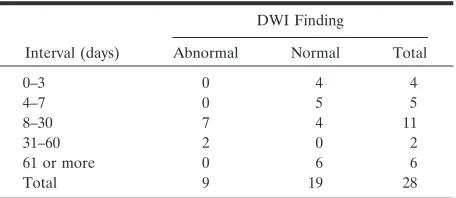
TABLE 2: Relationship between DWI abnormality and interval after hemorrhage onset
Interval (days)
DWI Finding
Abnormal Normal Total
0–3 0 4 4
4–7 0 5 5
8–30 7 4 11
31–60 2 0 2
61 or more 0 6 6
[image:3.585.307.535.255.354.2]normality than in patients with no diffusion abnor-mality (P⫽.040).
Follow-up MR images were obtained in two pa-tients in the abnormal DW imaging group and in two patients in the normal DW imaging group. In
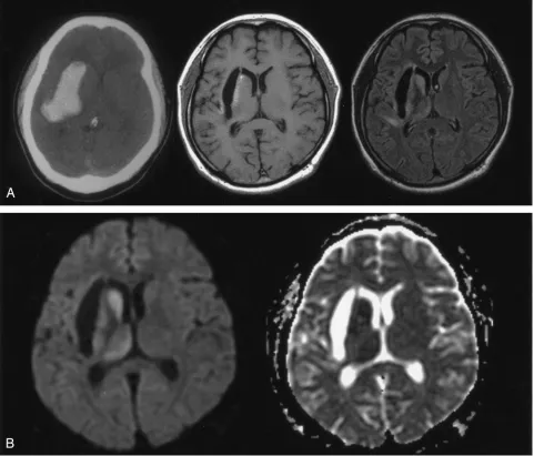
abnor-mal DW imaging group, a significant reduction in volume was noted in the caudate (19 –34%) and thal-FIG 2. Patient 1. A 33-year-old man with early chronic intracerebral hematoma on MR images obtained 40 days after symptom onset. A, CT scan (left) 12 days after symptom onset shows a hyperattenuating hematoma primarily in the right external capsule between the lateral putamen and the insular cortex. T1-weighted image (middle) shows a hypointense cavitary lesion in the right external capsule. FLAIR image (right) reveals subtle hyperintensity of the deep gray matter in the caudate head, putamen, thalamus, and part of the insular cortex.
[image:4.585.54.535.59.470.2]B, Diffusion-weighted image (left) shows obvious hyperintensity in the caudate head, putamen, and thalamus, which are separate from the hematoma itself. No other diffusion abnormality is seen. ADC map (right) shows decreased ADC in the caudate, putamen, and thalamus; ADC ratios relative to contralateral gray matter were 0.75, 0.79, and 0.80, respectively. This suggests that the diffusion abnormality is impaired intracellular diffusion rather than a T2 shine-through effect.
TABLE 4: Relationship between DWI abnormality and ADC change
Location and DWI Finding
ADC
(10⫺6mm/s) ADC Ratio PValue
Caudate .000
Abnormal (n⫽8) 582⫾135 0.76⫾0.10 Normal (n⫽19) 857⫾152 1.03⫾0.18
Putamen .000
Abnormal (n⫽8) 612⫾68 0.79⫾0.07 Normal (n⫽19) 821⫾151 1.04⫾0.13
Thalamus .028
[image:4.585.307.536.575.699.2]Abnormal (n⫽5) 650⫾68 0.85⫾0.11 Normal (n⫽22) 820⫾75 1.00⫾0.07
TABLE 3: Relationship between DWI abnormality and location of the hemorrhage
Location
DWI Finding
Abnormal Normal Total
Lateral 9 7 16
Posterolateral 0 10 10
Anterior 0 2 2
[image:4.585.56.283.586.664.2]FIG 3. Patient 8. A 66-year-old man with late subacute intracerebral hematoma on MR images obtained 30 days after symptom onset. A, T1-weighted image (top left) shows hyperintensity of the hematoma mainly in the left external capsule area. Hematoma also extends into a part of the posterior angle of the putamen and insular cortex. T2-weighted image (top right) shows the hematoma as a hyperintense area circumscribed by a hypointense rim; this suggests subacute hematoma. Subtle hyperintensity is noted in the ipsilateral caudate head and putamen. DW image (bottom left) shows obvious hyperintensity in the caudate and putamen and mild hyperintensity in the thalamus. ADC map (bottom right) shows decreased ADC in the caudate, part of the putamen and medial-posterior thalamus; ADC ratios relative to the contralateral gray matter were 0.59, 0.61, and 0.87, respectively.
amus (16 –18%). On the contrary, the normal DW imaging group showed minimal volume reduction (2– 4% in the caudate and 5–7% in the thalamus).
Discussion
In the present study, hyperintensity of the deep gray matter was found on DW imaging in nine (32%) of 28 patients with striatocapsular hemorrhage. These DW imaging abnormalities were found exclusively in the external capsular hemorrhage in the subacute stages. On follow-up, the deep gray matter with diffusion ab-normality showed significant atrophic change.
Interestingly, all patients with DW imaging abnor-malities had ICH in the external capsule between the insular cortex and the lateral margin of the putamen. Although the external capsule is not part of the basal ganglia, hemorrhage of the external capsule has been long regarded as the lateral type of striatocapsular hemorrhage (2). The hemorrhage mainly involving external capsule results in an ellipsoid mass of maxi-mal anteroposterior diameter along the white matter pathways. This type does not directly affect the puta-men but often compresses the middle portion of the putamen (2). Despite their relatively large hematoma, patients with the lateral type presented with a benign clinical course and were therefore treated medically (2). Why these diffusion abnormalities were seen ex-clusively in patients with the external capsular hem-orrhage is unclear.
Diffusion abnormality separated from ICH re-sulted in the atrophy of the affected area on follow-up imaging study. The resultant atrophy of the previous diffusion abnormality suggests structural change in the affected area. These characteristics of DW imag-ing abnormality in the deep gray matter in cases of striatocapsular hemorrhage suggest possible underly-ing mechanisms for the diffusion abnormality after hemorrhage, such as secondary transneuronal degen-eration (13, 14), ischemic change by direct compres-sion (15–17), and inflammatory reaction (18, 19).
Neuronal damage caused by a focal brain lesion influences function and morphology in regions intact after the primary injury (13, 14, 20 –23). The classic forms of neuronal degeneration are wallerian and transneuronal. Wallerian degeneration is afferent ax-onal degeneration occurring after injury to the axon or cell body; this type does not extend to other neu-rons. On the contrary, transneuronal degeneration involves both afferent and efferent neurons through synaptic connections (13, 14). Cerebral infarction in the territory of the middle cerebral artery can cause transneuronal degeneration in the substantia nigra and the thalamus, which have transsynaptic connec-tions with the caudoputamen and the cerebral cortex, respectively (24, 25–27). In this study, diffusion ab-normalities after external capsular hemorrhage were observed in the substantia nigra and thalamus, as well as in the striatum. We can speculate that a mechanism analogous to that of transneuronal degeneration may be behind the diffusion abnormality remote from ICH.
Major afferents to the striatum are derived from
the cerebral cortex, thalamus, and substantia nigra (28). Major efferents from the striatum terminate at the globus pallidus and substantia nigra. Although the structure of external capsule is not entirely known, it is thought to contain corticostriate fibers connecting the cerebral cortex and the striatum (28). Regarding the striatum, antegrade neuronal degeneration of the corticostriate fibers has not been reported, while ret-rograde degeneration is reported in the external cap-sule after the necrosis of the striatum (27). As both antegrade and retrograde transneuronal degenera-tion are possible after any neuronal injury, external capsular lesion may induce antegrade neuronal de-generation involving the striatum.
Unlike the caudate and the putamen, the thalamus and the substantia nigra do not contain a direct nection with the external capsule. The thalamus con-tains thalamostriate fibers to the striatum; these may be the target of retrograde neuronal degeneration due to striatal injury (14). The substantia nigra has both afferent and efferent connections to the stria-tum. Therefore, both antegrade and retrograde neu-ronal degeneration of the substantia nigra may be possible after striatal injury.
Although decreased ADC in secondary neuronal degeneration is unclear, axonal swelling (28) and cel-lular swelling of astrocytic endplate and neurons (24, 29) could be the major causes of impaired intracellu-lar diffusion resulting in decreased ADC.
Another possible reason for the diffusion abnor-mality may be inflammation induced by intracerebral hematoma (18, 19). ICH causes adjacent parenchy-mal inflammation within hours of its occurrence; this is initiated by the adherence by leukocytes to dam-aged brain endothelia and their subsequent entry in to the brain (18). Although levels of most inflamma-tory molecules are increased immediately after ICH, one report (19) describes an increase in some inflam-matory molecules at a week after ICH. This might explain the delayed diffusion abnormality after ICH. Nevertheless, the inflammation cannot explain the diffusion abnormality in the thalamus and midbrain, which are separated from the initial hematoma.
In cases of external capsular hemorrhage, diffusion abnormality in the ipsilateral deep gray matter may mimic an acute ischemic lesion due to another cause. No clinical evidence suggests any new acute ischemic insult, and the distribution of sites of diffusion abnor-mality indicates that decreased ADC in the deep gray matter is not caused by any incidental acute infarction not associated with ICH. Therefore, the DW imaging abnormality in this study was not due to acute infarc-tion, but rather, a delayed injury accompanying the hemorrhage.
Our study had some limitations. First, histopatho-logic correlation with diffusion abnormality was lack-ing. Animal experiments may be helpful in widening our knowledge on diffusion abnormality and its mech-anism after ICH. Second, this study was not prospec-tive but retrospecprospec-tive, and we obtained follow-up MR images in only a limited number of patients. Prospec-tive study with serial observation may be required to determine the clinical relevance of diffusion abnor-mality after external capsular ICH.
Conclusion
Diffusion abnormalities in the ipsilateral gray mat-ter afmat-ter exmat-ternal capsular ICH are uncommon and leave residual localized atrophy. Secondary neuronal degeneration may be suggested as a plausible mech-anism. Diffusion abnormality of the caudoputamen or thalamus should not be misdiagnosed as new-onset infarction on follow-up MR imaging in patients with external capsular hemorrhage.
References
1. Labovitz DL, Sacco RL.Intracerebral hemorrhage: update.Curr Opin Neurol2001;14:103–108
2. Chung CS, Caplan LR, Yamamoto Y, et al.Striatocapsular hem-orrhage.Brain2000;123:1850 –1862
3. Schellinger PD, Jansen O, Fiebach JB, Hacke W, Sartor K. A standardized MRI stroke protocol: comparison with CT in hyper-acute intracerebral hemorrhage.Stroke1999;30:765–768 4. Atlas SW, DuBois P, Singer MB, Lu D.Diffusion measurements in
intracranial hematomas: implications for MR imaging of acute stroke.AJNR Am J Neuroradiol2000;21:1190 –1194
5. Kang BK, Na DG, Ryoo JW, Byun HS, Roh HG, Pyeun YS.
Diffusion-weighted MR imaging of intracerebral hemorrhage. Ko-rean J Radiol2001;2:183–191
6. Wiesmann M, Mayer TE, Yousry I, Hamann GF, Bruckmann H.
Detection of hyperacute parenchymal hemorrhage of the brain
using echo-planar T2*-weighted and diffusion-weighted MRI.Eur Radiol2001;11:849 – 853
7. Carhuapoma JR, Wang PY, Beauchamp NJ, Keyl PM, Hanley DF, Barker PB.Diffusion-weighted MRI and proton MR spectroscopic imaging in the study of secondary neuronal injury after intracere-bral hemorrhage.Stroke2000;31:726 –732
8. Carhuapoma JR, Barker PB, Hanley DF, Wang P, Beauchamp NJ.
Human brain hemorrhage: quantification of perihematoma edema by use of diffusion-weighted MR imaging.AJNR Am J Neuroradiol 2002;23:1322–1326
9. Karibe H, Shimizu H, Tominaga T, Koshu K, Yoshimoto T. Diffu-sion-weighted magnetic resonance imaging in the early evaluation of corticospinal tract injury to predict functional motor outcome in patients with deep intracerebral hemorrhage. J Neurosurg 2000;92:58 – 63
10. Pierpaoli C, Barnett A, Pajevic S, et al.Water diffusion changes in Wallerian degeneration and their dependence on white matter architecture.Neuroimage2001;13:1174 –1185
11. Forbes KP, Pipe JG, Heiserman JE.Diffusion-weighted imaging provides support for secondary neuronal damage from intraparen-chymal hematoma.Neuroradiology2003;45:363–367
12. Kamal AK, Dyke JP, Katz JM, et al.Temporal evolution of diffu-sion after spontaneous supratentorial intracranial hemorrhage.
AJNR Am J Neuroradiol2003;24:895–901
13. Ogawa T, Okudera T, Inugami A, et al.Degeneration of the ipsi-lateral substantia nigra after striatal infarction: evaluation with MR imaging.Radiology1997;204:847– 851
14. Ogawa T, Yoshida Y, Okudera T, Noguchi K, Kado H, Uemura K.
Secondary thalamic degeneration after cerebral infarction in the middle cerebral artery distribution: evaluation with MR imaging.
Radiology1997;204:255–262
15. Qureshi AI, Hanel RA, Kirmani JF, Yahia AM, Hopkins LN.
Cerebral blood flow changes associated with intracerebral hemor-rhage.Neurosug Clin N Am2002;13:355–370
16. Nath FP, Jenkins A, Mendelow AD, Graham DI, Teasdale GM.
Early hemodynamic changes in experimental intracerebral hemor-rhage.J Neurosurg1986;65:697–703
17. Bullock R, Brock-Utne J, van Dellen J, Blake G.Intracerebral hemorrhage in a primate model: effect on regional cerebral blood flow.Surg Neurol1988;29:101–107
18. Del Bigio MR, Yan HJ, Buist R, et al.Experimental intracerebral hemorrhage in rats. Magnetic resonance imaging and histopatho-logic correlates.Stroke1996;27:2312–2319
19. Power C, Henry S, Del Bigio MR, et al.Intracerebral hemorrhage induces macrophage activation and matrix metalloproteinase.Ann Neurol2003;53:731–742
20. Nagasawa H, Kogure K.Exo-focal postischemic neuronal death in the rat brain.Brain Res1990;524:196 –202
21. Iizuka H, Sakatani K, Young W.Neural damage in the rat thala-mus after cortical infarcts.Stroke1990;21:790 –794
22. Fujie W, Kirino T, Tomukai N, Iwasawa T, Tamuwa A.Progressive shrinkage of the thalamus following middle cerebral artery occlu-sion in rats.Stroke1990;21:1485–1488
23. Nakane M, Tamura A, Nagaoka K, Hirakawa K.MR detection of secondary changes remote from ischemia: preliminary observa-tions after occlusion of the middle cerebral artery in rats.AJNR Am J Neuroradiol1997;18:945–950
24. Abe O, Nakane M, Aoki S, et al.MR imaging of postischemic neuronal death in the substantia nigra and thalamus following middle cerebral artery occlusion in rats. NMR Biomed 2003;16:152–159
25. Nakane M, Teraoka A, Asato R, Tamura A.Degeneration of the ipsilateral substantia nigra following cerebral infarction in the striatum.Stroke1992;23:328 –332
26. Forno LS.Reaction of the substantia nigra to massive basal gan-glia infarction.Acta Neuropathol (Berl)1983;62:96 –102
27. Herrero MT, Barcia C, Navarro JM.Functional anatomy of thal-amus and basal ganglia.Child Nerv Syst2002;18:386 – 404 28. Williams PL, Warwick R.Gray’s Anatomy. 36th ed. Edinburgh,
Churchill Livingstone; 1980:1002–1044
29. Zhao F, Kuroiwa T, Miyasaka N, et al.Characteristic changes in T(2)-value, apparent diffusion coefficient, and ultrastructure of substantia nigra evolving exofocal postischemic neuronal death in rats.Brain Res2001;895:238 –244
30. Mazumdar A, Mukherjee P, Miller JH, Malde H, McKinstry RC.